. 2007 Dec;14(12):1347-56.
doi: 10.1016/j.chembiol.2007.11.006.

A comprehensive profile of brain enzymes that hydrolyze the endocannabinoid 2-arachidonoylglycerol

Affiliations

A comprehensive profile of brain enzymes that hydrolyze the endocannabinoid 2-arachidonoylglycerol

Jacqueline L Blankman et al. Chem Biol. 2007 Dec.

Abstract

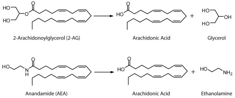
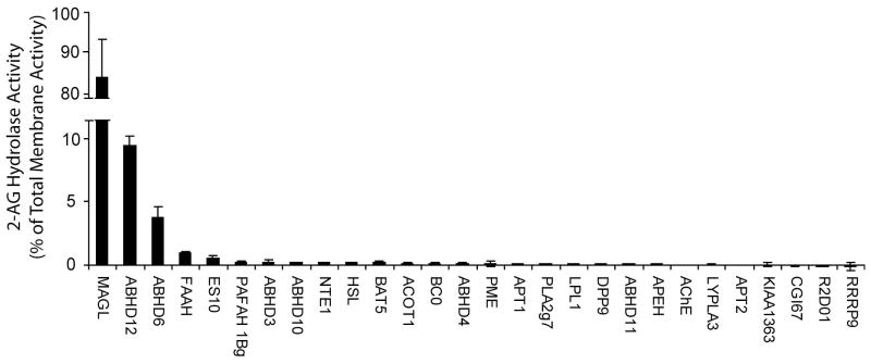
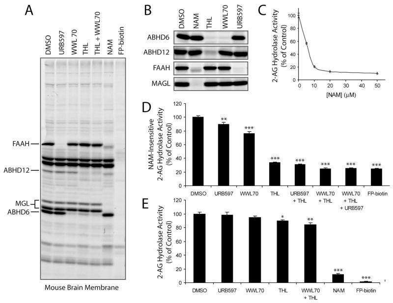
Endogenous ligands for cannabinoid receptors ("endocannabinoids") include the lipid transmitters anandamide and 2-arachidonoylglycerol (2-AG). Endocannabinoids modulate a diverse set of physiological processes and are tightly regulated by enzymatic biosynthesis and degradation. Termination of anandamide signaling by fatty acid amide hydrolase (FAAH) is well characterized, but less is known about the inactivation of 2-AG, which can be hydrolyzed by multiple enzymes in vitro, including FAAH and monoacylglycerol lipase (MAGL). Here, we have taken a functional proteomic approach to comprehensively map 2-AG hydrolases in the mouse brain. Our data reveal that approximately 85% of brain 2-AG hydrolase activity can be ascribed to MAGL, and that the remaining 15% is mostly catalyzed by two uncharacterized enzymes, ABHD6 and ABHD12. Interestingly, MAGL, ABHD6, and ABHD12 display distinct subcellular distributions, suggesting that they may control different pools of 2-AG in the nervous system.


Figures

Figure 1
Figure 1
Structures of 2-arachidonoylglycerol (2-AG), anandamide (AEA) and their hydrolysis products.
Figure 2
Figure 2
Mouse brain 2-AG hydrolase activity is completely inhibited by the activity-based proteomic probe FP-biotin. The 2-AG hydrolase activity of membrane and soluble mouse brain fractions was reduced by >98% following treatment with FP-biotin (5 μM, 1 hr). 2-AG hydrolase activity was measured under the following conditions: 50 mM Tris-HCl, pH 7.5, 0.05 μg protein/mL, 100 μM 2-AG, 200 μL reaction volume, 10 min, room temperature. Results represent the average values ± standard errors of the mean (SEM) for 3 independent experiments.
Figure 3
Figure 3
Recombinant expression of mouse brain serine hydrolases. COS-7 cells transiently transfected with mouse brain serine hydrolase cDNAs were labeled with FP-rhodamine (2 μM, 1 hr), separated by SDS-PAGE, and analyzed by in-gel fluorescence scanning to confirm expression of active enzymes. Expression efficiency of active enzyme was calculated from the integrated fluorescence intensities of the asterisked bands. Fluorescent gel show in grayscale.
Figure 4
Figure 4
The relative 2-AG hydrolase activities for mouse brain membrane serine hydrolases. SH-transfected cell homogenates were assayed for 2-AG hydrolase activity (50 mM Tris-HCl, pH 7.5, 0.05 μg protein/mL, 100 μM 2-AG, 10 min, room temperature), and these values were normalized to account for differences in enzyme expression in transfected cells. To determine the relative contribution of each enzyme to total brain membrane 2-AG hydrolase activity, the results were further normalized based on mouse brain expression levels for each serine hydrolase, as estimated by their average spectral count values from the ABPP-MudPIT analysis (Table 1) corrected for the number of theoretical tryptic peptides per enzyme. Results represent the average values ± SEM of 2 independent experiments for 2 separate transfections per enzyme (n = 4).
Figure 5
Figure 5
Effects of inhibitors of MAGL, ABHD12 and ABHD6 on brain membrane 2-AG hydrolase activity. (A) The selectivity profiles of NAM (50 μM), WWL70 (10 μM), THL (20 μM), URB597 (10 μM), and FP-biotin (5 μM) as judged by competitive ABPP analysis [1 hr pre-incubation with inhibitors, followed by 1 hr labeling with FP-rhodamine (2 μM)] in the mouse brain membrane proteome (A) and transfected COS-7 proteomes (B). Note that MAGL migrates as two distinct protein bands in the brain proteome, consistent with previous findings [23, 48]. (C) NAM treatment (0 – 50 μM, 1 hr) inhibited up to 85% of the 2-AG hydrolase activity of the mouse brain membrane proteome. (D) Effects of inhibitors of ABHD12, ABHD6, and FAAH [with THL (20 μM), WWL70 (10 μM), and URB597 (10 μM), respectively] on the “NAM-resistant” 2-AG hydrolase activity of the mouse brain membrane proteome. Assays were conducted in proteomic samples pre-treated with NAM (50 μM, 10 min) to block MAGL activity. (E) Effects of inhibitors on total brain membrane 2-AG hydrolase activity. For C-E, results represent the average ± SEM of 3–5 individual experiments. *, p < 0.05; **, p < 0.01; ***, p < 0.001 for inhibited versus control (DMSO) treated samples.
Figure 6
Figure 6
MAGL, ABHD12 and ABHD6 exhibit distinct subcellular distributions. (A) The distribution of enzymes in the membrane and soluble fractions of transfected COS-7 cells, as judged by ABPP analysis. PNGaseF treatment revealed that ABHD12, but not MAGL or ABHD6, is a glycoprotein, indicating a luminal/extracellular orientation for this enzyme. (B) Cartoon model representing the predicted orientations of the principal 2-AG hydrolases in mouse brain.

Comment in

Similar articles

Cited by

References

    1. Piomelli D. The molecular logic of endocannabinoid signalling. Nat Rev Neurosci. 2003;4:873–884. - PubMed
    1. Di Marzo V, Bisogno T, De Petrocellis L. Endocannabinoids and Related Compounds: Walking Back and Forth between Plant Natural Products and Animal Physiology. Chemistry & Biology. 2007;14:741–756. - PubMed
    1. Hohmann AG, Suplita RL, Bolton NM, Neely MH, Fegley D, Mangieri R, Krey JF, Walker JM, Holmes PV, Crystal JD, Duranti A, Tontini A, Mor M, Tarzia G, Piomelli D. An endocannabinoid mechanism for stress-induced analgesia. Nature. 2005;435:1108–1112. - PubMed
    1. Di Marzo V, Goparaju SK, Wang L, Liu J, Batkai S, Jarai Z, Fezza F, Miura GI, Palmiter RD, Sugiura T, Kunos G. Leptin-regulated endocannabinoids are involved in maintaining food intake. Nature. 2001;410:822–825. - PubMed
    1. Marsicano G, Wotjak CT, Azad SC, Bisogno T, Rammes G, Cascio MG, Hermann H, Tang J, Hofmann C, Zieglgansberger W, Di Marzo V, Lutz B. The endogenous cannabinoid system controls extinction of aversive memories. Nature. 2002;418:530–534. - PubMed

Publication types

MeSH terms