. 2008 Feb 15;586(4):1077-87.
doi: 10.1113/jphysiol.2007.147942. Epub 2007 Dec 20.

GTP up-regulated persistent Na+ current and enhanced nociceptor excitability require NaV1.9

Affiliations

GTP up-regulated persistent Na+ current and enhanced nociceptor excitability require NaV1.9

Johan A R Ostman et al. J Physiol. .

Abstract

Persistent tetrodotoxin-resistant (TTX-r) sodium currents up-regulated by intracellular GTP have been invoked as the site of action of peripheral inflammatory mediators that lower pain thresholds, and ascribed to the Na(V)1.9 sodium channel. Here we describe the properties of a global knock-out of Na(V)1.9 produced by replacing exons 4 and 5 in SCN11A with a neomycin resistance cassette, deleting the domain 1 voltage sensor and introducing a frameshift mutation. Recordings from small (< 25 microm apparent diameter) sensory neurones indicated that channel loss eliminates a TTX-r persistent current. Intracellular dialysis of GTP-gamma-S did not cause an up-regulation of persistent Na(+) current in Na(V)1.9-null neurones and the concomitant negative shift in voltage-threshold seen in wild-type and heterozygous neurones. Heterologous hNa(V)1.9 expression in Na(V)1.9 knock-out sensory neurones confirms that the human clone can restore the persistent Na(+) current. Taken together, these findings demonstrate that Na(V)1.9 underlies the G-protein pathway-regulated TTX-r persistent Na(+) current in small diameter sensory neurones that may drive spontaneous discharge in nociceptive nerve fibres during inflammation.


Figures

Figure 1
Figure 1. Construct and genotyping
A, schematic diagram of the NaV1.9 knock-out allele. The figure shows the size and location of the homologous arms. Also shown is the part of the NaV1.9 gene replaced by a neomycin cassette. B, Southern blot confirmation of the targeting of the NaV1.9 gene. An external 5′ probe identifies the wild-type band and the knock-out bands. C, analysis of NaV1.9 mRNA from DRG of KO mice using RT-PCR. Using PCR primers flanking the deleted exons (filled arrow primer pairs) reveals a shorter band. Using a primer in exon 5 (open arrow primer pairs) produces a product in the wild-type DRG but not in the knock-out DRG. This indicates that the neomycin cassette is spliced out and that exons 3 and 6 are spliced together in the knock-out mouse. D, sequencing RT-PCR products reveals that the knock-out mRNA lacks exons 4 and 5 (in bold); the deleted part starts with the end of exon 3 and ends with the beginning of exon 6.
Figure 2
Figure 2. Lack of GTP up-regulated, TTX-r persistent current in NaV1.9 knock-out neurones
A, separate mean current–voltage (I–V) relation for TTX-r currents for Hetero (•, mean –s.e.m.) and WT neurones (open circles, mean +s.e.m.), which are not different. B, mean current–voltage relation for TTX-r currents evoked in pooled NaV1.9 Hetero (n = 7) and WT (n = 12) neurones (•±s.e.m.). Data from the Hetero neurone shown in Fig. 3 are also presented as an example I–V (^) with the maximum amplitude scaled to match the average maximum value. Apparent reversal of mean I–V relation more negative than ENa is mainly due to the presence of residual K+ currents. C, mean current–voltage (I–V) relation for TTX-r currents evoked in NaV1.9 KO neurones (•, n = 22), consistent with NaV1.8 only contributing to the inward current. None of the neurones studied showed any persistent current that was up-regulated by GTP-γ-S. Data from a typical individual recording are also presented as an example I–V (^) with the maximum amplitude scaled to match the average maximum value. D, current amplitudes at negative potentials in WT-Hetero neurones (•) and in KO neurones (^). Current amplitudes, and derived current densities, show significant differences where currents at −30 mV in WT-Hetero are significantly larger than in KO (#P < 0.04, Student/s t test). The current density is significantly more negative at −40 and −50 mV in pooled WT-Hetero data than in KO (*P < 0.03 at −40, P < 0.02 at −50 mV, Student/s t test). Current density data at −50 mV is shown as a bar graph (inset), *P < 0.02.
Figure 3
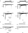
Figure 3. GTP up-regulated current present in heterozygote but not in knock-out
Example recordings of TTX-r Na+ currents in a sensory neurone from an NaV1.9 Hetero, compared with a KO. A, left-hand panels, Na+ currents recorded from Hetero neurone immediately on attaining the whole-cell configuration at the start of voltage-clamp recording (upper), and after 3.5 min (lower). The voltage step to −30 mV and current evoked at the same potential are indicated with a thick trace. The TTX-r current was seen to increase in amplitude, notably within the most negative potential range, because of the addition of a persistent current. Right-hand panels, Na+ currents recorded from an NaV1.9 KO neurone over a similar time (upper, immediately on attaining the whole-cell configuration; lower, after 4 min). In this recording no current is activated more negative than −30 mV. The current was generated by NaV1.8 operating alone, and also undergoes an increase in amplitude. B, in order to view the up-regulated current in isolation, an off-line digital subtraction was performed, where the current traces recorded at 0 min were subtracted from currents recorded at 3.5 min in Hetero (left) and at 4 min in KO (right). The up-regulated persistent Na+ current is clearly evoked over a potential range more negative than that associated with NaV1.8, and only in the Hetero neurone.
Figure 4
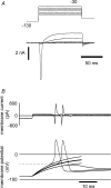
Figure 4. Measurement of voltage-threshold in NaV1.9 knock-out neurones
A, example currents with quasi-physiological solutions recorded in voltage-clamp from a NaV1.9 KO neurone. Transient Na+ current began to be recruited at −40 mV without there being any evidence for a persistent inward current operating at more negative potentials. A delayed-rectifier, voltage-dependent K+ current typically activated close to −50 mV. B, in the same neurone as in A, the total transmembrane current (both applied and endogenous active) and membrane potential responses are shown top and bottom panels, respectively. The subthreshold membrane potential changes are fitted with single exponentials. The voltage-threshold is defined as that value of potential where there is a clear deviation from a passive response subsequently leading to an action potential. This is shown as the dotted line (in this example, −40 mV). After the moment voltage threshold is reached, active inward current is also apparent in the membrane current records, and these two indicators may be simultaneous.
Figure 5
Figure 5. Action potential voltage thresholds in NaV1.9 knock-out neurones and the effect of intracellular GTP-γ-S
A, in current-clamp experiments, the intracellular dialysis of GTP-γ-S did not cause a substantial change in voltage threshold in NaV1.9 KO neurones (•) in recordings lasting between 1.5 and 31 min. In both Hetero (▵) and WT (^) littermates (n = 12 and 18, respectively), there was a significant skew in the response to GTP-γ-S, with a fraction of the neurones showing a negative shift in threshold greater than 2 s.d. from the mean KO value (for recording times, see Methods). B, 5 neurones lying beyond −2 s.d. indicate that the WT-Hetero population can be described as two populations, those that underwent threshold change (n = 5) and that are therefore different from KO, and those that did not (n = 25). The mean threshold change in these 5 neurones is significantly different from both the KO and other WT-Hetero neurones (P = 0.02; P < 0.02 Student/s t test). C, simultaneous recordings of membrane potential and total transmembrane current near the start of whole-cell recording (left) and a few minutes later (right). The depolarizing current steps generate subthreshold and just supra-threshold responses in each case (thin and thick traces), the voltage threshold becoming more negative and action potential latency becoming longer with GTP-γ-S dialysis, even finally occurring after the offset of applied depolarization (indicated by the arrow). D, results of a voltage-clamp experiment on the same neurone, recorded after the data shown in C, and using the same solutions. A clear persistent inward current is seen operating at −50 mV (thick trace), which was not activated at −80 mV. Further depolarization activates a transient Na+ current and an inactivating K+ current (or A-current), *. The functional importance of the A-current in this particular neuron is reflected in the brief hyperpolarizing afterpotential following the action potential in C.
Figure 6
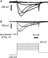
Figure 6. Transfection of hNaV1.9 results in the restoration of TTX-r persistent current in NaV1.9 knock-out neurones
Example currents recorded in voltage-clamp following intranuclear injection (A) and electroporation (B) of small diameter neurones cultured from NaV1.9 KO mice (total n = 5). Currents elicited in the presence of TTX have the appropriate negative voltage dependence and slow kinetics previously described for persistent Na+ current. In B, a transient TTX-r current is recruited close to −30 mV, consistent with activation of NaV1.8, presumed endogenous to the neurone.

Comment in

  • Nav1.9, G-proteins, and nociceptors.
    Waxman SG, Estacion M. Waxman SG, et al. J Physiol. 2008 Feb 15;586(4):917-8. doi: 10.1113/jphysiol.2007.149922. J Physiol. 2008. PMID: 18287383 Free PMC article. No abstract available.

References

    1. Amaya F, Wang H, Costigan M, Allchorne AJ, Hatcher JP, Egerton J, Stean T, Morisset V, Grose D, Gunthorpe MJ, Chessell IP, Tate S, Green PJ, Woolf CJ. The voltage-gated sodium channel NaV1.9 is an effector of peripheral inflammatory pain hypersensitivity. J Neurosci. 2006;26:12852–12860. - PMC - PubMed
    1. Baker MD, Bostock H. Low-threshold persistent sodium current in rat large dorsal root ganglion neurones in culture. J Neurophysiol. 1997;77:1503–1513. - PubMed
    1. Baker MD. Protein kinase C mediates up-regulation of tetrodotoxin-resistant, persistent Na+ current in rat and mouse sensory neurones. J Physiol. 2005;567:851–867. - PMC - PubMed
    1. Baker MD, Chandra SY, Ding Y, Waxman SG, Wood JN. GTP-induced tetrodotoxin-resistant Na+ current regulates excitability in mouse and rat small diameter sensory neurones. J Physiol. 2003;548:373–382. - PMC - PubMed
    1. Black JA, Waxman SG. Molecular identities of two tetrodotoxin-resistant sodium channels in corneal axons. Exp Eye Res. 2002;75:193–199. - PubMed

Publication types

MeSH terms

LinkOut - more resources