. 2008 Feb 15;586(4):1089-103.
doi: 10.1113/jphysiol.2007.145383. Epub 2007 Dec 20.

Conduction velocity is regulated by sodium channel inactivation in unmyelinated axons innervating the rat cranial meninges

Affiliations

Conduction velocity is regulated by sodium channel inactivation in unmyelinated axons innervating the rat cranial meninges

Roberto De Col et al. J Physiol. .

Abstract

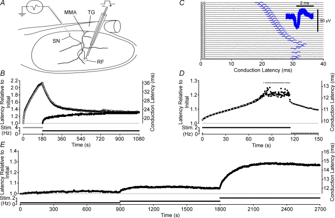
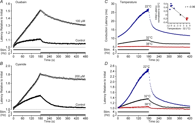
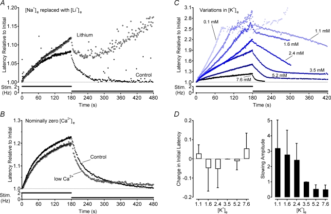
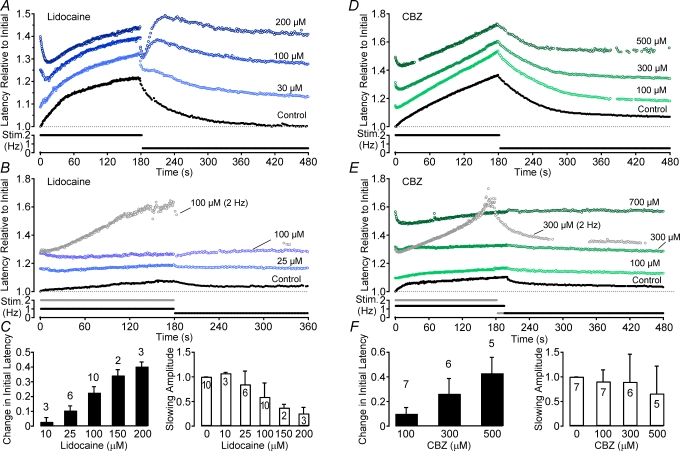
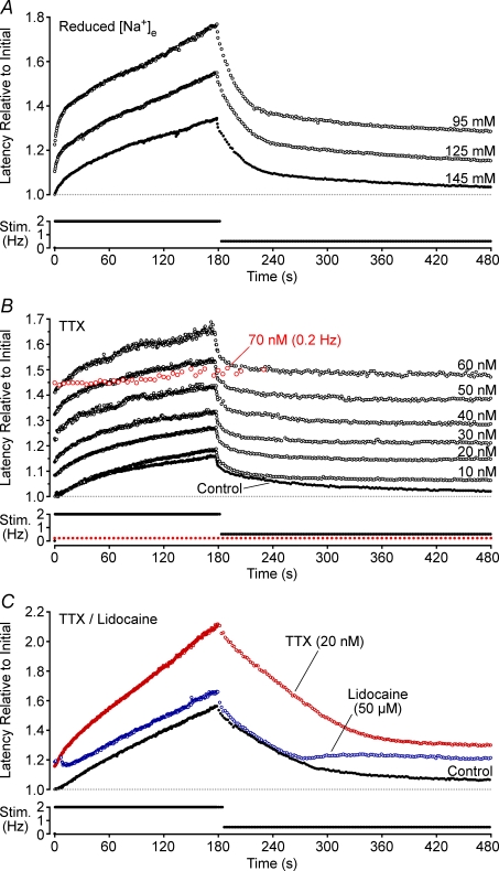
Axonal conduction velocity varies according to the level of preceding impulse activity. In unmyelinated axons this typically results in a slowing of conduction velocity and a parallel increase in threshold. It is currently held that Na(+)-K(+)-ATPase-dependent axonal hyperpolarization is responsible for this slowing but this has long been equivocal. We therefore examined conduction velocity changes during repetitive activation of single unmyelinated axons innervating the rat cranial meninges. In direct contradiction to the currently accepted postulate, Na(+)-K(+)-ATPase blockade actually enhanced activity-induced conduction velocity slowing, while the degree of velocity slowing was curtailed in the presence of lidocaine (10-300 microm) and carbamazepine (30-500 microm) but not tetrodotoxin (TTX, 10-80 nm). This suggests that a change in the number of available sodium channels is the most prominent factor responsible for activity-induced changes in conduction velocity in unmyelinated axons. At moderate stimulus frequencies, axonal conduction velocity is determined by an interaction between residual sodium channel inactivation following each impulse and the retrieval of channels from inactivation by a concomitant Na(+)-K(+)-ATPase-mediated hyperpolarization. Since the process is primarily dependent upon sodium channel availability, tracking conduction velocity provides a means of accessing relative changes in the excitability of nociceptive neurons.


Figures

Figure 1
Figure 1. Features of activity-induced changes in conduction latency
Deriving from somata in the trigeminal ganglion (TG), the spinosus nerve (SN) branches from the trigeminal nerve and enters the skull recurrently accompanying the medial meningeal artery (MMA) briefly before branching to innervate the meninges (A). A loose patch recording from the SN allows action potentials (C, inset) in single axons to be discerned by a consistency of spike shape (C, 100 spikes overlaid) and a constant latency response to electrical stimulation in their receptive field (RF). Repetitive electrical stimulation alters conduction velocity, measured as latency, in a frequency-dependent manner. For each stimulus frequency a corresponding steady-state conduction latency is approached from both an initially shorter latency (•, B) as well as from longer latencies, induced with a brief period of higher frequency stimulation (^, B). When stimulated at a sufficiently high rate, conduction latency progressively increases until a point is reached at which some stimuli fail to evoke an action potential response (grey symbols indicate no response, D; lower sweeps in C). The open symbols on every sixteenth sweep in D correspond to the raw data sweeps shown in C. As failures begin to occur the variance of the response latency increases (C and D) and we have termed this point the ‘critical deficit’ latency, a latency beyond which conduction is stochastic (see Discussion). For frequencies of stimulation below those bringing the fibre to the critical deficit latency, the relationship between stimulus frequency and steady-state conduction latency is monotonic positive (E).
Figure 2
Figure 2. Effect of pharmacological Na+–K+-ATPase blockade and temperature on activity-induced changes in conduction velocity
Direct Na+–K+-ATPase blockade with ouabain (100 μm, A) as well as indirect blockade via disruption of mitochondrial ATP production with cyanide (200 μm, B) both result in an increase in conduction velocity changes produced by repetitive stimulation. Increases and decreases in temperature affect the absolute conduction velocity (C and inset) as well as the relative change in latency induced by activity (D). The effect of temperature alone on activity-induced changes is clear after normalization for the temperature effect on initial latency (D).
Figure 3
Figure 3. Effects of extracellular ion concentration on activity-induced changes in conduction velocity
Na+ was completely removed from the extracellular perfusing solution by replacement with an equimolar amount of Li+ (A) and the extracellular concentration of Ca2+ was reduced by the addition of 1.5 mm EGTA to the perfusate (B). Changes in latency in both cases are shown relative to the initial latency under control conditions. The effect of changes in extracellular K+ concentration were normalized to the initial latency at that concentration (C). Changes in extracellular K+ were osmotically balanced by removal or addition of an equimolar amount of Na+. Group means show the effect of each K+ concentration on both initial latency relative to that for 3.5 mm K+ and the amplitude of activity-induced slowing (D).
Figure 5
Figure 5. Effects of the anti-convulsant carbamazepine (CBZ) and the local anaesthetic lidocaine on activity-induced changes in conduction latency
Different doses of CBZ and lidocaine were added to the solution and the electrical stimulation protocol was carried out at each dose. For both compounds low doses result in an increase in the amplitude of slowing during a 180 s period of stimulation at 2 Hz (A and D). As the dose increases an initial speeding of conduction latency becomes apparent. For lower frequencies of stimulation it is possible to increase the dose of both lidocaine and CBZ so that no slowing of conduction latency is observed in response to electrical stimulation (B and E). However, if the stimulation frequency is increased, it is still possible to induce activity-induced conduction velocity slowing as shown for one trial in 300 μm CBZ (E) and one trial in 100 μm lidocaine (B). Both the stimulation profile and the response to this higher rate of stimulation are shown in grey. Group data for lidocaine (C) and CBZ (F) are shown according to the dose-dependent effect on the initial latency (filled bars) and the amplitude of activity-induced slowing (open bars). Changes in latency are shown relative to the initial latency under control conditions.
Figure 4
Figure 4. Effects of reduced extracellular Na+ and the sodium channel blockers TTX and lidocaine on activity-induced changes in conduction latency
Data from four individual axons is shown, respectively, in each panel. Extracellular Na+ concentration was reduced by replacement with an equimolar amount of choline (A). B shows the effect of increasing doses of TTX on activity-induced slowing (10–70 nm), as well as the block of conduction by TTX at 70 nm (open grey symbols). In this example, TTX blocks conduction at 70 nm while at 60 nm changes in conduction latency produced by activity exceed the TTXs latency. In C, TTX (20 nm) and lidocaine (50 μm) produced a similar shift in base latency for this axon allowing a direct comparison of the effects of the two substances. Changes in latency are shown relative to the initial latency under control conditions.

Comment in

References

    1. Akopian AN, Sivilotti L, Wood JN. A tetrodotoxin-resistant voltage-gated sodium channel expressed by sensory neurons. Nature. 1996;379:257–262. - PubMed
    1. Andres KH, von During M, Muszynski K, Schmidt RF. Nerve fibres and their terminals of the dura mater encephali of the rat. Anat Embryol (Berl) 1987;175:289–301. - PubMed
    1. Barrett EF, Barrett JN. Intracellular recording from vertebrate myelinated axons: mechanism of the depolarizing afterpotential. J Physiol. 1982;323:117–144. - PMC - PubMed
    1. Beck PW, Handwerker HO, Zimmermann M. Nervous outflow from the cat/s foot during noxious radiant heat stimulation. Brain Res. 1974;67:373–386. - PubMed
    1. Blair NT, Bean BP. Roles of tetrodotoxin (TTX) -sensitive Na+ current, TTX-resistant Na+ current, and Ca2+ current in the action potentials of nociceptive sensory neurons. J Neurosci. 2002;22:10277–10290. - PMC - PubMed

Publication types

MeSH terms

LinkOut - more resources