. 2008 Feb;36(3):1026-36.
doi: 10.1093/nar/gkm667. Epub 2007 Dec 20.

HU binds and folds single-stranded DNA

Affiliations

HU binds and folds single-stranded DNA

Dmitri Kamashev et al. Nucleic Acids Res. 2008 Feb.

Abstract

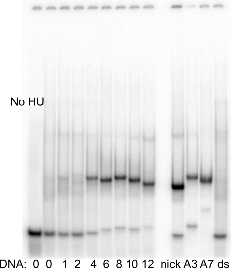
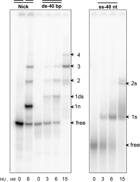
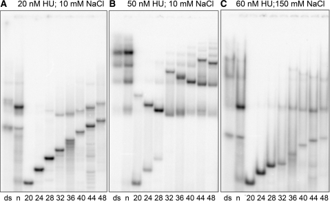
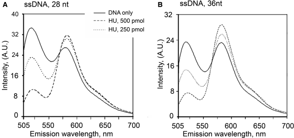
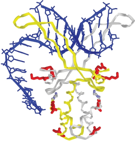
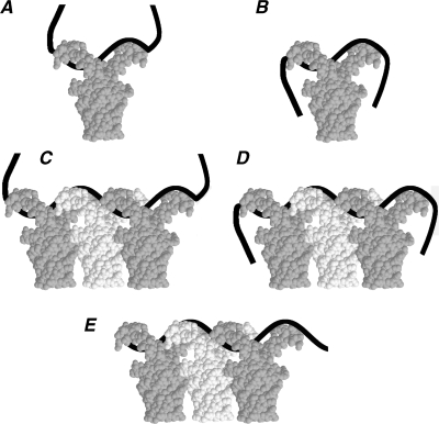
The nucleoid-associated protein HU plays an important role in bacterial nucleoid organization and is involved in numerous processes including transposition, recombination and DNA repair. We show here that HU binds specifically DNA containing mismatched region longer than 3 bp as well as DNA bulges. HU binds single-stranded DNA (ssDNA) in a binding mode that is reminiscent but different from earlier reported specific HU interactions with double-helical DNA lesions. An HU dimer requires 24 nt of ssDNA for initial binding, and 12 nt of ssDNA for each additional dimer binding. In the presence of equimolar amounts of HU dimer and DNA, the ssDNA molecule forms an U-loop (hairpin-like) around the protein, providing contacts with both sides of the HU body. This mode differs from the binding of the single-strand-binding protein (SSB) to ssDNA: in sharp contrast to SSB, HU binds ssDNA non-cooperatively and does not destabilize double-helical DNA. Furthermore HU has a strong preference for poly(dG), while binding to poly(dA) is the weakest. HU binding to ssDNA is probably important for its capacity to cover and protect bacterial DNA both intact and carrying lesions.


Figures

Figure 1.
Figure 1.
HU binding to DNA mismatches and bulges in stringent conditions (high salt). HU protein (50 nM) was mixed with a dsDNA containing in its middle a mismatched region of varying size (from 1 to 12) as indicated (left) or with dsDNA containing an insert of three or seven adenines (lanes A3 and A7). DsDNA (ds) or the duplex containing a nick (nick) were used as control. All the DNAs were 40-nt long and originated from sequence ‘X’. Samples were incubated in 200 mM NaCl and run in 95 mM Tris–borate. Lane no HU is the 40-mer dsDNA in the absence of HU.
Figure 2.
Figure 2.
HU binding to ssDNA, dsDNA and nick in low salt conditions. HU protein, at the concentration indicated in nM on the bottom, was mixed with end-labeled DNA, 40-bp long dsDNA, the same DNA with a nick in the middle and the 40 nt ssDNA, at 10 mM NaCl and analyzed by polyacrylamide gel buffered with 18 mM Tris–borate. All DNAs are originated of the sequence ‘X’. Arrows ‘1n’ and ‘1ds’ mark the bands corresponding to HU dimer–nick (1:1) and HU–dsDNA (1:1) complexes, respectively; arrows ‘2’, ‘3’ and ‘4’ mark the bands corresponding to two, three and four HU dimers bound to nick or dsDNA, while arrows ‘1s’ and ‘2s’ mark the bands corresponding to one or two HU dimers bound to ssDNA, respectively. Position of the free DNA is marked as ‘free’.
Figure 3.
Figure 3.
HU binding to ssDNA of various lengths. Binding of labeled DNA to HU protein was analyzed by polyacrylamide gel electrophoresis. DNA samples were: dsDNA (ds), nick (n), both 36-bp long; and ssDNA of sequence ‘D’ with the length varying from 20 to 48 nt (indicated at the bottom in nt). HU concentration was 20 nM (A), 50 nM (B) and 60 nM (C). Depending on the salt conditions, the gel was buffered with 18 mM Tris–borate for 10 mM NaCl (panels A and B), or with 55 mM Tris–borate for 150 mM NaCl (C).
Figure 4.
Figure 4.
Fluorescence resonance energy transfer between 5′ fluorescein and 3′- tetramethyl rhodamine (FAM) for 28-nt-long (Panel A) and 36-nt-long (Panel B) ssDNA is induced by HU binding. To 1 µM of oligonucleotides (solid lines) was added HU in (dimer:DNA) ratio 1:2 (dashed lines) and 1:1 (dotted line). On the right, the fluorescence signal of the donor (circles) and of the acceptor [IA/(ID+IA), squares] is shown for different ratios of HU and ssDNA.
Figure 5.
Figure 5.
Complex of HU dimer with 3′-overhang. A hypothetical conformation obtained by homology modeling based on the X-ray structure of HU–DNA complex (34,45). The coordinates of the central part, including the protein and the double-helical DNA, are close to those in the original PDB entry 1p51. The double-helical DNA fragment (in blue) was first built in a fiber canonical B-DNA conformation with the base-pair sequence corresponding to the PDB entry. The abasic site originally present in this DNA segment was removed and the structure was energy minimized with all relevant atom positions restrained to X-ray coordinates. The 3′-overhang was built by continuing the double-helical DNA with an AT-alternating sequence of appropriate length in the initial conformation corresponding to a single strand of a canonical B-DNA. The whole structure was next energy minimized with X-ray restraints as well as with a few additional restraints corresponding to putative contacts of the ssDNA segment with the charged residues (in red) on the surface of HU core, discussed in our previous studies (35,36).
Figure 6.
Figure 6.
The putative model of HU:ssDNA binding in complexes of different stoichiometry. The ssDNA is shown as a black band. The HU dimers are shown in white and gray for clarity. Plates A–B and plates C–E display sketches of complexes of a ssDNA strand with one and three HU, respectively. HU forms two types of complexes with ssDNA. Complexes of the first type have low affinity and low gel mobility (plates A and C). Complexes of the second type are more compact and have higher affinity and gel mobility (plates B and D). The conformation of the ssDNA part, which is not covered by HU protein, explains the difference between the high- and low-affinity complexes. Plate E represents the complex formed by ssDNA absorbed on HU oligomers so that the DNA extremities are hidden by the protein.

References

    1. Rouviere-Yaniv J, Gros F. Characterization of a novel, low-molecular-weight DNA-binding protein from Escherichia coli. Proc. Natl Acad. Sci. USA. 1975;72:3428–3432. - PMC - PubMed
    1. Rouviere-Yaniv J, Yaniv M, Germond JE. E. coli DNA binding protein HU forms nucleosomelike structure with circular double-stranded DNA. Cell. 1979;17:265–274. - PubMed
    1. Drlica K, Rouviere-Yaniv J. Histone like proteins of bacteria. Microbiol. Rev. 1987;51:301–319. - PMC - PubMed
    1. Rouviere-Yaniv J. Localization of the HU protein on the Escherichia coli nucleoid. Cold Spring Harb. Symp. Quant. Biol. 1978;42:439–447. - PubMed
    1. Wery M, Woldringh CL, Rouviere-Yaniv J. HU-GFP and DAPI co-localize on the Escherichia coli nucleoid. Biochimie. 2001;83:193–200. - PubMed

Publication types