Comparative Study
. 2008 Feb;18(2):261-71.
doi: 10.1101/gr.6687808. Epub 2007 Dec 20.

Controlled exchange of chromosomal arms reveals principles driving telomere interactions in yeast

Affiliations
Comparative Study

Controlled exchange of chromosomal arms reveals principles driving telomere interactions in yeast

Heiko Schober et al. Genome Res. 2008 Feb.

Abstract

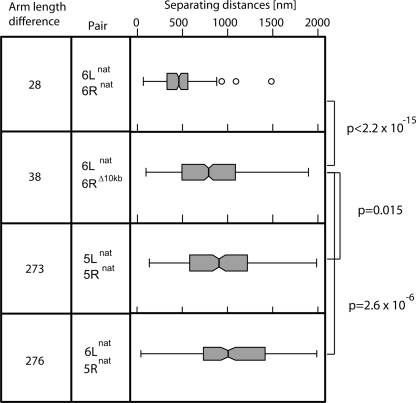
The 32 telomeres in the budding yeast genome cluster in three to seven perinuclear foci. Although individual telomeres and telomeric foci are in constant motion, preferential juxtaposition of some telomeres has been scored. To examine the principles that guide such long-range interactions, we differentially tagged pairs of chromosome ends and developed an automated three-dimensional measuring tool that determines distances between two telomeres. In yeast, all chromosomal ends terminate in TG(1-3) and middle repetitive elements, yet subgroups of telomeres also share extensive homology in subtelomeric coding domains. We find that up to 21 kb of >90% sequence identity does not promote telomere pairing in interphase cells. To test whether unique sequence elements, arm length, or chromosome territories influence juxtaposition, we reciprocally swapped terminal domains or entire chromosomal arms from one chromosome to another. We find that the distal 10 kb of Tel6R promotes interaction with Tel6L, yet only when the two telomeres are present on the same chromosome. By manipulating the length and sequence composition of the right arm of chr 5, we confirm that contact between telomeres on opposite chromatid arms of equal length is favored. These results can be explained by the polarized Rabl arrangement of yeast centromeres and telomeres, which promote to telomere pairing by allowing contact between chromosome arms of equal length in anaphase.


Figures

Figure 1.
Figure 1.
In vivo 4D fluorescence microscopy of telomeric foci. (A) Time-lapse 3D microscopy on haploid budding yeast in interphase carrying an integrated Rap1-GFP fusion under its own promoter. Microscopy was performed as described in the Methods, with stacks taken at 20-sec intervals over 90 min. Shown are 3D reconstructions from a typical 6-min series. Regions of maximal Rap1-GFP intensity were detected using the spot detection tool from the Imaris software from Bitplane, allowing each focus to be differentially labeled. When two foci fuse, they adopt one color, allowing us to score fusion and fission events. (B) Live time-lapse 3D microscopy and focus detection are as in A, but of a haploid yeast cell in interphase expressing Sir3-GFP under its own promoter. 3D stacks were taken at 30-sec intervals. (C) A kymograph of time-lapse imaging of Sir3-GFP as in B but spanning 30 min. The X-axis represents time. (Arrows) Branch or fusion points of Sir3-GFP foci. See Supplemental Movie 2 for rotation of the kymograph.
Figure 2.
Figure 2.
Dynamics of telomeric clusters relative to a single telomere. (A) 3D reconstruction of a yeast nucleus expressing Rap1-YFP, which labels all telomeres, and LacI-CFP, which recognizes a lacO array inserted on Tel14L (GA-2558). The larger view is rotated to allow visualization in the form of two other versions using Imaris software. The position of Tel14L (green) is scored relative to the Rap1 foci (red). (c) Tel14L colocalizing with a Rap1 focus, (ad) Tel14L adjacent to a Rap1 focus, (ap) Tel14L apart from Rap1 foci. Two-hundred-twenty nuclei reconstituted in 3D were analyzed, and in 70% of the nuclei Tel14L was either colocalizing or adjacent to the Rap1 focus. (B) Time-lapse imaging of the same yeast strain presented in A (GA-2558). 3D image stacks were taken at 30-sec intervals, deconvolved, and are shown in a 3D reconstitution. The top view is shown, and the juxtaposition of Tel14L to Rap1 was scored and indicated as in A. Continuous juxtaposition can be seen over 5 min.
Figure 3.
Figure 3.
Extensive homology does not confer interaction in telomeric foci. (A) 3D image stacks were acquired at 0.2-μm spacing along the Z-axis of the indicated yeast strains having targeted integration of lacO and tetO arrays and expressing LacI-CFP and TetR-YFP. Image stacks were analyzed by the SpotDistance plug-in of ImageJ and are represented as a box plot. The notch around the median represents ±5%. Outliers are defined as 1.5 times the Inter Quartile Range (IQR) (open circles). (B) (Left panel) Sketch of various telomere pairs represented as lines with TG repeats (black box), Y′ elements (open blue box), STR elements (open red box), and X core elements (green) indicated to the left. The shading between telomeres indicates >90% identity. (Right panel) Distance distributions between the telomeres in the left panel represented as box plots. The strains and number of cells analyzed per pair are as follows: (panel 1) GA-2686, n = 282, pair: 10L–10L; (panel 2) GA-2685, n = 315, pair: 9L–10L; (panel 3) GA-2753, n = 331, pair 14L–6L; (panel 4) GA-2691, n = 437, pair: 10L–16L; (panel 5) GA-2687, n = 367, pair: 9L–14R; (panel 6) GA-3268, n = 286, pair: 14R–10L; (panel 7) GA-2731, n = 91 pair: 14L–16L; (panel 8) GA-958, n = 231, pair: 6L–6R.
Figure 4.
Figure 4.
Truncation of Tel6R impairs Tel6R–Tel6L interaction. Distance distribution box plots were performed and are represented as in Fig. 3 for the indicated pairs of telomeres. The relevant strains and numbers of cells analyzed are as follows: (panel 1) GA-958, n = 231, pair: 6L–6R; (panel 2) GA-3742, n = 368, pair: 6L–6RΔ10kb; (panel 3) GA-3708, n = 202, pair: 5L–5R natural; (panel 4) GA-957, n = 180, pair: 6L–5R. The indicated P-values compare the likelihood that the two indicated patterns are different, and was calculated in R using the two-sample Kolmogorov–Smirnov test.
Figure 5.
Figure 5.
Reciprocal exchange of telomeres and chromosome arms in yeast. (A) Depiction of native chr 5 (red) and chr 6 (blue). (B,C) Overview of the telomere swap (B) and chromosome arm swap (C) (see Supplemental Fig. 5 for detailed explanation). Black bars underneath the chromosomes indicate the annealing positions of the probes used for Southern hybridization. (D) Total intact genomic DNA in the form of chromosomes was isolated from the indicated strains before and after the exchange (swap). Chromosomes were separated by CHEF gel electrophoresis and stained with ethidium bromide. Southern blot analysis was performed with probes indicated in (AC). (Lane 1) GA-3146, (lane 2) GA-1607, (lane 3) GA-1459, (lane 4) GA-1095, (lane 5) GA-3747, (lane 6) GA-3746, (lane 7) GA-3749, (lane 8) GA-3748, (lane 9) GA-3747, (lane 10) GA-3746, (lane 11) GA-3749, (lane 12) GA-3748.
Figure 6.
Figure 6.
Equal chromosomal arm length and colinearity promote telomere interaction. (A) Distance distributions were determined between the indicated lacO and tetO tags inserted in isogenic strains. The results are represented as box plots as in Fig. 3. A representation of the analyzed chromosome is indicated as folded in a Rabl configuration. The relevant pairs of loci analyzed are as follows: (1) Tel6L–Tel6R (GA-958, n = 231); (2) Tel6L–Tel6R5Rsubtelo (GA-1094, n = 194); (3) Tel6L–Tel5R6Rsubtelo (GA-1095, n = 265); (4) Tel6L–Tel5R (GA-957, n = 180). (B) Superposition of a brightfield image with a maximal projection of fluorescence image stacks is shown for anaphase cells bearing differentially tagged telomeres as indicated. Bar, 2 μm. (Upper left panel) A cell bearing lacO and tetO tags at Tel6L and Tel6R, respectively (GA-958), (upper right panel) Tel5L and Tel5R (GA-3746), (lower right panel) Tel6L and Tel5R (GA-957), (lower left panel) probable configuration of chr 6 in anaphase. (C) Quantitative analysis of Tel6L–Tel 6R (GA-958, n = 36), Tel5L–Tel5R (GA-3746, n = 33), and Tel6L–Tel5R (GA-957, n = 30) in anaphase and telophase cells. The two-sample Kolmogorov–Smirnov test confirmed that the differences between the distributions are significant. (D) As in A, but box plots are shown for the following strains resulting from chromosome arm exchange: (1) Tel5L–Tel5R (GA-3746, n = 307); (2) Tel5L–ARS514 (GA-3747, n = 320); (3) Tel5L–ARS5146Rsubtelo (GA-3749, n = 211); (4) Tel5L–Tel6R5Rarm (GA-3748, n = 296); (5) Tel5L–ARS514 (GA-3689) in cells expressing either LexA-Ku80 or LexA-Sir4, which then bind ARS514 bearing 8× LexA binding sites.
Figure 7.
Figure 7.
The Rabl configuration in telophase can influence telomere juxtaposition. Shown is a model suggesting how the Rabl organization of chromosomes in late anaphase and telophase might influence telomere–telomere clustering into perinuclear foci in interphase. The drawing indicates how arm length and metacentricity lead to late anaphase juxtaposition. An unknown sequence- or structure-recognizing factor may further promote interaction between Tel6R and Tel6L.

References

    1. Andrulis E.D., Neiman A.M., Zappulla D.C., Sternglanz R., Neiman A.M., Zappulla D.C., Sternglanz R., Zappulla D.C., Sternglanz R., Sternglanz R. Perinuclear localization of chromatin facilitates transcriptional silencing. Nature. 1998;394:592–595. - PubMed
    1. Burgess-Beusse B., Farrell C., Gaszner M., Litt M., Mutskov V., Recillas-Targa F., Simpson M., West A., Felsenfeld G., Farrell C., Gaszner M., Litt M., Mutskov V., Recillas-Targa F., Simpson M., West A., Felsenfeld G., Gaszner M., Litt M., Mutskov V., Recillas-Targa F., Simpson M., West A., Felsenfeld G., Litt M., Mutskov V., Recillas-Targa F., Simpson M., West A., Felsenfeld G., Mutskov V., Recillas-Targa F., Simpson M., West A., Felsenfeld G., Recillas-Targa F., Simpson M., West A., Felsenfeld G., Simpson M., West A., Felsenfeld G., West A., Felsenfeld G., Felsenfeld G. The insulation of genes from external enhancers and silencing chromatin. Proc. Natl. Acad. Sci. 2002;99:16433–16437. (Suppl. 4) - PMC - PubMed
    1. Bystricky K., Heun P., Gehlen L., Langowski J., Gasser S.M., Heun P., Gehlen L., Langowski J., Gasser S.M., Gehlen L., Langowski J., Gasser S.M., Langowski J., Gasser S.M., Gasser S.M. Long-range compaction and flexibility of interphase chromatin in budding yeast analyzed by high-resolution imaging techniques. Proc. Natl. Acad. Sci. 2004;101:16495–16500. - PMC - PubMed
    1. Bystricky K., Laroche T., van Houwe G., Blaszczyk M., Gasser S.M., Laroche T., van Houwe G., Blaszczyk M., Gasser S.M., van Houwe G., Blaszczyk M., Gasser S.M., Blaszczyk M., Gasser S.M., Gasser S.M. Chromosome looping in yeast: Telomere pairing and coordinated movement reflect anchoring efficiency and territorial organization. J. Cell Biol. 2005;168:375–387. - PMC - PubMed
    1. Celniker S.E., Drewell R.A., Drewell R.A. Chromatin looping mediates boundary element promoter interactions. Bioessays. 2007;29:7–10. - PubMed

Publication types

MeSH terms

LinkOut - more resources