. 2008 Mar 15;111(6):3236-44.
doi: 10.1182/blood-2007-10-117812. Epub 2007 Dec 20.

Somatic inactivation of the PHD2 prolyl hydroxylase causes polycythemia and congestive heart failure

Affiliations

Somatic inactivation of the PHD2 prolyl hydroxylase causes polycythemia and congestive heart failure

Yoji Andrew Minamishima et al. Blood. .

Abstract

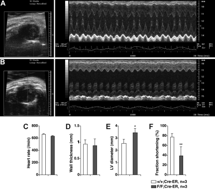
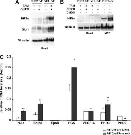
Pharmacologic activation of the heterodimeric HIF transcription factor appears promising as a strategy to treat diseases, such as anemia, myocardial infarction, and stroke, in which tissue hypoxia is a prominent feature. HIF accumulation is normally linked to oxygen availability because an oxygen-dependent posttranslational modification (prolyl hydroxylation) marks the HIFalpha subunit for polyubiquitination and destruction. Three enzymes (PHD1, PHD2, and PHD3) capable of catalyzing this reaction have been identified, although PHD2 (also called Egln1) appears to be the primary HIF prolyl hydroxylase in cell culture experiments. We found that conditional inactivation of PHD2 in mice is sufficient to activate a subset of HIF target genes, including erythropoietin, leading to striking increases in red blood cell production. Mice lacking PHD2 exhibit premature mortality associated with marked venous congestion and dilated cardiomyopathy. The latter is likely the result of hyperviscosity syndrome and volume overload, although a direct effect of chronic, high-level HIF stimulation on cardiac myocytes cannot be excluded.


Figures

Figure 1
Figure 1
Creation of conditional PHD2 allele. (A) Targeting strategy for mouse PHD2. NEO cassette flanked by 2 Frt sites (▵) is excised by fliplase to create floxed allele. Exons 2 and 3 flanked by 2 LoxP sites (▴) are excised by Cre recombinase to create a null allele. DT-A indicates diphtheria toxin-A used to select against nonhomologous recombinants; ■, exon; and □, untranslated region (UTR). Selected restriction sites are shown: EcoRI (RI), HindIII (H), BamHI (B), SpeI (Sp), and NotI (N). One-sided arrows = PCR primers used for genotyping. 2-sided arrows indicate BamHI fragment detected by Southern Blot with indicated probe. (B) Southern blot analysis of tail genomic DNA, digested with BamHI, from mice with indicated genotypes using probe shown in panel A. In the bottom panel, mice contained EIIA-Cre transgene where indicated. (C,D) RT-PCR (C) and immunoblot analysis (D) of MEFs with indicated genotypes. *The faint band is probably a background band, as PHD2 mRNA is undetectable in these cells. (E) Immunoblot analysis of PHD2 flox/flox (F/F);Cre-ER MEFs after treatment with 4-hydroxy tamoxifen (4-OHT; 200 nM) for the indicated time period. (F) Immunoblot analysis of kidney and liver from PHD2 flox/flox;Cre-ER mice after tamoxifen treatment.
Figure 2
Figure 2
Gross phenotypes of mice after conditional inactivation of PHD2. (A) Body weights of 10-week-old female mice with indicated genotypes after treatment with tamoxifen at E17.5 and at 3 weeks of age. (B) Appearance of ears (top row) and skin (bottom row). Note vascular dilation and erythema in PHD2 flox/flox;Cre-ER mice (right column). (C) Kaplan-Meier survival curves for mice with indicated genotypes. Error bars indicate 1 SD.
Figure 3
Figure 3
PHD2 loss causes severe polycythemia. (A) Hematologic parameters and (B) serum erythropoietin (Epo) levels in 10-week-old PHD2+/+;Cre-ER (+/+) and PHD2 flox/flox;Cre-ER (F/F) mice after treatment with tamoxifen at E17.5 and at 3 weeks of age. (C) Real-time RT-PCR analysis for the indicated mRNAs from PHD2 conditional knockout (F/F) mice treated as in panels A and B. *P < .05; **P < .01. Error bars indicate 1 SD.
Figure 4
Figure 4
Induction of erythropoietic mRNAs in mice lacking PHD2. Real-time RT-PCR analysis for indicated mRNAs in erythroid lineage cells (A), kidneys (B), and small intestines (C) from 12-week-old PHD2 flox/flox;Cre-ER (−) and PHD2 flox/flox;Cre-ER (+) mice after treatment with tamoxifen at E17.5 and at 3 weeks of age. *P < .05; **P < .01. Error bars indicate 1 SD.
Figure 5
Figure 5
Histologic and cardiac alterations after conditional inactivation of PHD2. (A,B) Gross appearance of abdomen in PHD2+/+;Cre-ER (A) and PHD2 flox/flox;Cre-ER (B) mice treated with tamoxifen at E17.5 and at 3 weeks of age. Note large retroperitoneal hematoma (asterisk) in PHD2 flox/flox;Cre-ER mouse (B). (C-L) Hematoxylin and eosin staining of PHD2+/+;Cre-ER (C,E,G,I,K) and PHD2 flox/flox;Cre-ER (D,F,H,J,L) treated with tamoxifen as in panels A and B. Sections are from spine (C,D), liver (E,F), kidney (G,H), lung (I,J), and heart (K,L). Note large retroperitoneal hematoma (arrowheads), severely dilated inferior vena cava (IVC; see also Figure S3), as well as evidence of hemorrhage in spinal canal (arrows). SC indicates spinal cord; V, vertebral body. *Aorta.
Figure 6
Figure 6
In vivo transthoracic echocardiography. Representative echocardiograms of 6 week old PHD2+/+;Cre-ER (A) and PHD2 flox/flox;Cre-ER (B) mice after Tamoxifen treatment as in Figure 4A,B. Short axis views (left panels) show evidence of left ventricular dilatation in PHD2 flox/flox mice. Representative M-mode views (right panels), in which a cross section of the short axis view is monitored over time, shows dilatation as well a depressed left ventricular function. Inactivation of PHD2 did not affect heart rate (C) or wall thickness (D), but increased end diastolic left ventricular diameter (E) and decreased fractional shortening (F). *P < .05; **P < .01. Error bars indicate 1 SD.
Figure 7
Figure 7
Cardiac HIF activity after PHD2 inactivation. (A,B) Immunoblot analysis of heart whole-cell extracts from 6-week-old (VHL flox/flox) or 8-week-old (PHD2 flox/flox) mice that did (+) or did not (−) express Cre-ER. Mice were treated with tamoxifen at E17.5 and at 3 weeks of age. (C) Real-time RT-PCR analysis for the indicated mRNAs in hearts from 26- to 33-day old PHD2 flox/flox;CreER (−) and PHD2 flox/flox;Cre-ER (+) mice after treatment with tamoxifen at E17.5 and at 3 weeks of age. *P < .05; **P < .01. Error bars indicate 1 SD.

Similar articles

Cited by

References

    1. Schofield CJ, Ratcliffe PJ. Oxygen sensing by HIF hydroxylases. Nat Rev Mol Cell Biol. 2004;5:343–354. - PubMed
    1. Kaelin WG., Jr The von Hippel-Lindau protein, HIF hydroxylation, and oxygen sensing. Biochem Biophys Res Commun. 2005;338:627–638. - PubMed
    1. Koivunen P, Hirsila M, Gunzler V, Kivirikko KI, Myllyharju J. Catalytic properties of the asparaginyl hydroxylase (FIH) in the oxygen sensing pathway are distinct from those of its prolyl 4-hydroxylases. J Biol Chem. 2004;279:9899–9904. - PubMed
    1. Bracken CP, Fedele AO, Linke S, et al. Cell-specific regulation of hypoxia-inducible factor (HIF)-1alpha and HIF-2alpha stabilization and transactivation in a graded oxygen environment. J Biol Chem. 2006;281:22575–22585. - PubMed
    1. Dayan F, Roux D, Brahimi-Horn MC, Pouyssegur J, Mazure NM. The oxygen sensor factor-inhibiting hypoxia-inducible factor-1 controls expression of distinct genes through the bifunctional transcriptional character of hypoxia-inducible factor-1alpha. Cancer Res. 2006;66:3688–3698. - PubMed

Publication types

MeSH terms

Substances