

Poly-N-acetyl glucosamine nanofibers regulate endothelial cell movement and angiogenesis: dependency on integrin activation of Ets1
- PMID: 18097146
- PMCID: PMC2769246
- DOI: 10.1159/000112544
Poly-N-acetyl glucosamine nanofibers regulate endothelial cell movement and angiogenesis: dependency on integrin activation of Ets1
Abstract
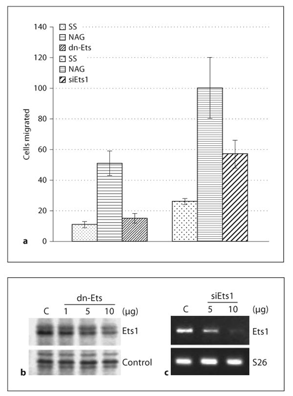
Poly-N-acetyl glucosamine (pGlcNAc) nanofiber-derived materials effectively achieve hemostasis during surgical procedures. Treatment of cutaneous wounds with pGlcNAc in a diabetic mouse animal model causes marked increases in cell proliferation and angiogenesis. We sought to understand the effect of the pGlcNAc fibers on primary endothelial cells (EC) in culture and found that pGlcNAc induces EC motility. Cell motility induced by pGlcNAc fibers is blocked by antibodies directed against alphaVbeta3 and alpha5beta1 integrins, both known to play important roles in the regulation of EC motility, in vitroand in vivo. pGlcNAc treatment activates mitogen-activated protein kinase and increases Ets1, vascular endothelial growth factor (VEGF) and interleukin 1 (IL-1) expression. pGlcNAc activity is not secondary to its induction of VEGF; inhibition of the VEGF receptor does not inhibit the pGlcNAc-induced expression of Ets1 nor does pGlcNAc cause the activation of VEGF receptor. Both dominant negative and RNA interference inhibition of Ets1 blocks pGlcNAc-induced EC motility. Antibody blockade of integrin results in the inhibition of pGlcNAc-induced Ets1 expression. These findings support the hypothesis that pGlcNAc fibers induce integrin activation which results in the regulation of EC motility and thus in angiogenesis via a pathway dependent on the Ets1 transcription factor and demonstrate that Ets1 is a downstream mediator of integrin activation.
Copyright 2007 S. Karger AG, Basel.
Figures


Similar articles
-
Synergistic platelet integrin signaling and factor XII activation in poly-N-acetyl glucosamine fiber-mediated hemostasis.Biomaterials. 2005 Sep;26(27):5433-43. doi: 10.1016/j.biomaterials.2005.01.023. Biomaterials. 2005. PMID: 15860200
-
Poly-N-acetyl glucosamine fibers accelerate hemostasis in patients treated with antiplatelet drugs.J Trauma. 2011 Aug;71(2 Suppl 1):S176-82. doi: 10.1097/TA.0b013e318225570d. J Trauma. 2011. PMID: 21814115
-
Functional overlap and cooperativity among alphav and beta1 integrin subfamilies during skin angiogenesis.J Invest Dermatol. 2003 Jun;120(6):1100-9. doi: 10.1046/j.1523-1747.2003.12236.x. J Invest Dermatol. 2003. PMID: 12787141
-
The alpha(1)beta(1) and alpha(2)beta(1) integrins provide critical support for vascular endothelial growth factor signaling, endothelial cell migration, and tumor angiogenesis.Am J Pathol. 2002 Jan;160(1):195-204. doi: 10.1016/s0002-9440(10)64363-5. Am J Pathol. 2002. PMID: 11786413 Free PMC article.
-
The Ets family contains transcriptional activators and repressors involved in angiogenesis.Int J Biochem Cell Biol. 2001 Apr;33(4):391-407. doi: 10.1016/s1357-2725(01)00025-5. Int J Biochem Cell Biol. 2001. PMID: 11312108 Review.
Cited by
-
pGlcNAc Nanofiber Treatment of Cutaneous Wounds Stimulate Increased Tensile Strength and Reduced Scarring via Activation of Akt1.PLoS One. 2015 May 8;10(5):e0127876. doi: 10.1371/journal.pone.0127876. eCollection 2015. PLoS One. 2015. PMID: 25955155 Free PMC article.
-
Successful treatment of two refractory venous stasis ulcers treated with a novel poly-N-acetyl glucosamine-derived membrane.BMJ Case Rep. 2012 Jul 9;2012:bcr0320126091. doi: 10.1136/bcr.03.2012.6091. BMJ Case Rep. 2012. PMID: 22778460 Free PMC article.
-
Integrin-dependent Akt1 activation regulates PGC-1 expression and fatty acid oxidation.J Vasc Res. 2012;49(2):89-100. doi: 10.1159/000332326. Epub 2012 Jan 13. J Vasc Res. 2012. PMID: 22249024 Free PMC article.
-
Engineered Biopolymeric Scaffolds for Chronic Wound Healing.Front Physiol. 2016 Aug 5;7:341. doi: 10.3389/fphys.2016.00341. eCollection 2016. Front Physiol. 2016. PMID: 27547189 Free PMC article. Review.
-
Solvation properties of N-acetyl-β-glucosamine: molecular dynamics study incorporating electrostatic polarization.J Comput Chem. 2011 Dec;32(16):3339-53. doi: 10.1002/jcc.21873. Epub 2011 Sep 7. J Comput Chem. 2011. PMID: 21898464 Free PMC article.
References
-
- Shiojima I, Walsh K. Role of Akt signaling in vascular homeostasis and angiogenesis. Circ Res. 2002;90:1243–1250. - PubMed
-
- Chen Z, Fisher RJ, Riggs CW, Rhim JS, Lautenberger JA. Inhibition of vascular endothelial growth factor-induced endothelial cell migration by ETS1 antisense oligonucleotides. Cancer Res. 1997;57:2013–2019. - PubMed
-
- Sementchenko VI, Watson DK. Ets target genes: past, present and future. Oncogene. 2000;19:6533–6548. - PubMed
-
- Vu TH, Werb Z. Matrix metalloproteinases: effectors of development and normal physiology. Genes Dev. 2000;14:2123–2133. - PubMed
Publication types
MeSH terms
Substances
Grants and funding
LinkOut - more resources
Full Text Sources
Other Literature Sources
Miscellaneous
