Review
. 2007 Winter;17(4):349-404.
doi: 10.1089/oli.2007.0097.

Bioconjugation of oligonucleotides for treating liver fibrosis

Affiliations
Review

Bioconjugation of oligonucleotides for treating liver fibrosis

Zhaoyang Ye et al. Oligonucleotides. 2007 Winter.

Abstract

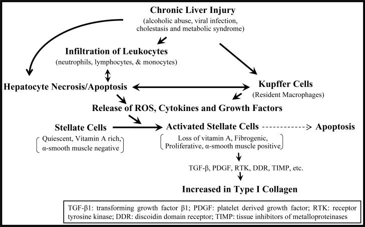
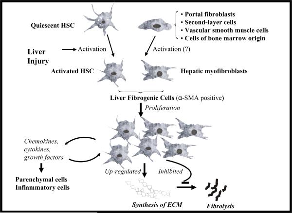
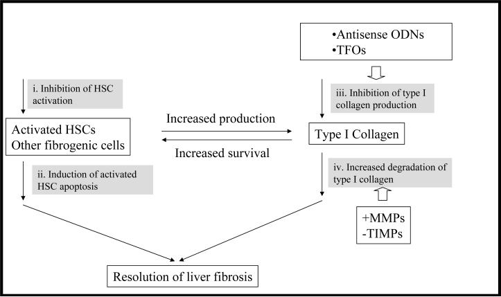
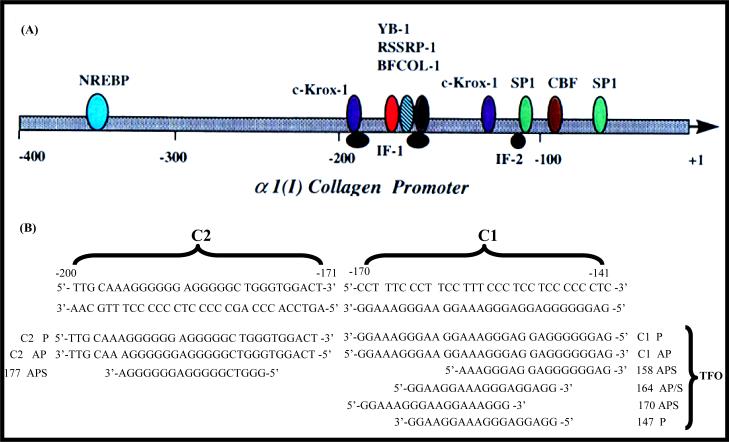
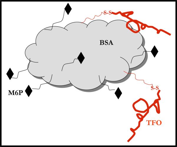
Liver fibrosis results from chronic liver injury due to hepatitis B and C, excessive alcohol ingestion, and metal ion overload. Fibrosis culminates in cirrhosis and results in liver failure. Therefore, a potent antifibrotic therapy is urgently needed to reverse scarring and eliminate progression to cirrhosis. Although activated hepatic stellate cells (HSCs) remain the principle cell type responsible for liver fibrosis, perivascular fibroblasts of portal and central veins as well as periductular fibroblasts are other sources of fibrogenic cells. This review will critically discuss various treatment strategies for liver fibrosis, including prevention of liver injury, reduction of inflammation, inhibition of HSC activation, degradation of scar matrix, and inhibition of aberrant collagen synthesis. Oligonucleotides (ODNs) are short, single-stranded nucleic acids, which disrupt expression of target protein by binding to complementary mRNA or forming triplex with genomic DNA. Triplex forming oligonucleotides (TFOs) provide an attractive strategy for treating liver fibrosis. A series of TFOs have been developed for inhibiting the transcription of alpha1(I) collagen gene, which opens a new area for antifibrotic drugs. There will be in-depth discussion on the use of TFOs and how different bioconjugation strategies can be utilized for their site-specific delivery to HSCs or hepatocytes for enhanced antifibrotic activities. Various insights developed in individual strategy and the need for multipronged approaches will also be discussed.


Figures

Figure 1
Figure 1. Events leading to liver fibrosis
During liver injury, infiltrating leukocytes (neutrophils, lymphocytes and monocytes) along with resident macrophages (Kupffer cells) release reactive oxygen species (ROS), growth factors and inflammatory cytokines, leading to activation of hepatic stellate cells (HSCs) into actively proliferating, α-smooth muscle actin-containing myofibroblast-like cells. The activated HSCs are the source of cytokines, chemotatics, and also secrete large amounts of type I collagen and other extracellular matrix (ECM) components. Apoptosis of activated HSCs is implicated in the spontaneous resolution of liver fibrosis.
Figure 2
Figure 2. Different liver fibrogenic cells
Due to liver injury, hepatic stellate cells (HSCs) undergo transformational change into myofibroblast-like activated HSCs, which are depleted of vitamin A, but rich in α-smooth muscle actin (α-SMA). There is considerable evidence supporting that HSCs are a major source of fibrogenic cells in the injured liver. However, contributions from other cell types including portal fibroblasts, second-layer cells located around centrolobular veins (CLVs), vascular smooth muscle cells and cells of bone marrow origin are also possible. These liver fibrogenic cells proliferate at the sites of liver injury, produce a variety of proinflammatory cytokines, chemokines and growth factors, synthesize extracellular matrix (ECM) proteins and inhibit their degradation, leading to fibrosis.
Figure 3
Figure 3. Treatment strategies for liver fibrosis
The common characteristics of liver fibrosis is that activated HSCs and other fibrogenic cells produce excess amount of type I collagen. Accumulation of type I collagen provides survival signal for activated HSCs. Therefore, strategies focusing on fibrogenic cells themselves as well as type I collagen regardless of etiology of liver injuries are attractive. Inhibition of the activation of HSCs (i) and induction of their apoptosis (ii) have attracted lots of attention. Gene silencing technologies have been utilized to directly inhibit the production of type I collagen (iii) at gene level. Inducing degradation of type I collagen (iv) can be realized by increasing the activity of matrix metalloproteinases (+MMPs) and inhibiting the activity of tissue inhibitor of metalloproteinases (−TIMPs). Decreased disposition of type I collagen and remodeling of extracellular matrix (ECM) accelerate apoptosis of activated HSCs.
Figure 4
Figure 4. Sequence of the rat α1(I) collagen promoter showing duplex targets C1 and C2 and the TFOs
(A) The schematic illustration of rat α1(I) collagen promoter region. Many transcription factors have been shown binding to this region and play key roles in regulation of the gene expression. (B) Phosphodiester and phosphorothioate triplex forming oligonucleotides (TFOs) are designed with different starting position corresponding to the type α1(I) collagen promoter sequence. These TFOs can be parallel or antiparallel to the target sequence. They are 18mer or 30mer. The ability of triplex formation can be different. P: parallel; AP: antiparallel; APS: antiparallel phosphorothioate. Reproduced with permission from Joseph et al. (1998) Nucleic Acids Res. 25(11):2182–8.
Figure 5
Figure 5. Rules of triplex formation
A third polynucleotide sequence can bind to double-stranded DNA at the major grove to form triplex structure via formation of Hoogsteen/reverse Hoogsteen hydrogen bonds. TFOs can only bind to polypurine strand of target DNA. TFOs can be either (G, A)-motif or (C, T)-motif. The (C, T)-motif involves the formation of C•G×C and T•A×T base triplets (• stands for Watson-Crick hydrogen bond; × stands for Hoogsteen hydrogen bond), upon binding of a (C, T)-containing TFO with a parallel orientation with respect to the purine strand (Hoogsteen hydrogen bonds). The (G, A)-motif involves the formation of C•G×G and T•A×A triplets, upon binding of a (G, A)-containing TFO in an antiparallel orientation with respect to the purine strand (reverse-Hoogsteen hydrogen bonds). A (G, T)-motif TFO is also permitted. The (G, T)-motif involves binding of a (G, T)-containing TFO, whose orientation depends on both the number of GpT or TpG steps in the third strand and on the length of G and T tracts.
Figure 6
Figure 6. Electrophoretic mobility shift assays showing triplex formation with parallel and antiparallel phosphodiester and phosphorothioate triplex forming oligonucleotides (TFOs) with C1 duplex
(A) Comparison between the TFOs with different directions relative to C1 duplex; (B) Comparison between TFOs with different start positions on the C1 duplex. C1 represents the sequence from −141 to −170 of α1(I) gene promoter, in which a polypurine sequence exists on the non-coding strand. Duplex concentration, 2 nM; TFO concentration (μM) in each triplex forming reaction is shown below the corresponding lane. T, triplex; D, duplex; P, parallel; AP, antiparrallel; APS, antiparrallel phosphorothioate; 164 AP/APS, TFO sequence corresponding to the region from −164 to −147; 147 P, TFO sequence corresponding to the region from −147 to −164; 158 APS, TFO sequence corresponding the region from −158 to −141; 170 APS, TFO sequence corresponding to the region from −170 to −153. Reproduced with permission from Joseph et al. (1998) Nucleic Acids Res. 25(11):2182–8.
Figure 7
Figure 7. Modifications of the TFOs
Different modifications have been developed to increase the stability and binding affinity of TFOs. Modifications can be on backbone and sugar moieties. More radical modification is to substitute the whole sugar structure, such as phosphorodiamidate morpholino oligonucleotides (PMOs) and peptide nuclei acids (PNAs). PO: phosphodiester; PS: phosphorothioate modification; PN: N3'→P5' phosphoramidate modification; MP: methyl phosphodiester; 2'-AE: 2'-O-aminoethyl (2'-AE); LNA: Locked nucleic acid (LNA).
Figure 8
Figure 8. Strategy for detecting triplex formation with genomic DNA using real-time PCR
Triplex formation can be detected using a real-time PCR based method. TFO is modified with psoralen at the 3' end for forming covalent attachment at triplex formation site. The triplex formation can be done with isolated genomic DNA or genomic DNA in isolated nuclei or live cells. After triplex formation reaction (i) and UVA irradiation at 366nm for 10 at ~3.8 mW/cm2 from 5 cm distance (ii), the DNA samples are subjected to a agarose gel electrophoresis (0.5%) and extracted for the gel (iii). The aliquots of extracted DNA samples are subjected to real-time PCR (iv). Two sets of primers are used: one set of primer for amplification of a control region and another for target region overlapping triplex forming site. The DNA with psoralen covalent modification is not substrate of PCR reaction. Inhibition of PCR reaction at the triplex formation site relates to the triplex formation.
Figure 9
Figure 9. Barriers to systemic delivery of ODNs
For ODNs to reach target cells from the site of administration there are four major barriers to overcome: instability against nucleases, non-specific tissue distribution, poor cellular uptake, and uncontrolled subcellular trafficking. ODNs are polyanions and have a relatively small molecular weight with around 10kDa. They are subjected to quick urine excretion. After uptake by cells, they are inefficient to escape from endosome/lysosomes to cytosol. For antisense ODNs, they need to be in cytosol and bind to mRNA; while TFOs have to enter nucleus and bind to genomic DNA.
Figure 10
Figure 10. Hepatic cellular localization and subcellular distribution of 33P-TFO after intravenous administration in rats at doses of 0.2 and 1 mg/kg
Different liver cells were isolated at 30 min post-injection by liver perfusion with a mixture of 0.5 mg/ml collagenase and 0.1 mg/ml pronase and fractionation on Nycodenz gradient. Fibrotic rats were induced by dimethyl nitrosamine (DMN). The amount of the TFO in each cell type is given as ng/mg cell protein (A) and % of the total liver recovery (B). Subcellular distribution of 33P-TFO in the liver at 2 and 4 h after intravenous administration in rats at doses of 1mg/kg. Highly purified nuclei were isolated from cytoplasm and cell debris using the sucrose gradient separation method. The purity and number of isolated nuclei were determined under microscopy by dilution in trypan blue solution (C). TFO distributions in the nuclei, cytoplasm and cell debris were given as % of the total liver recovery (D). Data are presented as the mean ± SD (n = 4). Reproduced with permission from Cheng et al. (2005) Mol Pharm. 2(3):206–217.
Figure 11
Figure 11
(A) Synthesis of TFO-Chol using thiocholesterol and bis-(5-nitro-2-pyridyl)-disulphide and bioconjugation via disulfide bond formation Reagent and conditions: (1) 5 ml of pyridine, stirring at room temperature for 2 h; and 2, 1500 μL of dimethylformamide (DMF), stirring under N2 protection at 40°C for 24 h. (B) Biodistribution of TFO-Chol after systematic administration of a mixture of 33P-TFO-Chol and TFO-Chol or a mixture of 33P-TFO and TFO at a dose of 0.2 mg/kg. At 30 min and 4 h after injection, blood was collected by cardiac puncture, and urine was collected from the bladder. The rats were sacrificed, tissues were collected, washed, and weighted, 150 mg of tissue was digested, and radioactivity was determined using a scintillation counter. Data are represented as the mean ± S.D. (n = 4). Reproduced with permission from Cheng et al. (2006) J Pharmacol Exp Ther. 317(2):797–805.
Figure 12
Figure 12. Synthetic polymer-based multicomponent carriers for delivery of ODNs
(A) Hydroxypropyl methacrylate (HPMA)-based copolymers. A phosphorothioate oligonucleotides (PS-ODN) was covalently linked to the HPMA copolymer via the degradable GFLG linker. The ODNs was labeled with fluorescein on the 3' end. The polymer backbone was labeled with Lissamine rhodamine B to allow independent visualization of the polymer and the ODN. Reproduced with permission from Jensen et al. (2002) Bioconjug Chem., 13(5), 975–984. (B) Polycefin, poly(β-L-malic acid) based copolymer. Percent values refer to the number of malyl moieties of PMLA that are conjugated with a given module (100%) total malyl content). The distribution of conjugates along the scaffold is assumed to be random. Abbreviations are: PMLA, poly(β-L-malic acid); PMLA-NHS, N-hydroxysuccinimidyl ester at pending carboxyl groups of PMLA; mPEG5000, methoxy poly(ethylene glycol) (5kDa); 2-MEA, 2-mercaptoethylamine; mAb OX-26, mouse monoclonal antibody to rat transferrin receptor; maleimide-PEG3400-maleimide, a bifunctional maleimide derivative of PEG (3400 Da); MORPH-AON-1, morpholino antisense oligonucleotide to laminin α4 chain; MORPH-AON-2, morpholino antisense oligonucleotide to laminin β1 chain. Reproduced with permission from Lee et al. (2006) Bioconjug Chem., 17(2), 317–326.
Figure 13
Figure 13. Schematic of a propose conjugate for site-specific delivery of TFOs to activated HSCs for treating liver fibrosis
Bovine serum albumin is used as the backbone, onto which targeting ligands, mannose 6-phosphate (M6P) can be attached with the NH2 of lysines. In addition, TFO molecules modified sulfhydryl functionalities can be conjugated with BSA via disulfide bond formation. The M6P/insulin-like growth factor II (M6P/IGF-II) receptor, which is expressed in particular upon HSCs during fibrosis, can be utilized for targeted delivery to activated HSCs. Disulfide bonds enable the intracellular release of TFOs upon taken up by HSCs. TFOs used are specific for type α1(I) collagen gene promoter and can inhibit the gene transcription. This conjugate has potential to be used as antifibrotic drugs.

Similar articles

Cited by

References

    1. Raghow R. The role of extracellular matrix in postinflammatory wound healing and fibrosis. Faseb J. 1994;8:823–31. - PubMed
    1. Anderson RN. Deaths: leading causes for 2000. Natl Vital Stat Rep. 2002;50:1–85. - PubMed
    1. Gines P, Cardenas A, Arroyo V, Rodes J. Management of cirrhosis and ascites. N Engl J Med. 2004;350:1646–54. - PubMed
    1. El-Serag HB. Hepatocellular carcinoma and hepatitis C in the United States. Hepatology. 2002;36:S74–83. - PubMed
    1. Befeler AS, Di Bisceglie AM. Hepatocellular carcinoma: diagnosis and treatment. Gastroenterology. 2002;122:1609–19. - PubMed

Publication types

MeSH terms