. 2008 Mar 1;44(5):847-55.
doi: 10.1016/j.freeradbiomed.2007.11.012. Epub 2007 Dec 4.

Catalytic inactive heme oxygenase-1 protein regulates its own expression in oxidative stress

Affiliations

Catalytic inactive heme oxygenase-1 protein regulates its own expression in oxidative stress

Qing S Lin et al. Free Radic Biol Med. .

Abstract

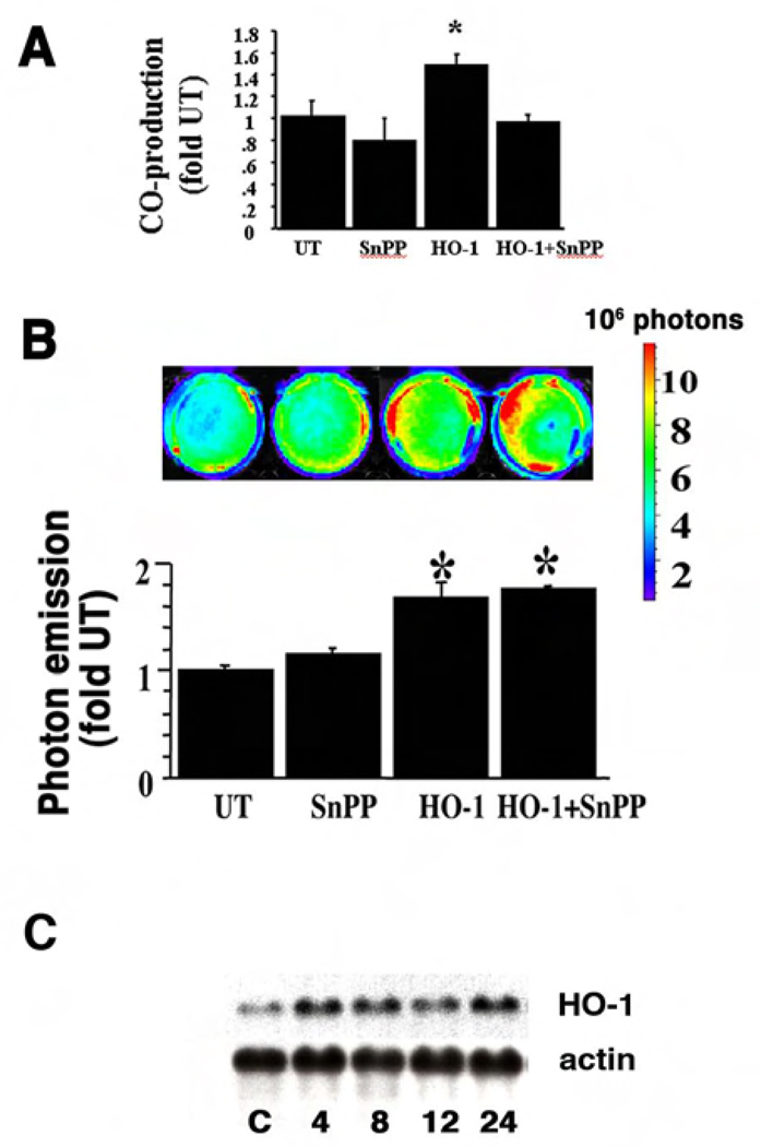
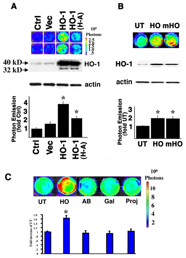
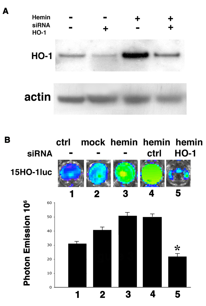
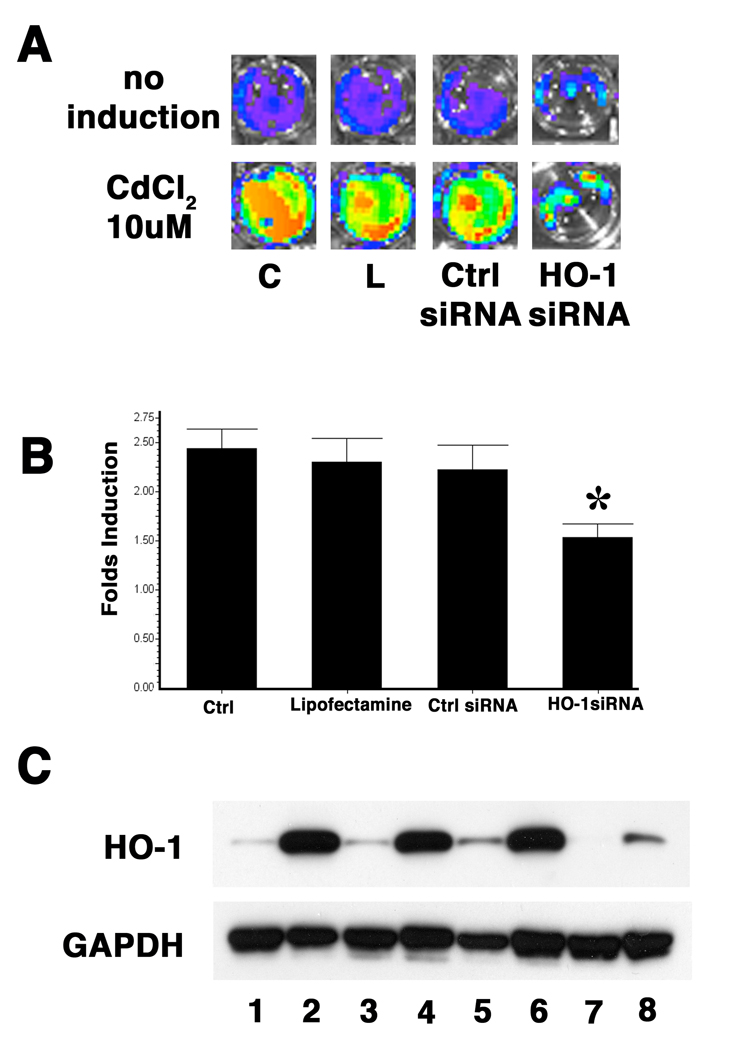
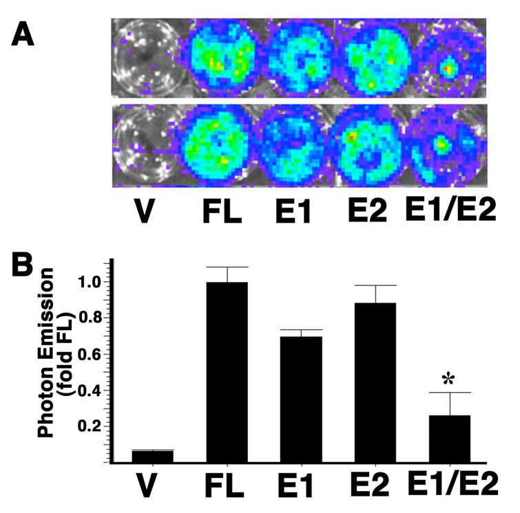
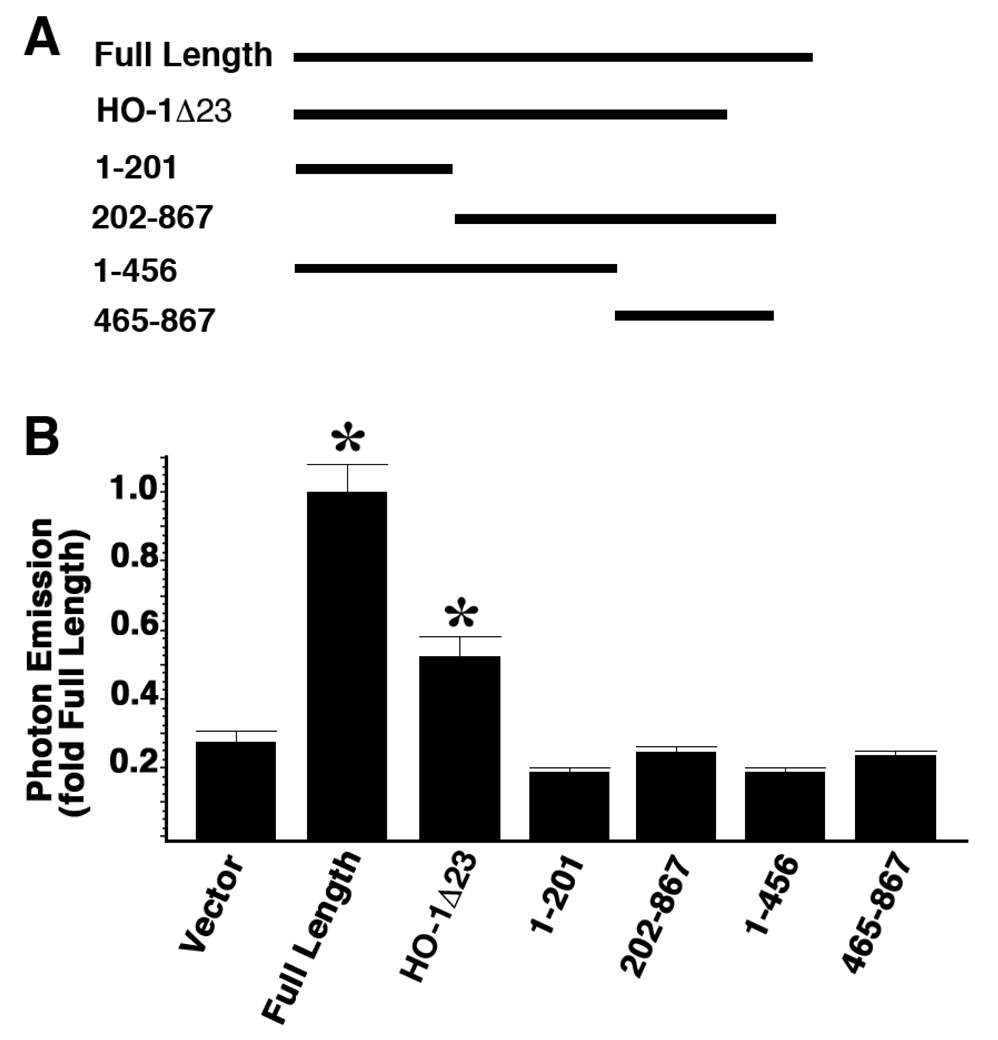
Heme oxygenase-1 (HO-1) catalyzes the degradation of heme and forms antioxidant bile pigments as well as the signaling molecule carbon monoxide. HO-1 is inducible in response to a variety of chemical and physical stress conditions to function as a cytoprotective molecule. Therefore, it is important to maintain the basal level of HO-1 expression even when substrate availability is limited. We hypothesized that the HO-1 protein itself could regulate its own expression in a positive feedback manner, and that this positive feedback was important in the HO-1 gene induction in response to oxidative stress. In cultured NIH 3T3 cells, transfection of HO-1 cDNA or intracellular delivery of pure HO-1 protein resulted in activation of a 15-kb HO-1 promoter upstream of luciferase as visualized by bioluminescent technology and increased HO-1 mRNA and protein levels. These effects were independent of HO activity because an enzymatically inactive mutant form of HO-1 similarly activated the HO-1 promoter and incubation with HO inhibitor metalloporphyrin SnPP did not affect the promoter activation. In addition, HO-1-specific siRNA significantly reduced hemin and cadmium chloride-mediated HO-1 induction. Furthermore, deletion analyses demonstrated that the E1 and E2 distal enhancers of the HO-1 promoter are required for this HO-1 autoregulation. These experiments document feed-forward autoregulation of HO-1 in oxidative stress and suggest that HO-1 protein has a role in the induction process. We speculate that this mechanism may be useful for maintaining HO-1 expression when substrate is limited and may also serve to up-regulate other genes to promote cytoprotection and to modulate cell proliferation.


Figures

Figure 1
Figure 1. Inhibition of HO activity did not affect the HO-1 promoter activation after HO-1 protein delivery
Cells were pre-incubated with SnPP, a potent inhibitor of HO activity. (A) HO activity in the cells where HO-1 protein was delivered (HO-1) with or without SnPP. (B) A representative image of photon emission and quantitative assessment of light intensity is shown. Values are the mean ± standard error of four separate determinations. Values are expressed as a fold of untransfected (UT) controls. *P<0.05 vs. UT. (C) A representative Northern blot illustrating HO-1 mRNA steady state levels 4 to 24 h after delivery of HO-1 protein is shown. The housekeeping gene β-actin demonstrates equal loading. C: control cells receiving Pro-ject reagent alone.
Figure 2
Figure 2. Inactive HO-1 protein also increases the HO-1 promoter activity
(A) 3T3-HO-1/luc cells were transiently transfected with pCMV vector, HO-1 cDNA (HO-1) and an enzymatically inactive HO-1 mutant in which Histidine 25 was substituted with Alanine (HO-1 H-A). A representative pseudoimage of photon emission is shown in the upper panel. Western blot analysis showed the 40 KD transfected HO-1 fusion proteins and the 32 KD endogenous HO-1 protein. Actin was used for loading control. Quantitation of the photon emission is shown in the lower panel. (B) Wildtype and inactive HO-1 proteins were delivered into 3T3-HO-1/luc cells. A representative pseudoimage of photon emission is shown in the upper panel. A representative Western blot of HO-1 immunoreactive protein is shown in the middle panel. Quantitation of the photon emission is shown in the lower panel. UT: untransfected; HO-1: Cells were HO-1 protein delivered; mutant HO-1: cells were an inactive mutant HO-1 (His25Ala) protein delivered. Values are expressed as a fold of untransfected controls and are the mean ± standard error of four separate determinations. *p < 0.05 vs. UT. (C) A pseudoimage of the photon emission from 3T3-HO-1/luc cells after delivery of HO-1 protein of 12 hours. Negative controls are cells that did not receive any protein (UT) or cells where other proteins such as galatosidase (Gal), a FITC-labeled immunoglobulin (AB) or Pro-ject alone (Proj) were delivered. Quantitative luminescence intensity is shown below. Values are the mean ± standard error of four independent measurements. *P<0.05 vs. UT.
Figure 3
Figure 3. HO-1 is required for hemin induced HO-1 promoter activation
3T3-HO-1/luc cells were transfected with siRNA targeting HO-1 gene or Cyclophillin B (Dharmacon). (A) Western blot analysis showing the repression of HO-1 expression by HO-1 siRNA. 72 hrs after siRNA transfection, cells were treated with or without 30 µM hemin for 24 hrs before harvesting. Whole cell lysates were separated on SDS gels and HO-1 protein levels were examined by Western Blot analysis. Actin was used for loading control. (B) Luciferase activity visualized via the IVIS. Note the significant decrease of light emission in cells transfected with HO-1 siRNA. siRNA against Cyclophilin B was used as non-specific siRNA control. 1: control 15HO-1luc cells; 2: 15HO-1luc cells with mock transfection; 3: 15HO-1luc cells with hemin treatment; 4: 15HO-1luc cells with control Cyclophilin B siRNA followed by hemin treatment; and 5: 15HO-1luc cells with HO-1 siRNA followed by hemin treatment. Quantitative analysis was presented at the lower panel as mean ± standard error. *P<0.05 vs. hemin with no siRNA (lane 3) or control siRNA (lane 4).
Figure 4
Figure 4. HO-1 is involved in Cadmium mediated HO-1 promoter induction
3T3-HO-1/luc cells were transfected with siRNA targeting HO-1 gene or Cyclophillin B (Darmacon). 72 hours after siRNA transfection, cells were treated with 10µM CdCl2 for 24 hours followed by analysis. (A) A representative pseudoimage of photon emission 24 hours after CdCl2 induction. C: control cells that were not transfected. L: cells transfected with lipofectamine 2000 only. Ctrl siRNA: cells transfected with Cyclophillin B siRNA. (B) Comparison of fold change of photon emissions after CdCl2 induction. C: control cells that were not transfected. L: cells transfected with lipofectamine 2000 only. Ctrl siRNA: cells transfected with Cyclophillin B siRNA. Values are expressed as a fold of untransfected controls and are the mean ± standard error of three separate experiments. *P<0.05 vs. cells transfected with control siRNA. (C) Western Blot analysis with anti-HO-1 antibody showing that the cadmium induction of HO-1 protein was blocked by HO-1 siRNA transfection. GAPDH expression was used as loading control. Lane 1, 2: control cells that were not transfected; lane 3, 4: cells transfected with lipofectamine 2000 only; lane 5, 6: cells transfected with Cyclophillin B siRNA; and land 7,8: cells transfected with HO-1 siRNA. Lane 1, 3, 5, 7: cells with no induction. Lane 2, 4, 6, 8: cells were induced with CdCl2.
Figure 5
Figure 5. HO-1 activates its own promoter through the distal E1 and E2 enhancers
NIH3T3 cells were co-transfected with HO-1 cDNA and different luciferase reporter constructs. V: vector containing only the luciferase reporter. FL: luciferase reporter controlled by the 15 kb full length HO-1 promoter. E1: luciferase reporter controlled by the 15 kb full length HO-1 promoter with mutation on the E1 enhancer. E2: luciferase reporter controlled by the 15 kb full length HO-1 promoter with mutation on the E2 enhancer. E1/E2: luciferase reporter controlled by the 15 kb full length HO-1 promoter with mutations on both the E1 and E2 enhancers. (A) A representative pseudoimage of photon emission 24 hours after transfections. (B) Quantitation of the photon emissions. Values are expressed as the mean ± standard error of three independent experiments with duplicate samples. *P<0.05 vs. FL
Figure 6
Figure 6. Intact HO-1 protein is required for maximal promoter activation but truncated HO-1 protein can also active the promoter
3T3-HO-1/luc cells were transfected with full length HO-1 cDNA as well as cDNAs with various truncations. Luciferase activities were measured with IVIS 48 hours after transfection. A: Schematic drawings of the cDNA constructs. Numbers represent the DNA base pairs in HO-1 coding region. B: Data represents fold of full length value and are mean ± standard error of three experiments with duplicate samples. Full Length: full length HO-1 cDNA; HO-1Λ23: HO-1 cDNA with 23 amino acids deleted from the C-terminal; 1–201, 202–867, 1–456, 465–867 represent different truncation isoforms. *p < 0.05 vs. vector.
Figure 7
Figure 7. HO-1 self-regulation does not require activation of p38 and ERK MAPK pathways
3T3-HO-1/luc cells were transfected with expression vector alone and full-length cDNA encoding wildtype and catalytic inactive forms of HO-1. 48 hours after transfection, cells were harvested and assayed for total p38, phosphorylated p38, total ERK and phosphorylated ERK by Western Blot analysis. Lane1: Cells treated with TNF-a, as positive control for p38 activation; lane 2: untreated 3TS-HO-1/luc cells; lane 3: cells transfected with vector alone; lane 4: cells transfected with wildtype full length HO-1 cDNA; Lane 5: cells transfected with enzymatically inactive HO-1 mutant in which Histidine 25 was substituted with Alanine (HO-1 H-A).

References

    1. Alam J, Killeen E, Gong P, Naquin R, Hu B, Stewart D, Ingelfinger JR, Nath KA. Heme activates the heme oxygenase-1 gene in renal epithelial cells by stabilizing Nrf2. Am J Physiol Renal Physiol. 2003;284:F743–F752. - PubMed
    1. Applegate LA, Luscher P, Tyrrell RM. Induction of heme oxygenase: a general response to oxidant stress in cultured mammalian cells. Cancer Res. 1991;51:974–978. - PubMed
    1. Keyse SM, Tyrrell RM. Heme oxygenase is the major 32-kDa stress protein induced in human skin fibroblasts by UVA radiation, hydrogen peroxide, and sodium arsenite. Proc Natl Acad Sci U S A. 1989;86:99–103. - PMC - PubMed
    1. Ricchetti GA, Williams LM, Foxwell BM. Heme oxygenase 1 expression induced by IL-10 requires STAT-3 and phosphoinositol-3 kinase and is inhibited by lipopolysaccharide. J Leukoc Biol. 2004;76:719–726. - PubMed
    1. Hori R, Kashiba M, Toma T, Yachie A, Goda N, Makino N, Soejima A, Nagasawa T, Nakabayashi K, Suematsu M. Gene transfection of H25A mutant heme oxygenase-1 protects cells against hydroperoxide-induced cytotoxicity. J Biol Chem. 2002;277:10712–10718. - PubMed

Publication types

MeSH terms