. 2008 Feb 1;375(5):1197-205.
doi: 10.1016/j.jmb.2007.11.036. Epub 2007 Nov 22.

Crystal structure at 2.8 A of Huntingtin-interacting protein 1 (HIP1) coiled-coil domain reveals a charged surface suitable for HIP1 protein interactor (HIPPI)

Affiliations

Crystal structure at 2.8 A of Huntingtin-interacting protein 1 (HIP1) coiled-coil domain reveals a charged surface suitable for HIP1 protein interactor (HIPPI)

Qian Niu et al. J Mol Biol. .

Abstract

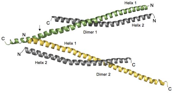
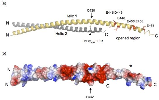
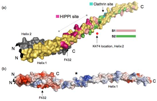
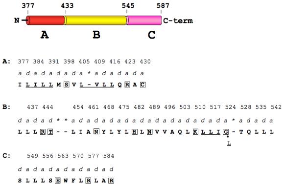
Huntington's disease is a genetic neurological disorder that is triggered by the dissociation of the huntingtin protein (htt) from its obligate interaction partner Huntingtin-interacting protein 1 (HIP1). The release of the huntingtin protein permits HIP1 protein interactor (HIPPI) to bind to its recognition site on HIP1 to form a HIPPI/HIP1 complex that recruits procaspase-8 to begin the process of apoptosis. The interaction module between HIPPI and HIP1 was predicted to resemble a death-effector domain. Our 2.8-A crystal structure of the HIP1 371-481 subfragment that includes F432 and K474, which is important for HIPPI binding, is not a death-effector domain but is a partially opened coiled coil. The HIP1 371-481 model reveals a basic surface that we hypothesize to be suitable for binding HIPPI. There is an opened region next to the putative HIPPI site that is highly negatively charged. The acidic residues in this region are highly conserved in HIP1 and a related protein, HIP1R, from different organisms but are not conserved in the yeast homologue of HIP1, sla2p. We have modeled approximately 85% of the coiled-coil domain by joining our new HIP1 371-481 structure to the HIP1 482-586 model (Protein Data Bank code: 2NO2). Finally, the middle of this coiled-coil domain may be intrinsically flexible and suggests a new interaction model where HIPPI binds to a U-shaped HIP1 molecule.


Figures

Figure 1
Figure 1
Orientation of HIP1 371-481 dimers in the crystal stabilizes the C-terminal region. Two dimers of HIP1 371-481 are arranged in an antiparallel “A” configuration. Helix 1 (green in Dimer 1) in each dimer shown is longer than Helix 2 (grey in Dimer 1) because of a crystal packing contact indicated by the arrow. At this point R380 and Y381 in Helix 1 of Dimer 2 (yellow) forms a pocket for D446 protruding from Helix 1 of Dimer 1 (green). The indicated lattice contact point faces away from the dimerization interfaces in the two dimers shown in the figure. The N- and C-termini of each helix are labeled N and C. The helices were rendered using PyMol [http://www.pymol.org]. Protein Expression and Purification: The plasmid encoding the N-terminal GST tagged HIP1 370-644 was kindly provided by the McPherson group. GST-HIP1 371-481 was made by altering the HIP1 cDNA to introduce a stop codon (TAG) at residue 482. Site-directed mutagenesis was performed according to the QuikChange mutagenesis protocol (Stratagene). The sequence was confirmed by DNA sequencing (IMBI, Indiana University) and then transformed into Rosetta 2 (DE3) pLysS cells (Novagen). The recombinant protein GST-HIP1 371-481 construct was expressed and purified as described previously with some modifications. Briefly, GST-HIP1 371-481 was over-expressed at 37°C in M9 minimum medium. After reaching an O.D. 600 of 0.5 units, selenomethionine (50 μg/ml final concentration) and IPTG (100μg/ml final concentration) were added to the bacterial culture. Cells were then incubated for 16 h at 30°C before being harvested and frozen for use. Cell pellets were thawed on ice and then resuspended gently in 45 ml of PBS (10 mM Na2HPO4, 1.8 mM KH2PO4 (pH 7.3), 140 mM NaCl, 2.7 mM KCl), supplemented with 0.25 ml of 1 M DTT, 0.25 ml of protease inhibitor cocktail (Sigma), and 2 ml of PMSF (17.4 mg/ml in 2-propanol) and incubated on ice for ∼15min. After sonication, 2.5 ml of 20% (v/v) Triton X-100 was added and the lysate was incubated at room temperature by rotating for ∼30 min. The crude bacterial lysate was cleared by centrifugation (19 k, 4°C) for 15 min and then the supernatant was mixed with ∼5 ml of glutathione Sepharose 4B (Amersham) resin suspended in PBS. This slurry was rocked gently at room temperature for 2 h before being transferred into a small column. The packed column was washed with 50 ml of PBS supplemented with 0.25 ml of 1M DTT until no more background protein was detected by staining with Coomassie brilliant blue. The GST-HIP1 371-481 was eluted from the column with 50 ml of 3 mg/ml L-glutathione (sigma) in (PBS) at pH 8.0 and dialyzed against the same buffer. The GST tag was removed by rocking the protein with sequencing grade thrombin (Novagen) at 37°C for 3 h. The digested sample was then dialyzed against 10 mM HEPES (pH 8.5), 20 mM Imidazole, 2 mM tris(2-carboxyethyl)-phosphine, 1% (v/v) glycerol. The protein was passed through an anion-exchange column (POROS 20 HQ) equilibrated in buffer A (10 mM HEPES, 20 mM imidazole, 2 mM tris(2-carboxyethyl)-phosphine, 1% (v/v) glycerol pH 8.5). The target protein was eluted with a linear gradient of buffer B (10 mM HEPES (pH 8.5), 20mM imidazole, 2 mM tris(2-carboxyethyl)-phosphine, 1% (v/v) glycerol, 250 mM NaCl). We confirmed the single selenomethionine substitution by electrospray mass spectroscopy. Crystallization and data collection: The protein was crystallized by the hanging drop method in reservoir buffer (15% PEG3350, 0.1 M succinate pH 7.0, 0.2 M potassium sodium tartrate, and 0.01 M NiCl2). The crystal grew in the tetragonal space group P43 (a=72.9, b=72.9, c=106.5, α=β=γ=90°), with two dimers in the asymmetric unit. The crystals were highly mosaic (∼2.9 degrees) along the c-axis. Exhaustive screening for alternative crystallization conditions or small molecule additives did not improve crystal quality. The crystals were looped out and quickly dipped in buffer containing ethylene glycol and then plunged in liquid nitrogen. The HIP 371-481 structure was solved by multi-wavelength anomalous dispersion (MAD). A 3-wavelength data set was obtained from a single crystal on beam line 4.2.2 at the Advanced Light Source, Lawrence Berkeley National Laboratory. The data were collected at 100 K in 1.0 degree oscillations using a NOIR-1 CCD detector. Wavelengths λ1 (1.0215Å) and λ2 (1.0213 Å) were at the peak and inflection, respectively, of the K-edge of selenium and λ3 (1.0373 Å) was a high energy remote. An inverse beam experiment was done at each wavelength, with E1=phi0 and E2=phi180 degrees, for a total oscillation of 90 degrees for each phi. The kappa angle was optimized to prevent overlaps in the long axis. Data were integrated and scaled using D*TREK. Phasing and refinement: The MAD data was phased using a Bayesian approach taking the high-energy remote (λ3) as the ‘native’ wavelength and the other two as ‘derivative’ wavelengths. The single selenium site in each monomer was found by SOLVE (http://www.solve.lanl.gov) and the experimental map was improved using RESOLVE . Model building was performed using O , the model was refined using CNS . Alternating rounds of positional, grouped B factor and simulated annealing were performed in reference to 2Fo-Fc and Fo- Fc maps and a bulk-solvent correction was applied near the end of refinement. The HIP1 371-481 structure refined against all the data from 30-2.8 Å with an R-factor of 26.5% and an Rfree of 32.4% at 2.8Å resolution.
Figure 2
Figure 2
(a) The opened region is highly negatively charged and is close to an interhelical disulfide bridge. Dimer 2 is shown and the colors of Helices 1 and 2 are the same as in Figure 1. There is a concentration of negatively charged residues in the open region (E445, D446, E448, E456, E458 and E465) indicated in red in Helix 1 (yellow). The C430 disulfide bridge between Helix 1 (grey) and Helix 2 (indicated by arrows) is directly upstream from the acidic opened region and is in the DDC430EF432LR stretch. The C-terminal end of Helix 2 is disordered and is not visible (see text). The ribbon model was created using PyMol [http://www.pymol.org]. (b) Surface potential analysis of HIP1 371-481 shows the opened region has a high negative potential (red surface marked by the asterisk). The position of the dimer is the same as in (a) and the arrow is F432 that is one residue away from the C430 disulfide bridge. The C430 disulfide bridge is in a region that forms an acidic ring around the HIP1 371-481 dimer (see red colored region around the arrow pointing to F432). The N- and C-termini in (a) and (b) are labeled N and C. The surface potentials (blue, positive; red, negative) were calculated using PyMol.
Figure 3
Figure 3
(a) Reassembled HIP1 coiled-coil domain using HIP1 371-481 (PDB code: 2QA7) and HIP1 482-586 (PDB code: 2NO2) models. Helix 1 of 2QA7 is in yellow and Helix 2 is in grey (ends just after F432, C-terminus labeled C). The N-terminus of 2QA7 has been cut slightly shorter for this figure. The C-terminal half of the assembled model is 2NO2, but only Helix 1 of this dimeric structure is shown in this figure for the sake of clarity. The surface bracketed by F432 and K474 indicated by the dashed line is a surface suitable for HIPPI binding. The position of K474 as it would be in Helix 2 (grey) is marked by the red dot. The residues forming the putative HIPPI binding surface between F432 and K474 are indicated in purple. The S1 (pink) and S2 (green) hydrophobic paths we described previously in 2NO2 are mapped out in the assembled HIP1 model to show that the HIPPI site is not in the dimerization interface, but is solvent exposed. The DLLRKN clathrin light chain-binding region (Clathrin site) is indicated in teal, and is barely visible because it is on the opposite face of Helix 1. The surface model was generated using PyMol [http://www.pymol.org]. (b) Surface potential analysis of the assembled model reveals the putative HIPPI site in (a) is highly basic (indicated by the asterisk) and is exposed to the solvent. This model is rotated relative to the model in (a) to show the basic region and is not on the same scale as the model in (a). The N- and C-termini are labeled N and C. The surface potentials (blue, positive; red, negative) were calculated using PyMol.
Figure 4
Figure 4
Hydrophobic core of the opened region of assembled HIP1 is disrupted by hydrophilic amino acids in a- and d-positions of the heptad repeat. The domain map of the assembled HIP1 model (371-587) is divided into coiled-coil regions A (red) and B (yellow) and an opened segment, designated region C (pink). N and C-term are the N- and C-terminal ends. Amino acids that occupy a- and d-positions (predicted by COILS 35) in regions A, B, and C are in bold capital letters and the a-position residue numbers are indicated above the a labels. Asterisks indicate skips in the heptad repeat flagged by COILS. Boxed amino acids in A, B, and C are disruptive to the hydrophobic core (see text). Region A: there are two coiled-coil stabilizing clusters (underlined, see text) separated by S398. Region B: the underlined LLI sequence may represent an altered coiled-coil cluster with glycine at position 524 instead of leucine. Changing G524 to L (indicated by the arrow) would create a stabilizing coiled-coil cluster (LLIL) seen in myosin .

References

    1. Nasir J, Floresco SB, O’Kusky JR, Diewert VM, Richman JM, Zeisler J, Borowski A, Marth JD, Phillips AG, Hayden MR. Targeted disruption of the huntington’s disease gene results in embryonic lethality and behavioral and morphological changes in heterozygotes. Cell. 1995;81:811–823. - PubMed
    1. Wanker EE, Rovira C, Scherzinger E, Hasenbank R, Walter S, Tait D, Colicelli J, Lehrach H. HIP-I: a huntingtin interacting protein isolated by the yeast two-hybrid system. Hum Mol Genet. 1997;6:487–95. - PubMed
    1. Gervais FG, Singaraja R, Xanthoudakis S, Gutekunst C-A, Leavitt BR, Metzler M, Hackam AS, Tam J, Vaillancourt JP, Houtzager V, Rasper DM, Roy S, Hayden MR, Nicholson DW. Recruitment and activation of caspase-8 by the huntingtin-interacting protein HIP-1 and a novel partner HIPPI. Nature Cell Biol. 2002;4:95–105. - PubMed
    1. Chen C-Y, Brodsky FM. Huntington-interacting protein 1 (Hip1) and Hip1-related protein (Hip1R) bind the conserved sequence of clathrin light chains and thereby influence clathrin assembly in vitro and actin distribution in vivo. J. Biol. Chem. 2005;280:6109–6117. - PubMed
    1. Mishra SK, Agostinelli NR, Brett TJ, Mizukami I, Ross TS, Traub LM. Clathrin- and AP-2-binding sites in HIP1 uncover a general assembly role for endocytic accessory proteins. J. Biol. Chem. 2001;276:46230–46236. - PubMed

Publication types

MeSH terms

Associated data