Review
. 2008 Mar 15;44(6):921-37.
doi: 10.1016/j.freeradbiomed.2007.11.008. Epub 2007 Nov 28.

Nonequilibrium thermodynamics of thiol/disulfide redox systems: a perspective on redox systems biology

Affiliations
Review

Nonequilibrium thermodynamics of thiol/disulfide redox systems: a perspective on redox systems biology

Melissa Kemp et al. Free Radic Biol Med. .

Abstract

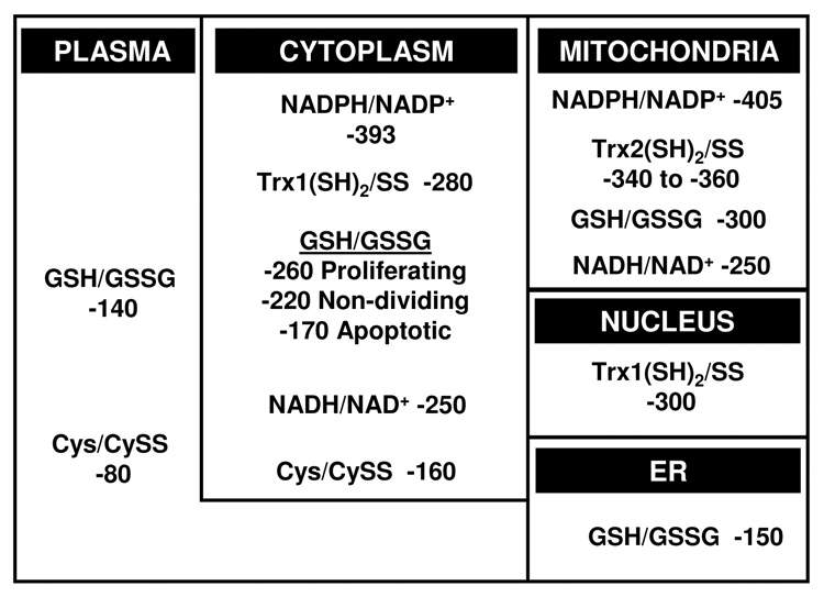
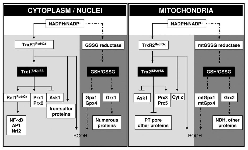
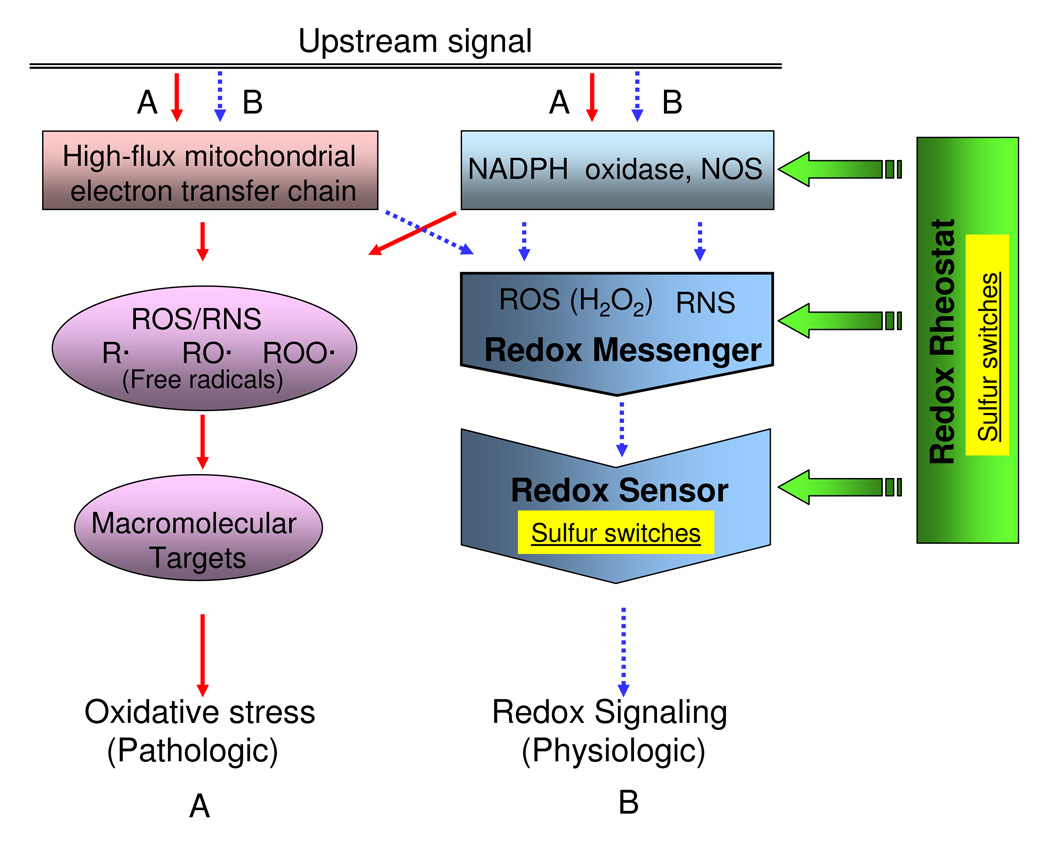
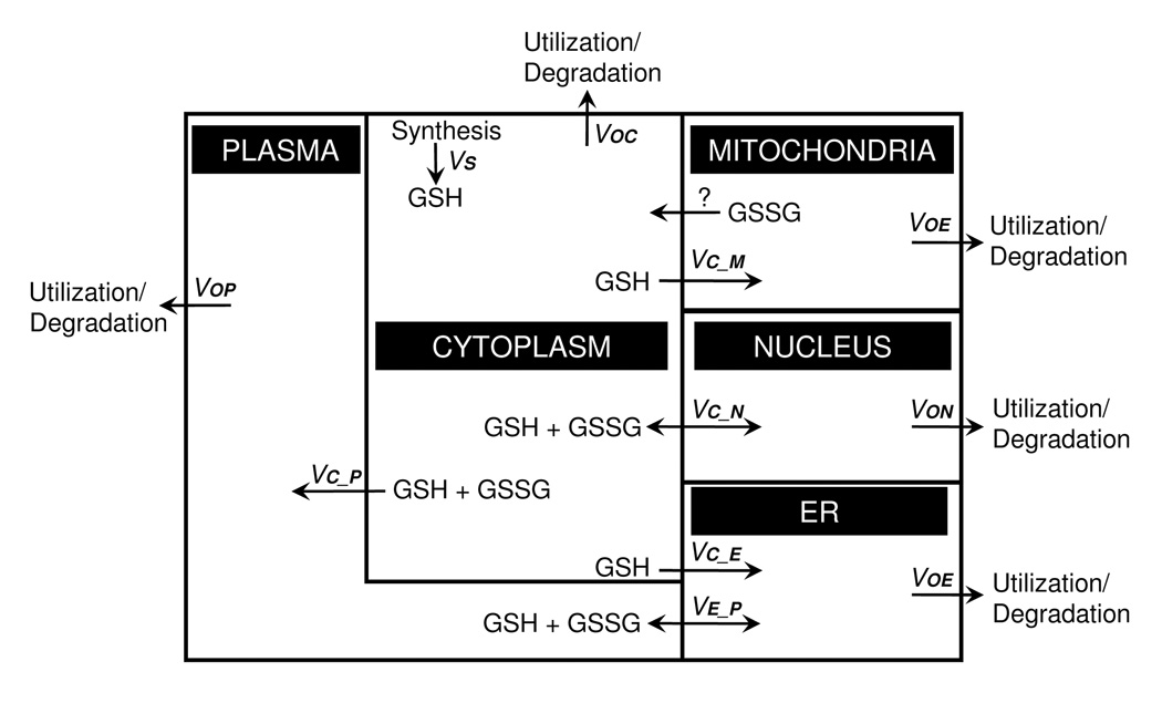
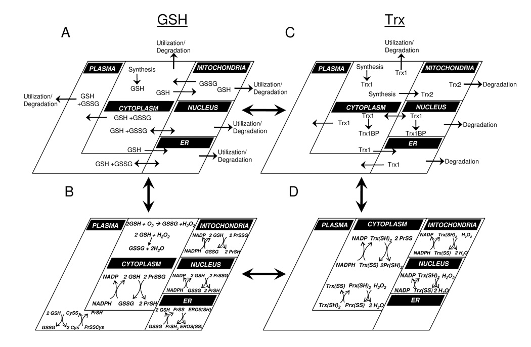
Understanding the dynamics of redox elements in biologic systems remains a major challenge for redox signaling and oxidative stress research. Central redox elements include evolutionarily conserved subsets of cysteines and methionines of proteins which function as sulfur switches and labile reactive oxygen species (ROS) and reactive nitrogen species (RNS) which function in redox signaling. The sulfur switches depend on redox environments in which rates of oxidation are balanced with rates of reduction through the thioredoxins, glutathione/glutathione disulfide, and cysteine/cystine redox couples. These central couples, which we term redox control nodes, are maintained at stable but nonequilibrium steady states, are largely independently regulated in different subcellular compartments, and are quasi-independent from each other within compartments. Disruption of the redox control nodes can differentially affect sulfur switches, thereby creating a diversity of oxidative stress responses. Systems biology provides approaches to address the complexity of these responses. In the present review, we summarize thiol/disulfide pathway, redox potential, and rate information as a basis for kinetic modeling of sulfur switches. The summary identifies gaps in knowledge especially related to redox communication between compartments, definition of redox pathways, and discrimination between types of sulfur switches. A formulation for kinetic modeling of GSH/GSSG redox control indicates that systems biology could encourage novel therapeutic approaches to protect against oxidative stress by identifying specific redox-sensitive sites which could be targeted for intervention.


Figures

Figure 1
Figure 1
Steady-state redox potentials for thiol/disulfide control nodes. A variety of methods have been used to estimate redox states of thiol/disulfide systems in different compartments. Most information is available for total cellular GSH/GSSG, which appears to largely represent cytoplasm. Rapidly proliferating cells typically have values which are 30 to 60 mV more reducing than non-dividing cells. Cells undergoing apoptosis become considerably more oxidized, mostly due to a loss of GSH. Cytoplasmic Trx1 is more reduced than GSH and is not affected by cell proliferation and is not oxidized until terminal stages of apoptosis. Cytoplasmic Cys/CySS is more oxidized than GSH/GSSG and varies independently. Trx2 and GSH/GSSG have a more reduced redox state than cytoplasmic counterparts. Nuclear Trx1 is more reduced than cytoplasmic Trx1 and is much more resistant to oxidation. The redox state of nuclear GSH/GSSG is not known, but ratios of PrSH/PrSSG for nuclear and cytoplasmic proteins indicates that the nuclear GSH pool is more reduced than in the cytoplasm. The extracellular pools of GSH/GSSG and Cys/CySS are considerably oxidized compared to the respective cytoplasmic pools.
Figure 2
Figure 2
GSH and Trx-dependent systems support different protein-dependent systems in cytoplasm and nuclei (left), and in mitochondria (right). Model is based upon observations that 1) Trx (Trx1 for cytoplasm and nuclei, Trx2 for mitochondria) and GSH systems function in parallel in peroxide metabolism, 2) steady-state redox potentials of NADPH/NADP, Trx1 (SH)2/(SS), GSH/GSSG in cytoplasm and nuclei, Trx2(SH)2/(SS), GSH/GSSG in mitochondria are maintained at different values, 3) GSH and Trxs have multiple functions in protection against oxidative stress and in redox control mechanisms. Thioredoxin reductase-1 (TrxR1) in cytoplasm and nuclei and TrxR2 in mitochondria also have multiple activities which include peroxide metabolism. Redox factor-1 (Ref-1), peroxiredoxin-1 (Prx1), Prx2 and Ask1 for Trx1-dependent proteins and Ask1, Prx3, and Prx5 for Trx2-dependent proteins are represented, which function in parallel with the GSH/GSSG-dependent proteins [glutathione peroxidase-1 (Gpx1), Gpx4, glutaredoxin (Grx1) for cytoplasm, mtGpx1, mtGpx4, and Grx2 for mitochondria]. TrxR1 and TrxR2-dependent proteins other than Trx1 and Trx2 could also exist, such as iron-sulfur proteins and cytochrome c (Cyt c). In this model, the steady-state redox values are maintained separately for each couple by the balance of NADPH-dependent reduction and peroxide-dependent oxidation rates. Each couple can thereby be used for different redox-control processes, and in the presence of excess peroxide generation rates, each can contribute to detoxification. Arrowheads represent flow of electrons while blunt-end lines represent redox-dependent interactions which could occur without electron transfer.
Figure 3
Figure 3
Sulfur switches can exist as "Redox Sensors" and as "Redox Rheostats" in generic redox circuitry models. Simplified models show (A) oxidative stress pathway (solid line) associated with high levels of ROS, RNS, and free radicals from mitochondrial dysfunction, NADPH oxidase activation, and nitric oxide synthase activation triggered by pathologic upstream signals. Redox signaling pathway (B, broken line) shows the process involving relatively low levels of diffusible reactive species associated with physiologic condition. In redox signaling, the small diffusible reactive species, termed "redox messengers" are generated by ROS/RNS sources and react with redox-sensitive macromolecules termed "redox sensors". These redox sensors are often sulfur switches, consisting of sulfur atoms within specific proteins. Sulfur switches also occur as regulatory elements, termed "redox rheostats", which are not needed to define the pathway but regulate its activity. The characteristics of these sulfur switches can differ because the redox sensors are integral components of pathways while the sulfur rheostats regulate activity.
Figure 4
Figure 4
A compartmental model of steady-state glutathione fluxes. Under homeostatic conditions, GSH transport must be balanced by degradation/utilization to balance the rate of cytosolic GSH synthesis. In the diagram, GSH influx to the ER, mitochondria and plasma is considered to be unidirectional, while the exocytosis of metabolites through the ER will release GSH and GSSG into the plasma at the same rate. Mitochondrial loss of GSH is poorly understood but could occur by unidirectional efflux of GSSG. Passive transport to the nucleus will be bidirectional and include GSH + GSSG. Salient features revealed by this formulation include: 1) an uncharacterized mechanism for loss of GSH or GSSG from mitochondria is needed to balance mitochondrial GSH/GSSG redox state, 2) non-equal partitioning of reductases and oxidases between the cytoplasm and nucleus could maintain a disequilibrium between compartments, 3) differential regulation of GSH and GSSG transport between compartments can determine differences in steady-state redox potential between compartments, and 4) the secretory pathway could provide a kinetically important route for electron transfer between cytoplasm and plasma.
Figure 5
Figure 5
Complex redox systems maps can be assembled from models for redox couples in different compartments. The steady-state flux model for GSH and GSSG (Panel A) can be combined with compartmental models for oxidation/reduction of GSH/GSSG (Panel B) to provide a more complex model including synthesis/degradation and transport along with redox processes. Similar models can be developed for thioredoxins (Panels C, D), and these can be combined with A and B to provide more complex descriptions of interacting redox systems. Such models can then be used to analyze cellular control of sulfur switches, linking redox changes to specific sensor and rheostat functions. In this simplified model, only GSH and Trx couples are illustrated; however, these concepts can be extended to NADPH/NADP+, peroxiredoxins, Cys/CySS and other redox couples. For illustrative purposes, peroxiredoxin reactions are only depicted in the cytosol, but are present in the nucleus and mitochondria as well.

Similar articles

Cited by

References

    1. Schafer FQ, Buettner GR. Redox environment of the cell as viewed through the redox state of the glutathione disulfide/glutathione couple. Free radical biology & medicine. 2001;30:1191–1212. - PubMed
    1. Hamdane D, Kiger L, Dewilde S, Green BN, Pesce A, Uzan J, Burmester T, Hankeln T, Bolognesi M, Moens L, Marden MC. The redox state of the cell regulates the ligand binding affinity of human neuroglobin and cytoglobin. The Journal of biological chemistry. 2003;278:51713–51721. - PubMed
    1. Jordan PA, Gibbins JM. Extracellular disulfide exchange and the regulation of cellular function. Antioxidants & redox signaling. 2006;8:312–324. - PubMed
    1. Kerblat I, Drouet C, Chesne S, Marche PN. Importance of thioredoxin in the proteolysis of an immunoglobulin G as antigen by lysosomal Cys-proteases. Immunology. 1999;97:62–68. - PMC - PubMed
    1. Reeves JP, Bailey CA, Hale CC. Redox modification of sodium-calcium exchange activity in cardiac sarcolemmal vesicles. The Journal of biological chemistry. 1986;261:4948–4955. - PubMed

Publication types