. 2008 Feb 1;314(1):127-36.
doi: 10.1016/j.ydbio.2007.11.018. Epub 2007 Nov 28.

A spatial bias for the origins of interneuron subgroups within the medial ganglionic eminence

Affiliations

A spatial bias for the origins of interneuron subgroups within the medial ganglionic eminence

Carl P Wonders et al. Dev Biol. .

Abstract

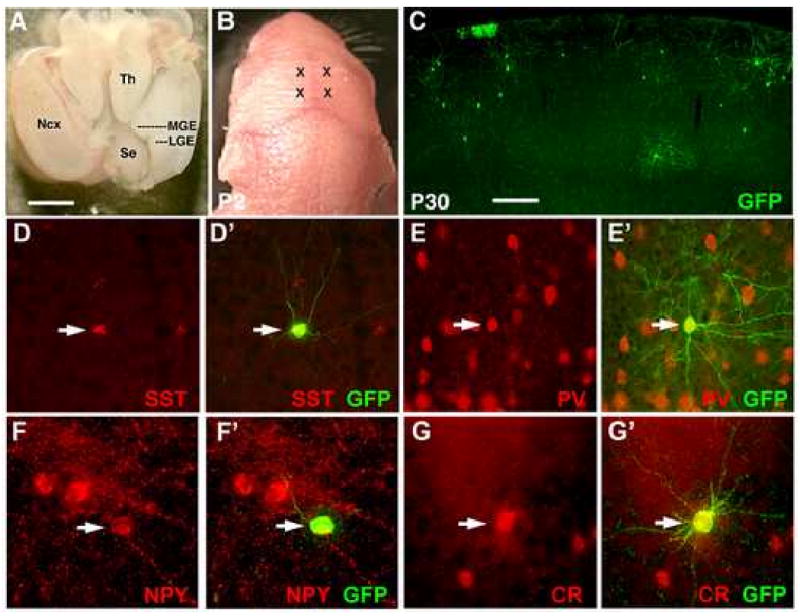
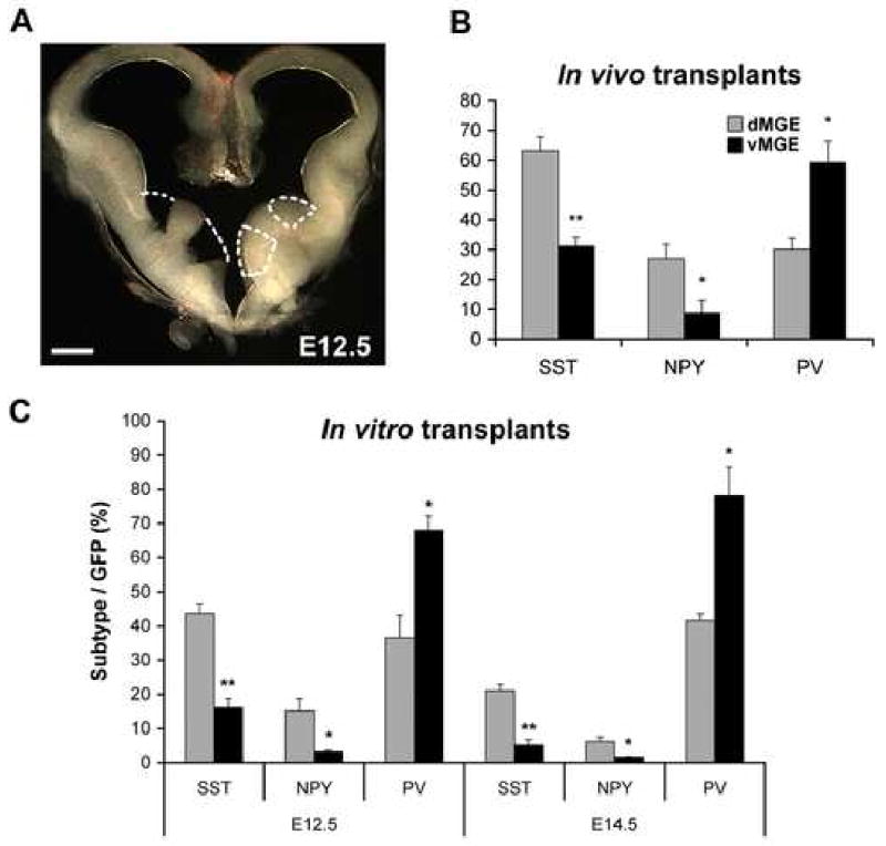
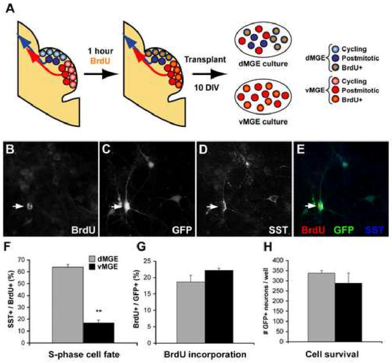
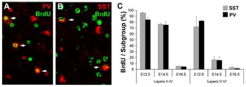
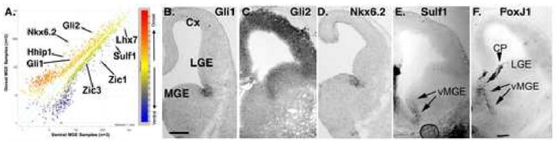
Although it is well established that the ventral telencephalon is the primary source of GABAergic cortical interneurons in rodents, little is known about the specification of specific interneuron subtypes. It is also unclear whether the potential to achieve a given fate is established at their place of origin or by signals received during their migration to or during their maturation within the cerebral cortex. Using both in vivo and in vitro transplantation techniques, we find that two major interneuron subgroups have largely distinct origins within the MGE. Somatostatin (SST)-expressing interneurons are primarily generated within the dorsal MGE, while parvalbumin (PV)-expressing interneurons primarily originate from the ventral MGE. In addition, we show that significant heterogeneity exists between gene expression patterns in the dorsal and ventral MGE. These results suggest that, like the spinal cord, neuronal fate determination in the ventral telencephalon is largely the result of spatially segregated, molecularly distinct microdomains arranged on the dorsal-ventral axis.


Figures

Figure 1
Figure 1. Cortical interneuron subgroup identity is determined within the MGE
(A) The brain of an E13.5 embryo as viewed from above. Rostral is to the panel bottom. To isolate the MGE, the neocortex (Ncx) was dissected away as shown for the right hemisphere. The sulcus between the MGE and LGE is extended slightly, and a cut made along the medial-lateral axis at the level of the thalamus (Th). The MGE can then be separated free from the remainder of the brain. (B) Two injection sites per hemisphere were made into each neonate (marked by X's). (C) By P30, transplanted GFP+ cells are found throughout the cortex, of which the majority has adopted cortical interneuron morphologies. (D-G) Transplanted cells co-express interneuron subgroup markers including somatostatin (D-D′), parvalbumin (E-E′), neuropeptide Y (F-F′), and calretinin (G-G′). Scale bar in (A) is 1mm, in (C) is 200μm.
Figure 2
Figure 2. A dorsal/ventral fate potential bias exists for MGE-derived progenitors transplanted both in vivo and in vitro
(A) The MGE of an E12.5 embryo dissected into dorsal and ventral domains as shown. Dissected domains were limited to the lighter-colored, periventricular region that is produced in these dark-field images by the higher cell packing density of the proliferative zone. Dissections of E13.5 and E14.5 MGE are performed similarly. (B) When E13.5 dorsal (d) and ventral (v) MGE progenitors are transplanted in vivo, both SST- and NPY-expressing interneurons are preferentially generated from dMGE transplants, while PV-expressing interneurons are preferentially generated from vMGE transplants. (C) At both E12.5 and E14.5, dMGE transplants also show a bias towards generating SST- and NPY-expressing interneurons, while the vMGE transplants preferentially give rise to PV-expressing interneurons when transplanted in vitro onto cortical feeders. Unpaired t-test: * p<0.05; ** p<0.005. Scale bar in (A) is 200μm.
Figure 3
Figure 3. Dorsal MGE cells labeled in S-phase 1 hr prior to transplantation in vitro show a strong bias towards SST+ interneuron generation
(A) Embryos are exposed to BrdU for 1 hour, allowing for the labeling of those cells in S-phase immediately prior to transplantation. This labeling makes it possible to separate “dMGE born” and “vMGE born” progenitors based on their expression of BrdU (orange). (B-E) Immunofluorescence labeling for BrdU (B), GFP (C), and SST (D) in transplant cultures grown for 10 DIV. (F) dMGE progenitors that had been in S-phase 1hr prior to transplant as labeled by an injection of BrdU (100 mg/kg) show a very pronounced bias towards SST interneuron generation compared to vMGE. (G-H) There is no significant difference in BrdU incorporation (% of cells that are BrdU+ at 10 DIV) or cell survival between dorsal and ventral MGE transplants. Unpaired t-test: * p<0.05; ** p<0.005.
Figure 4
Figure 4. SST- and PV-expressing interneuron subgroups are generated contemporaneously during development
The cortex of P30 mice, exposed to BrdU at E12.5, E14.5, or E16.5 as previously published (Xu et al., 2004), was processed for immunofluorescence for BrdU and either parvalbumin (A; PV) or somatostatin (B; SST) with arrows indicating co-expression. (C) The percentages of SST/PV co-labeling with BrdU are not significantly different at any of the ages studied in either the superficial (Layers II-IV) or deep (Layers V-VI) cortex.
Figure 5
Figure 5. An array based approach to identify genes enriched in the dorsal or ventral MGE
(A) The plot shows results averaged from 3 separately prepared, non-amplified RNA samples of dorsal or ventral MGE (dMGE, vMGE), run on the Affimatrix 430 2.0 array. Distance from the origin reflects strength of signal. Distance from the diagonal line is proportion to relative difference between dorsal and ventral signals for that RNA such that RNAs above the line are enriched in the dMGE, whereas RNAs below the line are enriched in the vMGE. (B-D) Several of the genes identified as enriched in the dMGE are known to be selectively expressed. Gli1 and Nkx6.2 expression in the dMGE have been shown to remain dependent on Shh signaling after initial patterning has been established (Xu et al., 2005). (E) Hedgehog Interacting Protein 1 (Hhip1) is also expressed in the dMGE along with another Shh signaling inhibitor, patched (Loulier et al., 2005). Hhip1 acts as an antagonist of Hedgehog signaling (Chuang et al., 2003), although it could conceivably also act to concentrate Hh proteins into a defined region where net Hh signaling could be enhanced (Saha and Schaffer, 2006).

References

    1. Alvarez-Dolado M, Calcagnotto ME, Karkar KM, Southwell DG, Jones-Davis DM, Estrada RC, Rubenstein JL, Alvarez-Buylla A, Baraban SC. Cortical inhibition modified by embryonic neural precursors grafted into the postnatal brain. J Neurosci. 2006;26:7380–9. - PMC - PubMed
    1. Anderson SA, Marin O, Horn C, Jennings K, Rubenstein JL. Distinct cortical migrations from the medial and lateral ganglionic eminences. Development. 2001;128:353–63. - PubMed
    1. Angevine JB, Sidman RL. Autoradiographic study of cell migration during histogenesis of cerebral cortex in the mouse. Nature. 1961;192:766–768. - PubMed
    1. Briscoe J, Chen Y, Jessell TM, Struhl G. A hedgehog-insensitive form of patched provides evidence for direct long-range morphogen activity of sonic hedgehog in the neural tube. Mol Cell. 2001;7:1279–91. - PubMed
    1. Butt SJ, Fuccillo M, Nery S, Noctor S, Kriegstein A, Corbin JG, Fishell G. The temporal and spatial origins of cortical interneurons predict their physiological subtype. Neuron. 2005;48:591–604. - PubMed

LinkOut - more resources