. 2008 Feb 8;376(1):23-34.
doi: 10.1016/j.jmb.2007.11.081. Epub 2007 Dec 4.

Cell type-specific cleavage of nucleocapsid protein by effector caspases during SARS coronavirus infection

Affiliations

Cell type-specific cleavage of nucleocapsid protein by effector caspases during SARS coronavirus infection

Claudia Diemer et al. J Mol Biol. .

Abstract

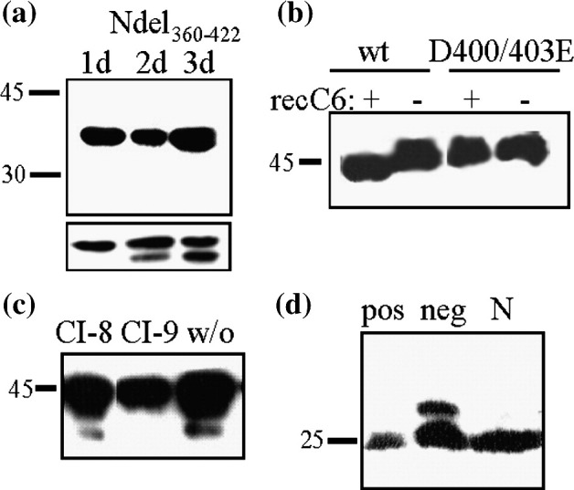
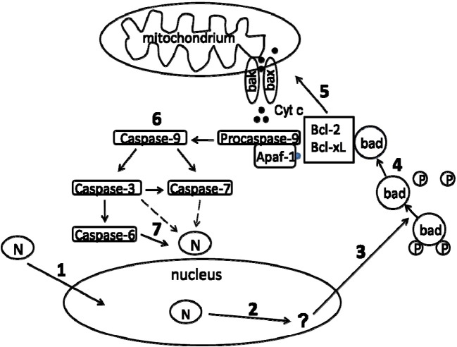
The epidemic outbreak of severe acute respiratory syndrome (SARS) in 2003 was caused by a novel coronavirus (CoV), designated SARS-CoV. The RNA genome of SARS-CoV is complexed by the nucleocapsid protein (N) to form a helical nucleocapsid. Besides this primary function, N seems to be involved in apoptotic scenarios. We show that upon infection of Vero E6 cells with SARS-CoV, which elicits a pronounced cytopathic effect and a high viral titer, N is cleaved by caspases. In contrast, in SARS-CoV-infected Caco-2 cells, which show a moderate cytopathic effect and a low viral titer, this processing of N was not observed. To further verify these observations, we transiently expressed N in different cell lines. Caco-2 and N2a cells served as models for persistent SARS-CoV infection, whereas Vero E6 and A549 cells did as prototype cell lines lytically infected by SARS-CoV. The experiments revealed that N induces the intrinsic apoptotic pathway, resulting in processing of N at residues 400 and 403 by caspase-6 and/or caspase-3. Of note, caspase activation is highly cell type specific in SARS-CoV-infected as well as transiently transfected cells. In Caco-2 and N2a cells, almost no N-processing was detectable. In Vero E6 and A549 cells, a high proportion of N was cleaved by caspases. Moreover, we examined the subcellular localization of SARS-CoV N in these cell lines. In transfected Vero E6 and A549 cells, SARS-CoV N was localized both in the cytoplasm and nucleus, whereas in Caco-2 and N2a cells, nearly no nuclear localization was observed. In addition, our studies indicate that the nuclear localization of N is essential for its caspase-6-mediated cleavage. These data suggest a correlation among the replication cycle of SARS-CoV, subcellular localization of N, induction of apoptosis, and the subsequent activation of caspases leading to cleavage of N.


Figures

Fig. 1
Fig. 1
SARS-CoV N processing in infected and transfected cell lines. Vero E6 (a) and Caco-2 (b) cells were infected with SARS-CoV and lysed 1, 2, or 3 days p.i. The lysates were subjected to immunoblot analysis using a polyclonal anti-N antiserum. SARS-CoV N was transiently transfected into Vero E6 (c), Caco-2 (d) A549 (e), or N2a (f) cells. Cells were lysed 1, 2, or 3 days after transfection, and lysates were analysed in immunoblot as described above. Identical results were obtained using additional polyclonal antisera (rabbit and human; data not shown). Molecular size markers are given on the left; arrows indicate the proteolytic cleavage product.
Fig. 2
Fig. 2
Effector caspases are mediating the N cleavage. Vero E6 cells were transiently transfected with SARS-CoV N as described above. Before harvesting, cells were treated with 10 mM NH4Cl (a and c), 2 μM LC (a and c), 100 μM z-VAD-FMK (CI) (a), 100 μM z-DEVD-FMK (CI-3) (b), or 100 μM z-VEID-FMK (CI-6) (c) or left untreated (w/o). For studies upon infection, Vero E6 cells were inoculated with SARS-CoV and treated with 100 μM z-VAD-FMK (CI) (d) or 100 μM z-VEID-FMK (CI-6) (e) or left untreated (w/o). Lysis occurred after treatment for 15 h, and lysates were analysed in immunoblot as described above. SARS-CoV-infected Vero E6 (Ve) and Caco-2 (Ca) cells or staurosporine-treated cells (co) were lysed 1 day p.i. and were subjected to immunoblot analysis using anti-cleaved lamin A antibodies (f).
Fig. 3
Fig. 3
Caspase-6 cleaves C-terminal residues 400 and 403 of N by activation of the intrinsic apoptotic pathway. A C-terminally truncated N mutant (Ndel360–422) was expressed in Vero E6 cells. Cells were lysed on days 1, 2, and 3 post-transfection and analysed in immunoblot (a). The inlet picture shows the situation for cells transfected in parallel with wt N. For in vitro caspase-6 cleavage assay, recombinant wt N and a double-substitution mutant (D400/403E) were each incubated with recombinant caspase-6 (+) or assay buffer (−). Cleavage of protein was investigated by immunoblotting (b). N-transfected Vero E6 cells were treated with 100 μM z-LEHD-FMK (CI-9) or 100 μM z-IETD-FMK (CI-8) or left untreated. Cell lysates were analysed in immunoblot (c). COS-1 cells were transfected with N or empty vector (neg) or were treated with staurosporine (pos) and lysed 3 days after transfection. Lysates were analysed using anti-Bad antibodies in immunoblot (d).
Fig. 4
Fig. 4
Nuclear and cytosolic fractions of N-transfected cells. Nuclear and cytosolic fractions of N-transfected Vero E6 (a), A549 (b), Caco-2 (c), and N2a (d) cells were separated at different time points [1 day (1d), 2 days (2d), and 3 days (3d)] post-transfection. The fractions were analysed with the polyclonal serum against N (left panel) in immunoblot. Anti-histone H3 (middle panel) and anti-Grb2 (right panel) antibodies were used as controls for purity of the nuclear and cytosolic fractions.
Fig. 5
Fig. 5
Subcellular localization of SARS-CoV N. For immunofluorescence assay, Vero E6 (a), A549 (b), Caco-2 (c), and N2a (d) cells were transiently transfected with SARS-CoV N. After permeabilization, N (red) was stained with a polyclonal serum against N and with cy3-conjugated goat anti-rabbit antibody (left panel) in the different cell lines. In the middle panel, the nuclei (blue) of cells were visualized by 4′,6-diamidino-2-phenylindole staining. The merge of both stainings is shown in the right panel.
Fig. 6
Fig. 6
Identification of the functional NLS of N. Schematic illustration of the three potential NLSs of SARS-CoV N (a) and that of the substitution mutant K257G,K258G,K262G (NLSII) of the NLS in the middle of N (b). Vero E6 cells were transiently transfected with the NLS substitution mutant (NLSII) and wt N (wt). The nuclear (n) and cytosolic (c) fractions were separated on days 2 and 3 after transfection and analysed in immunoblot (c). Vero E6 cells were transiently transfected with NLSII-Grb2 or Grb2, and nuclei (n) and cytosol (c) were fractionated on day 2 after transfection and analysed in immunoblot (d). COS-1 cells were transiently transfected with N or NLSII. After lysis, the samples were analysed with anti-Bad (e) or anti-N (f) (for loading control) antibodies by immunoblotting.
Fig. 7
Fig. 7
Hypothetical model for correlation of nuclear localization and caspase-mediated cleavage of N in lytic SARS-CoV infection. N translocates into the nucleus (1) and may activate gene expression or interact with nuclear components there (2), resulting in the de-phosphorylation of Bad (3). In this form, Bad is enabled to interact with Bcl-2 and Bcl-XL (4). This interaction releases Bax and Bak from pro-survival Bcl-2 proteins, enabling insertion of Bax and Bak in the mitochondrial membrane. The resulting membrane permeabilization facilitates cytochrome c efflux (5), which provokes the activation of pro-caspase-9 by formation of apoptosome. Once caspase-9 is activated, the caspase cascade occurs and effector caspases (i.e., caspase-3, caspase-6, and caspase-7) are activated (6). Finally, N is cleaved by caspase-6 (7), but cleavage of N may additionally be caused by caspase-3 or caspase-7.

References

    1. Drosten C., Gunther S., Preiser W., van der W.S., Brodt H.R., Becker S. Identification of a novel coronavirus in patients with severe acute respiratory syndrome. N. Engl. J. Med. 2003;348:1967–1976. - PubMed
    1. Ksiazek T.G., Erdman D., Goldsmith C.S., Zaki S.R., Peret T., Emery S. A novel coronavirus associated with severe acute respiratory syndrome. N. Engl. J. Med. 2003;348:1953–1966. - PubMed
    1. Ziebuhr J. Molecular biology of severe acute respiratory syndrome coronavirus. Curr. Opin. Microbiol. 2004;7:412–419. - PMC - PubMed
    1. Marra M.A., Jones S.J., Astell C.R., Holt R.A., Brooks-Wilson A., Butterfield Y.S. The genome sequence of the SARS-associated coronavirus. Science. 2003;300:1399–1404. - PubMed
    1. Rota P.A., Oberste M.S., Monroe S.S., Nix W.A., Campagnoli R., Icenogle J.P. Characterization of a novel coronavirus associated with severe acute respiratory syndrome. Science. 2003;300:1394–1399. - PubMed