. 2008 Jan;172(1):194-202.
doi: 10.2353/ajpath.2008.070597. Epub 2007 Dec 21.

Prolactin drives estrogen receptor-alpha-dependent ductal expansion and synergizes with transforming growth factor-alpha to induce mammary tumors in males

Affiliations

Prolactin drives estrogen receptor-alpha-dependent ductal expansion and synergizes with transforming growth factor-alpha to induce mammary tumors in males

Lisa M Arendt et al. Am J Pathol. 2008 Jan.

Abstract

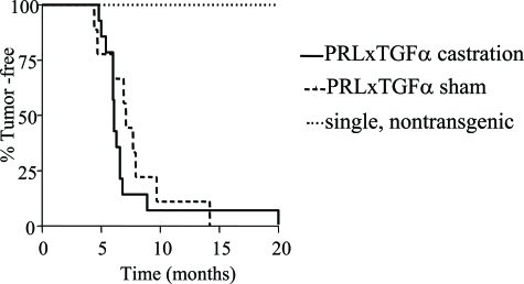
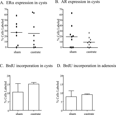
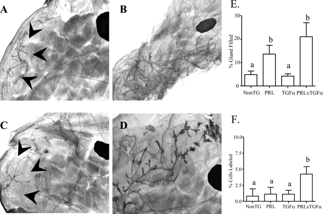
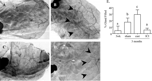
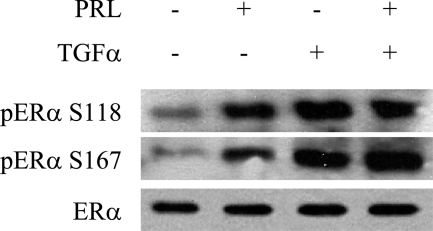
Male breast cancer is rare and has been the focus of limited research. Although the etiology is unclear, conditions increasing circulating prolactin (PRL), as well as estrogen, increase the risk of tumorigenesis. We modeled exposure to elevated PRL in transgenic mice, using the mammary-selective, estrogen-insensitive promoter neu-related lipocalin (NRL), to drive PRL expression. Male NRL-PRL mice did not develop mammary tumors. However, in cooperation with the well-characterized oncogene transforming growth factor-alpha (TGF-alpha), PRL induced mammary tumors in 100% of male bitransgenic mice. Similar to disease in human males, these tumors expressed variable levels of estrogen receptor-alpha (ER-alpha) and androgen receptors. However, carcinogenesis was not responsive to testicular steroids because castration did not alter latency to tumor development or tumor ER-alpha expression. Interestingly, both NRL-TGF-alpha/PRL and NRL-PRL males demonstrated increased ductal development, which occurred during puberty, similar to female mice. This outgrowth was diminished in NRL-PRL males treated with ICI 182,780, suggesting that PRL enhances ER-mediated growth. Treatment of MCF-7-derived cells with PRL increased phosphorylation of ER-alpha at residues implicated in unliganded ER-alpha activity. Together, these studies suggest that PRL expands the pool of cells susceptible to tumorigenesis, which is then facilitated by PRL and TGF-alpha cross talk. Activation of ER-alpha is one mechanism by which PRL may contribute to breast cancer and points to other therapeutic strategies for male patients.


Figures

Figure 1
Figure 1
Expression pattern of NRL promoter, mammary neoplasias, preneoplastic lesions, and steroid receptor expression. A: hPAP was uniformly expressed in mammary epithelial cells of transgenic male NRL-hPAP mice. Higher magnification of epithelial cells from gland of transgenic male NRL-hPAP exposed to BCIP substrate and counterstained with nuclear fast red. B: Glands from wild-type male mice did not stain positively for hPAP. C: Simple papillary epithelial lining of macrocyst from gland of NRL-TGF-α/PRL male. D: Squamous adenocarcinoma, with fibrous capsule and keratin deposition from gland of NRL-TGF-α/PRL male. E: Preneoplastic mammary intraepithelial neoplasia (MIN) from gland of NRL-TGF-α/PRL male. F: Adenosis surrounding region of normal ducts from gland of NRL-TGF-α/PRL male. Note hyperplastic glandular structures containing eosinophilic secretions. G: ER-α+ macrocyst. H: ER-α macrocyst. I: AR+ macrocyst. J: AR macrocyst. K: pERK1/2 expression in epithelial cells lining macrocyst. Original magnifications: ×25 (A, B); ×100 (C); ×200 (D–F); ×400 (G–K); ×600 (inset).
Figure 2
Figure 2
Castration did not affect tumor latency in NRL-TGF-α/PRL male mice. Bitransgenic, single transgenic, and nontransgenic male mice underwent castration or sham surgery at 3 months of age and were monitored for tumor development. End stage was defined when tumors reached 1.5 cm in diameter. The latencies were compared by Kaplan-Meier analysis, and differences were detected using the Mantel-Haenszel test. All bitransgenic male mice developed mammary tumors with no significant difference in latency (P = 0.14). No castrated and sham-treated single transgenic and nontransgenic littermates developed mammary lesions.
Figure 3
Figure 3
Effect of castration on ER-α and AR expression in macrocysts and proliferation in mammary lesions of NRL-TGF-α/PRL male mice. Percentage of ER-α+ (A) and AR+ (B) cells present in mammary macrocysts. Each symbol represents a single macrocyst. Proliferation in macrocysts (C) and adenosis lesions (D) was determined as described in Materials and Methods and expressed as mean ± SD.
Figure 4
Figure 4
Representative whole mounts from inguinal glands of males of the different genotypes. A: Nontransgenic. B: NRL-PRL. C: NRL-TGF-α. D: NRL-TGF-α/PRL. The large dark oval in each mammary gland is a lymph node, and arrowheads mark boundaries of ductal elongation. E: Quantitation of ductal elongation into mammary fat pad of male mice. F: Proliferation rates as determined by BrdU labeling in glands of male mice. Glands were collected from single and nontransgenic males at 1 year of age or after macrocyst development in bitransgenic males, and prepared and evaluated as described in Materials and Methods. Data are expressed as mean ± SD. Different lowercase letters denote statistical differences among the different lines as determined by the Kruskal-Wallis test followed by Mann-Whitney post test (P < 0.05). Original magnifications, ×10.
Figure 5
Figure 5
Ductal elongation is significantly inhibited in NRL-PRL males treated with ICI 182,780. A: Rudimentary ductal growth in glands of 3-week-old NRL-PRL male. B: Glands from 3-month-old NRL-PRL males demonstrated ductal elongation during puberty. C: Ductal elongation was significantly enhanced in glands of 3-month-old NRL-PRL males castrated at 3 weeks of age. D: Ductal elongation was significantly inhibited in glands of 3-month-old NRL-PRL males after weekly injections of ICI 182,780 starting at 3 weeks of age. E: Quantitation of ductal elongation into mammary fat pad as described in Materials and Methods. Data are expressed as mean ± SD. Different lowercase letters denote statistical differences among the different lines as determined by the Kruskal-Wallis test followed by Mann-Whitney post test (P < 0.05). Original magnifications: ×20 (A); ×10 (B–D).
Figure 6
Figure 6
PRL and TGF-α induce phosphorylation of ER-α. Representative immunoblot of MCF-7-derived cells treated with either 4 nmol/L PRL or 5.5 nmol/L TGF-α, which induced increased levels of S118 and S167 phosphorylated ER-α compared to vehicle.

References

    1. American Cancer Society Atlanta: American Cancer Society; Cancer Facts and Figures, 2006–2007. 2007
    1. Nahleh ZA. Hormonal therapy for male breast cancer: a different approach for a different disease. Cancer Treat Rev. 2006;32:101–105. - PubMed
    1. Krause W. Male breast cancer—an andrological disease: risk factors and diagnosis. Andrologia. 2004;36:346–354. - PubMed
    1. Giordano SH. A review of the diagnosis and management of male breast cancer. Oncologist. 2005;10:471–479. - PubMed
    1. O’Malley C, Shema S, White E, Glaser S. Incidence of male breast cancer in California, 1988–2000: racial/ethnic variation in 1759 men. Breast Cancer Res Treat. 2005;93:145–150. - PubMed

Publication types