. 2008 Mar;190(5):1531-8.
doi: 10.1128/JB.01660-07. Epub 2007 Dec 21.

LuxG is a functioning flavin reductase for bacterial luminescence

Affiliations

LuxG is a functioning flavin reductase for bacterial luminescence

Sarayut Nijvipakul et al. J Bacteriol. 2008 Mar.

Abstract

The luxG gene is part of the lux operon of marine luminous bacteria. luxG has been proposed to be a flavin reductase that supplies reduced flavin mononucleotide (FMN) for bacterial luminescence. However, this role has never been established because the gene product has not been successfully expressed and characterized. In this study, luxG from Photobacterium leiognathi TH1 was cloned and expressed in Escherichia coli in both native and C-terminal His6-tagged forms. Sequence analysis indicates that the protein consists of 237 amino acids, corresponding to a subunit molecular mass of 26.3 kDa. Both expressed forms of LuxG were purified to homogeneity, and their biochemical properties were characterized. Purified LuxG is homodimeric and has no bound prosthetic group. The enzyme can catalyze oxidation of NADH in the presence of free flavin, indicating that it can function as a flavin reductase in luminous bacteria. NADPH can also be used as a reducing substrate for the LuxG reaction, but with much less efficiency than NADH. With NADH and FMN as substrates, a Lineweaver-Burk plot revealed a series of convergent lines characteristic of a ternary-complex kinetic model. From steady-state kinetics data at 4 degrees C pH 8.0, Km for NADH, Km for FMN, and kcat were calculated to be 15.1 microM, 2.7 microM, and 1.7 s(-1), respectively. Coupled assays between LuxG and luciferases from P. leiognathi TH1 and Vibrio campbellii also showed that LuxG could supply FMNH- for light emission in vitro. A luxG gene knockout mutant of P. leiognathi TH1 exhibited a much dimmer luminescent phenotype compared to the native P. leiognathi TH1, implying that LuxG is the most significant source of FMNH- for the luminescence reaction in vivo.


Figures

FIG. 1.
FIG. 1.
Map diagramming the construction of the P. leiognathi-luxG knockout.
FIG. 2.
FIG. 2.
Pairwise alignment of the sequences of LuxG from P. leiognathi TH1 isolated in the present study (isolated_luxG) and LuxG previously reported (accession number AAA25621). The conserved residues are indicated by asterisk marks (*). Boldface letters represent the residues identical to those of E. coli Fre. Based on the structure of E. coli Fre (15), the flavin reductase can be divided into two domains; the N-terminal domain that binds flavin (indicated by “F”) and the C-terminal domain that probably binds NAD(P)H (indicated by “N”). Based on the structure of Fre (15), the secondary structure elements are shown above the text (β strands are indicated by arrows and α helices are indicated by solid blocks).
FIG. 3.
FIG. 3.
SDS-PAGE (15%) analysis of the purification of recombinant LuxG and His6-tagged LuxG. Lanes 1 to 3 (LuxG): 1, crude extract; 2, after purification by DEAE-Sepharose chromatography; 3, after purification by Sephacryl S-200 chromatography. Lane 4 shows the results for His6-tagged LuxG after purification by Ni+-Sepharose chromatography. The molecular size markers were phosphorylase b (97.4 kDa), bovine serum albumin (66.2 kDa), hen egg white ovalbumin (45 kDa), carbonic anhydrase (31 kDa), soybean trypsin inhibitor (21.5 kDa), and hen egg white lysozyme (14.4 kDa). The subunit molecular mass of LuxG was calculated to be ∼26 kDa.
FIG. 4.
FIG. 4.
SDS-PAGE analysis of the purification of recombinant LuxAB. Lanes: 1, crude extract; 2, ammonium sulfate fraction (35 to 60%); 3, purified by DEAE-Sepharose chromatography; 4, extract purified by Sephacryl-200 chromatography. P. leiognathi luciferase is a heterodimeric protein with the α subunit (∼40.5 kDa) and β subunit (∼38 kDa).
FIG. 5.
FIG. 5.
Steady-state kinetics of the LuxG reaction. A primary double-reciprocal plot of initial rates of the His6-tagged LuxG reactions versus various concentrations of NADH and FMN. Each line represents e/V values at a fixed concentration of FMN. Assay reactions were performed in 50 mM Tris-Cl (pH 8), 10% (vol/vol) glycerol, 1 mM DTT, and 100 nM concentrations of His6-tagged LuxG with various concentrations of FMN as indicated (1 to 40 μM) and NADH (5 to 200 μM) using a stopped-flow apparatus at 4°C under aerobic conditions. The Dalziel parameters (7, 9) were determined: φ[NADH] = 8.97 μM s, φ[FMN] = 1.60 μM s, and φo = 0.59 s.
FIG. 6.
FIG. 6.
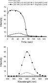
In vitro luminescence kinetics of coupled reactions with LuxAB and His6-tagged LuxG. The reaction was composed of 400 nM P. leiognathi LuxAB (•) or V. campbellii LuxAB (□) (34), 100 μM dodecanal, 20 μM FMN, and 200 μM NADH in 50 mM Tris-Cl buffer (pH 8.0), 10% (vol/vol) glycerol, and 1 mM DTT in the presence or absence (○) of 400 nM His6-tagged LuxG under aerobic conditions. (Upper panel) Light emitted at 490 nm was observed with time in the spectrofluorometer with no excitation light. (Lower panel) Emission spectra of light emitted from the same reactions of the upper panel.
FIG. 7.
FIG. 7.
Comparison of the luminescence of P. leiognathi-luxG knockout and of native P. leiognathi TH1. Both strains were cultured on the same LB agar plate at 28°C for ∼16 h. The picture was taken in the dark by using a Canon Powershot A620 digital camera with an exposure time of 30 s and an aperture of 4.0. This result indicates that the luxG knockout strain was much less luminescent that the native strain.
FIG. 8.
FIG. 8.
Proposed kinetic mechanism of the LuxG reaction.

Similar articles

Cited by

References

    1. Alexeyev, M. F. 1999. The pKNOCK series of broad-host-range mobilizable suicide vectors for gene knockout and targeted DNA insertion into the chromosome of gram-negative bacteria. BioTechniques 26824-826, 828. - PubMed
    1. Andrews, S. C., D. Shipley, J. N. Keen, J. B. Findlay, P. M. Harrison, and J. R. Guest. 1992. The hemoglobin-like protein (HMP) of Escherichia coli has ferrisiderophore reductase activity and its C-terminal domain shares homology with ferredoxin NADP+ reductases. FEBS Lett. 302247-252. - PubMed
    1. Ast, J. C., H. Urbanczyk, and P. V. Dunlap. 2007. Natural merodiploidy of the lux-rib operon of Photobacterium leiognathi from coastal waters of Honshu, Japan. J. Bacteriol. 1896148-6158. - PMC - PubMed
    1. Bruns, C. M., and P. A. Karplus. 1995. Refined crystal structure of spinach ferredoxin reductase at 1.7 Å resolution: oxidized, reduced and 2′-phospho-5′-AMP bound states. J. Mol. Biol. 247125-145. - PubMed
    1. Chaiyen, P., C. Suadee, and P. Wilairat. 2001. A novel two-protein component flavoprotein hydroxylase. Eur. J. Biochem. 2685550-5561. - PubMed

Publication types

MeSH terms

LinkOut - more resources