. 2008 Jan;233(1):94-105.
doi: 10.3181/0704-RM-113.

Mechanistic analysis of electroporation-induced cellular uptake of macromolecules

Affiliations

Mechanistic analysis of electroporation-induced cellular uptake of macromolecules

David A Zaharoff et al. Exp Biol Med (Maywood). 2008 Jan.

Abstract

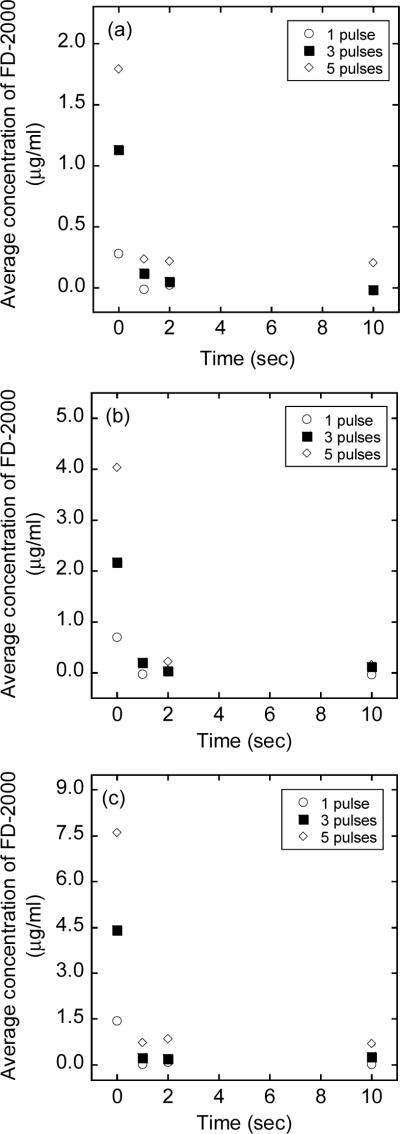
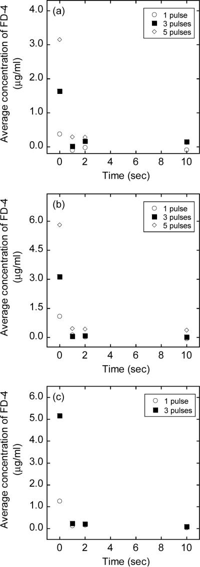
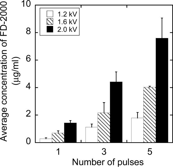
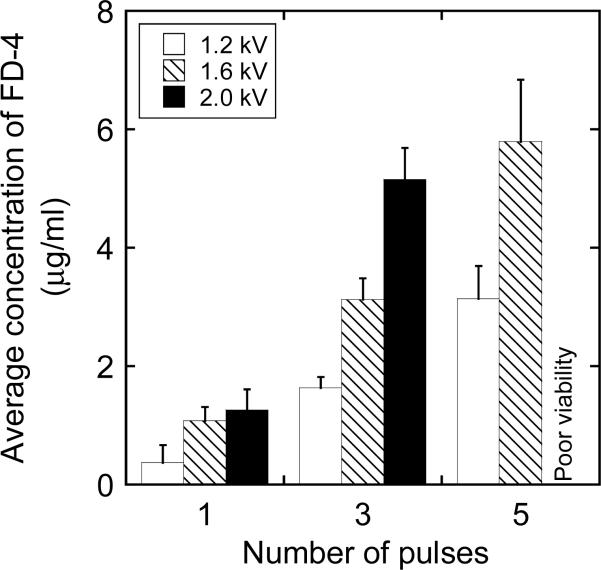
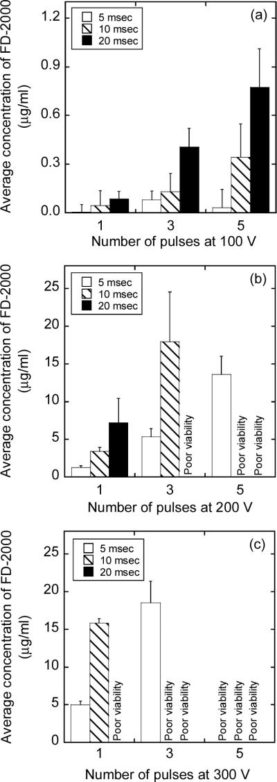
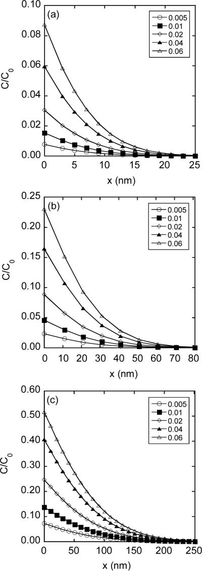
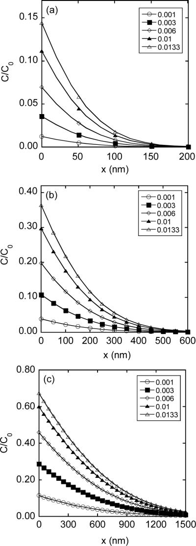
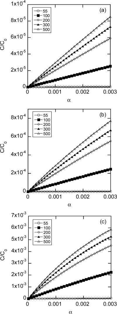
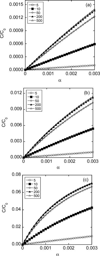
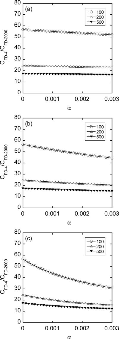
Pulsed electric field has been widely used as a nonviral gene delivery platform. The delivery efficiency can be improved through quantitative analysis of pore dynamics and intracellular transport of plasmid DNA. To this end, we investigated mechanisms of cellular uptake of macromolecules during electroporation. In the study, fluorescein isothiocyanate-labeled dextran (FD) with molecular weight of 4,000 (FD-4) or 2,000,000 (FD-2000) was added into suspensions of a murine mammary carcinoma cell (4T1) either before or at different time points (ie, 1, 2, or 10 sec) after the application of different pulsed electric fields (in high-voltage mode: 1.2-2.0 kV in amplitude, 99 microsec in duration, and 1-5 pulses; in low-voltage mode: 100-300 V in amplitude, 5-20 msec in duration, and 1-5 pulses). The intracellular concentrations of FD were quantified using a confocal microscopy technique. To understand transport mechanisms, a mathematical model was developed for numerical simulation of cellular uptake. We observed that the maximum intracellular concentration of FD-2000 was less than 3% of that in the pulsing medium. The intracellular concentrations increased linearly with pulse number and amplitude. In addition, the intracellular concentration of FD-2000 was approximately 40% lower than that of FD-4 under identical pulsing conditions. The numerical simulations predicted that the pores larger than FD-4 lasted <10 msec after the application of pulsed fields if the simulated concentrations were on the same order of magnitude as the experimental data. In addition, the simulation results indicated that diffusion was negligible for cellular uptake of FD molecules. Taken together, the data suggested that large pores induced in the membrane by pulsed electric fields disappeared rapidly after pulse application and convection was likely to be the dominant mode of transport for cellular uptake of uncharged macromolecules.


Figures

Figure 1
Figure 1
Average concentrations of FD-2000 in 4T1 cells exposed to high voltage, short duration pulses with an amplitude of (a) 1.2 kV, (b) 1.6 kV, or (c) 2.0 kV. The number of pulses was 1, 3, or 5; and the pulse duration was 99 μsec in all cases. FD-2000 was added into cell suspensions either immediately before the exposure, which is indicated as time to be at zero, or at different time points (i.e., 1, 2, or 10 sec) after the exposure. Each data point presents of the mean of measurements in three repeated experiments.
Figure 2
Figure 2
Average concentrations of FD-4 in 4T1 cells exposed to pulsed electric fields. The experimental conditions were identical to those for FD-2000 shown in Figure 1, except that FD-4 instead of FD-2000 was added into cell suspensions. Each data point presents of the mean of measurements in three repeated experiments.
Figure 3
Figure 3
Average concentration of FD-2000 in 4T1 cells exposed to pulsed electric fields. The experimental conditions were identical to those shown in Figure 1, except that FD-2000 was always added into cell suspensions immediately before the exposure. The error bar represents the standard deviation of data from three repeated experiments.
Figure 4
Figure 4
Average concentration of FD-4 in 4T1 cells exposed to pulsed electric fields. The experimental conditions were identical to those shown in Figure 2, except that FD-4 was always added into cell suspensions immediately before the exposure. For cells treated with 5 pulses at 2.0 kV each, the intracellular concentrations were not measured because the cell viability was estimated to be less than 10%. The error bar represents the standard deviation of data from three repeated experiments.
Figure 5
Figure 5
Average concentrations of FD-2000 in 4T1 cells exposed to low voltage, long duration pulses with an amplitude of (a) 100 V, (b) 200 V, or (c) 300 V. The number of pulses was 1, 3, or 5; and the pulse duration was 5, 10, or 20 msec. FD-2000 was added into cell suspensions immediately before the exposure. The intracellular concentrations were not quantified if the cell viability was < 10% as indicated by “poor viability”. The error bar represents the standard deviation of data from three repeated experiments.
Figure 6
Figure 6
Spatial distribution of intracellular concentration of FD-2000 for different pore opening periods (T): (a) T = 0.1 msec; (b) T = 1 msec; and (c) T = 10 msec. The concentration profiles normalized by the concentration in pulsing medium (C0) were simulated using the mathematical model described in the Materials and Methods section. At the permeabilized membrane, x = 0. The concentration profiles depended on the parameter hδ, where h is the ratio of membrane permeability versus diffusion coefficients and δ is the membrane thickness. The maximum range of hδ was estimated by using Equation 8, in which λ was assumed to be zero, D0/D = 1/0.05, and the value of α was between 0 and 0.3% (see the Materials and Methods section). As a result, hδ ≤ 0.06. It was varied from 0.005 to 0.06 in the simulation.
Figure 7
Figure 7
Spatial distribution of intracellular concentration of FD-4 for different pore opening periods (T): (a) T = 0.1 msec; (b) T = 1 msec; and (c) T = 10 msec. The concentration profiles were normalized by the concentration in pulsing medium (C0). The value of hδ was varied from 0.001 to 0.0133 (see the legend of Figure 6).
Figure 8
Figure 8
Average intracellular concentrations of FD-2000 as a function of the fractional area of pores in the permeabilized membrane (α) for different pore opening periods (T): (a) T = 0.1 msec; (b) T = 1 msec; and (c) T = 10 msec. The average concentrations were normalized by the concentration in pulsing medium (C0), which depended on average pore diameter ranging from 55 to 500 nm in the simulation.
Figure 9
Figure 9
Average intracellular concentrations of FD-4 as a function of the fractional area of pores in the permeabilized membrane (α) for different pore opening periods (T): (a) T = 0.1 msec; (b) T = 1 msec; and (c) T = 10 msec. The average concentrations were normalized by the concentration in pulsing medium (C0), which depended on average pore diameter ranging from 5 to 500 nm in the simulation.
Figure 10
Figure 10
Ratios of intracellular concentrations between FD-4 and FD-2000 as a function of the fractional area of pores in the permeabilized membrane cap (α) for different pore opening periods (T): (a) T = 0.1 msec; (b) T = 1 msec; and (c) T = 10 msec. The ratios depended on average pore diameter that was chosen to be 100, 200, or 500 nm in the simulation.

References

    1. Verma IM, Somia N. Gene therapy -- promises, problems and prospects. Nature. 1997;389:239–242. - PubMed
    1. Kay MA, Glorioso JC, Naldini L. Viral vectors for gene therapy: the art of turning infectious agents into vehicles of therapeutics. Nat Med. 2001;7:33–40. - PubMed
    1. Wang Y, Yuan F. Delivery of viral vectors to tumor cells: extracellular transport, systemic distribution, and strategies for improvement. Ann Biomed Eng. 2006;34:114–127. - PubMed
    1. Zaharoff DA, Barr RC, Li CY, Yuan F. Electromobility of plasmid DNA in tumor tissues during electric field-mediated gene delivery. Gene Ther. 2002;9:1286–1290. - PubMed
    1. Zaharoff DA, Yuan F. Effects of pulse strength and pulse duration on in vitro DNA electromobility. Bioelectrochemistry. 2004;62:37–45. - PubMed

Publication types

Substances