

Comparison of X-chromosome inactivation patterns in multiple tissues from human females
- PMID: 18156436
- PMCID: PMC5489244
- DOI: 10.1136/jmg.2007.055244
Comparison of X-chromosome inactivation patterns in multiple tissues from human females
Abstract
Background: X-chromosome inactivation (XCI) is the mechanism by which gene dosage uniformity is achieved between female mammals with two X chromosomes and male mammals with a single X chromosome, and is thought to occur randomly. For molecular genetic testing, accessible tissues (eg blood) are commonly studied, but the relationship with inaccessible tissues (eg brain) is poorly understood. For accessible tissues to be informative for genetic analysis, a high degree of concordance of genetic findings among tissue types is required.
Objective: To determine the relationship among multiple tissues within females at different ages (fetus to 82 years).
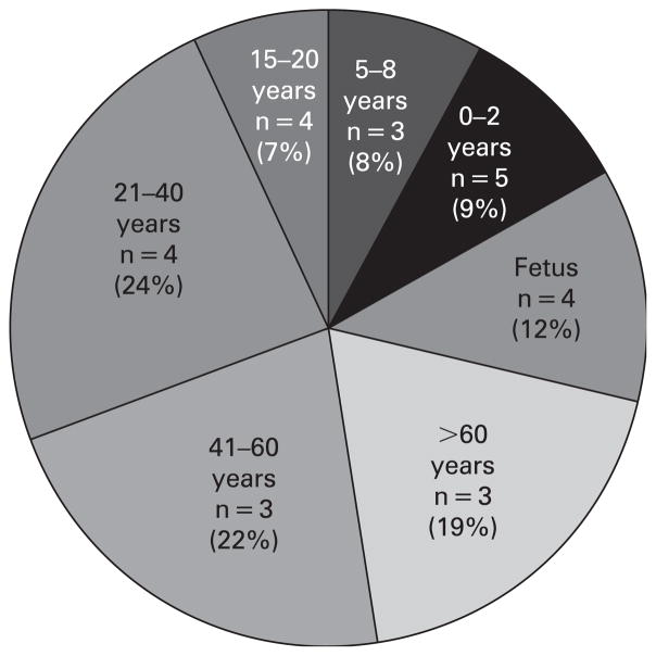
Methods: XCI patterns were analysed using the polymorphic androgen receptor (AR) gene assay. DNA was isolated from 26 different human females without history of malignancy, using 34 autopsy tissues representing the three embryonic germ layers.
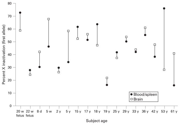
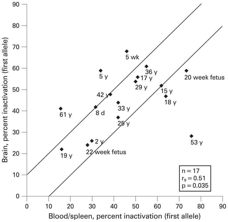
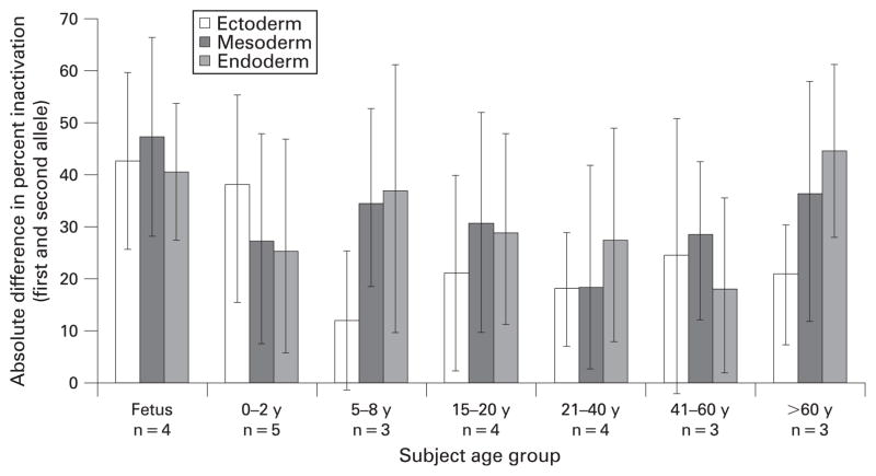
Results: 33 of the 280 tissue samples analysed from 13 of the 26 females showed skewed XCI values (>80:20%). Average XCI value was not significantly different among the tissues, but a trend for increasing XCI variability was observed with age in blood and other tissues studied (eg the SD for all tissues studied for the 0-2 years group was 9.9% compared with 14.8% in the >60 years group). We found a significant correlation (r(s) = 0.51, p = 0.035) between XCI values for blood and/or spleen and brain tissue, and in most other tissues representing the three embryonic germ layers.
Conclusions: In our study, XCI data were comparable among accessible (eg blood) and inaccessible tissues (eg brain) in females at various ages, and may be useful for genetic testing. A trend was seen for greater XCI variability with increasing age, particularly in older women (>60 years).
Conflict of interest statement
Figures
References
-
- Lyon MF. Gene action in the X-chromosome of the mouse (Mus musculus L.) Nature. 1961;190:372–3. - PubMed
-
- Avner P, Heard E. X-chromosome inactivation: counting, choice and initiation. Nat Rev Genet. 2001;2:59–67. - PubMed
-
- Bacher CP, Guggiari M, Brors B, Augui S, Clerc P, Avner P, Eils R, Heard E. Transient colocalization of X-inactivation centres accompanies the initiation of X inactivation. Nat Cell Biol. 2006;8:293–9. - PubMed
-
- Hatakeyama C, Anderson CL, Beever CL, Penaherrera MS, Brown CJ, Robinson WP. The dynamics of X-inactivation skewing as women age. Clin Genet. 2004;6:327–32. - PubMed
Publication types
MeSH terms
Substances
Grants and funding
LinkOut - more resources
Full Text Sources
Other Literature Sources
Research Materials