. 2008 Jan;31(1):1-6.
doi: 10.1097/CJI.0b013e31815c193f.

T-cell receptor gene therapy of established tumors in a murine melanoma model

Affiliations

T-cell receptor gene therapy of established tumors in a murine melanoma model

John D Abad et al. J Immunother. 2008 Jan.

Abstract

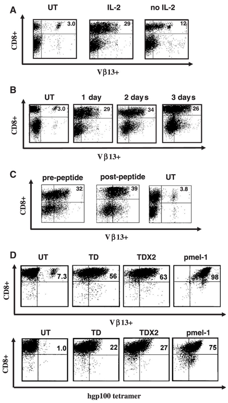
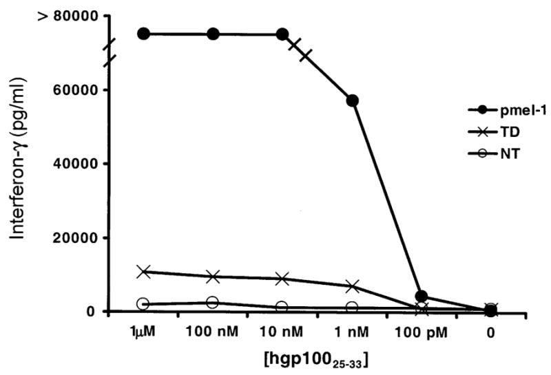
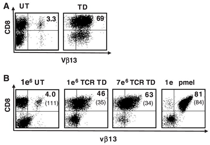
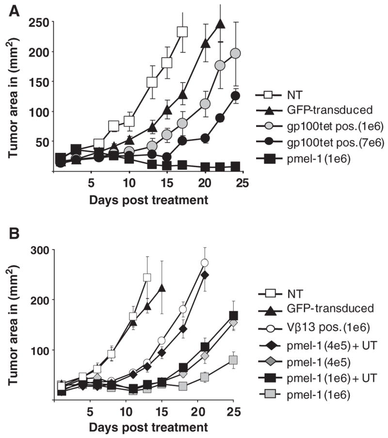
Adoptive cell transfer therapy using tumor-infiltrating lymphocytes for patients with metastatic melanoma has demonstrated significant objective response rates. One major limitation of these current therapies is the frequent inability to isolate tumor-reactive lymphocytes for treatment. Genetic engineering of peripheral blood lymphocytes with retroviral vectors encoding tumor antigen-specific T-cell receptors (TCRs) bypasses this restriction. To evaluate the efficacy of TCR gene therapy, a murine treatment model was developed. A retroviral vector was constructed encoding the pmel-1 TCR genes targeting the B16 melanoma antigen, gp100. Transduction of C57BL/6 lymphocytes resulted in efficient pmel-1 TCR expression. Lymphocytes transduced with this retrovirus specifically recognized gp100-pulsed target cells as measured by interferon-gamma secretion assays. Upon transfer into B16 tumor-bearing mice, the genetically engineered lymphocytes significantly slowed tumor development. The effectiveness of tumor treatment was directly correlated with the number of TCR-engineered T cells administered. These results demonstrated that TCR gene therapy targeting a native tumor antigen significantly delayed the growth of established tumors. When C57BL/6 lymphocytes were added to antigen-reactive pmel-1 T cells, a reduction in the ability of pmel-1 T cell to treat B16 melanomas was seen, suggesting that untransduced cells may be deleterious to TCR gene therapy. This model may be a powerful tool for evaluating future TCR gene transfer-based strategies.


Figures

FIGURE 1
FIGURE 1
TCR transduction of mouse splenocytes. A, Murine C57BL/6 splenocytes were left untransduced (UT), transduced with retrovirus encoding the pmel-1 TCR in the presence or absence of IL-2 and 24 hours later were analyzed by flow cytometry for TCR Vβ13 staining. B, Following TCR transduction, samples of in vitro IL-2 cultured splenocytes were analyzed on 3 consecutive days for TCR gene expression (measured by Vβ13 staining). C, Transduced cells were stimulated with gp100-specific peptide 2 days posttransduction, and analyzed 3 days poststimulation for Vβ13 expression by FACS. D, After CD8+ enrichment through negative selection murine splenocytes were transduced with pmel-1 TCR vector (TD). Twenty-four hours later, a second retroviral TCR transduction was performed (TD × 2). The levels of Vβ13+ transgene expression and gp100 tetramer binding in comparison to pmel-1 T cells was evaluated by flow cytometry. Data shown are representative of 3 independent experiments.
FIGURE 2
FIGURE 2
Function of transduced splenocytes. C57BL/6 splenocytes were transduced 1 time (TD) and co-cultured with hgp10025–33-pulsed MCA-205 cells for 24 hours. The amount of interferon-γ release was analyzed by ELISA. Untransduced (NT), and transgenic pmel-1 T cells (pmel) were used as negative and positive controls, respectively.
FIGURE 3
FIGURE 3
Persistence of TCR-transduced splenocytes after in vivo transfer. A, (In vitro) Murine C57BL/6 splenocytes were left untransduced (UT), or transduced (TD) with retrovirus encoding the pmel-1 TCR and 24 hours later analyzed by flow cytometry for TCR Vβ13 staining. B, (In vivo) The total number of CD8+gp100 tetramer+ transduced cells was calculated and 1 × 10⁶ or 7 × 10⁶ CD8+ gp 100 tetramer+ cells were transferred into 5 Gy irradiated, B16 tumor bearing C57BL/6 mice with concurrent fowlpox virus expressing hgp 100 vaccination and twice daily IL-2 administration for 3 days. Five days posttransfer, the peripheral blood of mice treated with pmel-1 TCR-transduced splenocytes was analyzed by flow cytometry. The mean fluorescence intensity of Vβ13 expression was as indicated in parentheses. A representative example of the FACS data is shown.
FIGURE 4
FIGURE 4
Delayed tumor growth following transfer of TCR-transduced splenocytes. A, C57BL/6 splenocytes were transduced and 1 × 10⁶ or 7 × 10⁶ CD8+ gp100 tetramer+ cells were transferred to B16 tumor bearing C57BL/6 mice. Animals received 5 Gy TBI with concurrent fowlpox virus expressing hgp100 vaccination and twice daily IL-2 administration for 3 days. As controls, animals were either left untreated (NT) or received GFP vector-transduced (GFP-transduced) splenocytes or transgenic pmel-1 T cells (pmel-1). Results of tumor area were the mean of measurement of 5 mice per group. The delay in tumor growth in mice that received 7 × 10⁶ CD8+ gp100 tetramer+ cells versus the same number of GFP vector-transduced cells was statistically significant (P=0.009). B, A cell mixing experiment was performed to mimic the administration of 1 × 10⁶ CD8+ Vβ13+-transduced cells. Using the same percentage of untransduced cells determined in the TCR-transduced population, untransduced C57BL/6 lymphocytes were added to 1 × 10⁶ or 4 × 10⁵ pmel-1 transgenic splenocytes (pmel-1+UT), and compared to pmel-1 without added untransduced cells (pmel-1). After 5 Gy TBI B16 tumor bearing C57BL/6 mice received cell transfer followed by fowlpox virus expressing hgp100 vaccination and twice daily IL-2 administration for 3 days. The addition of untransduced cells to pmel-1 splenocytes had a significant negative effect on tumor treatment (1 × 10⁶ pmel-1, P=0.009 and with 4 × 10⁵ pmel-1, P=0.014). Tumor area is presented as the mean of 5 mice per group.

References

    1. Dudley ME, Wunderlich JR, Robbins PF, et al. Cancer regression and autoimmunity in patients after clonal repopulation with antitumor lymphocytes. Science. 2002;298:850–854. - PMC - PubMed
    1. Dudley ME, Wunderlich JR, Yang JC, et al. Adoptive cell transfer therapy following non-myeloablative but lymphodepleting chemotherapy for the treatment of patients with refractory metastatic melanoma. J Clin Oncol. 2005;23:2346–2357. - PMC - PubMed
    1. Sadelain M, Riviere I, Brentjens R. Targeting tumours with genetically enhanced T lymphocytes. Nat Rev Cancer. 2003;3:35–45. - PubMed
    1. Schumacher TN. T-cell-receptor gene therapy. Nat Rev Immunol. 2002;2:512–519. - PubMed
    1. Hughes MS, Yu YY, Dudley ME, et al. Transfer of a TCR gene derived from a patient with a marked antitumor response conveys highly active T-cell effector functions. Hum Gene Ther. 2005;16:457–472. - PMC - PubMed

Publication types

MeSH terms