. 2008 Jan 23;27(2):350-60.
doi: 10.1038/sj.emboj.7601952. Epub 2007 Dec 20.

Cooperative control of striated muscle mass and metabolism by MuRF1 and MuRF2

Affiliations

Cooperative control of striated muscle mass and metabolism by MuRF1 and MuRF2

Christian C Witt et al. EMBO J. .

Abstract

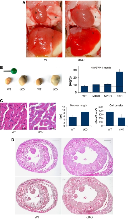
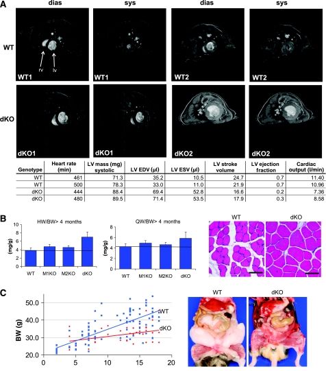
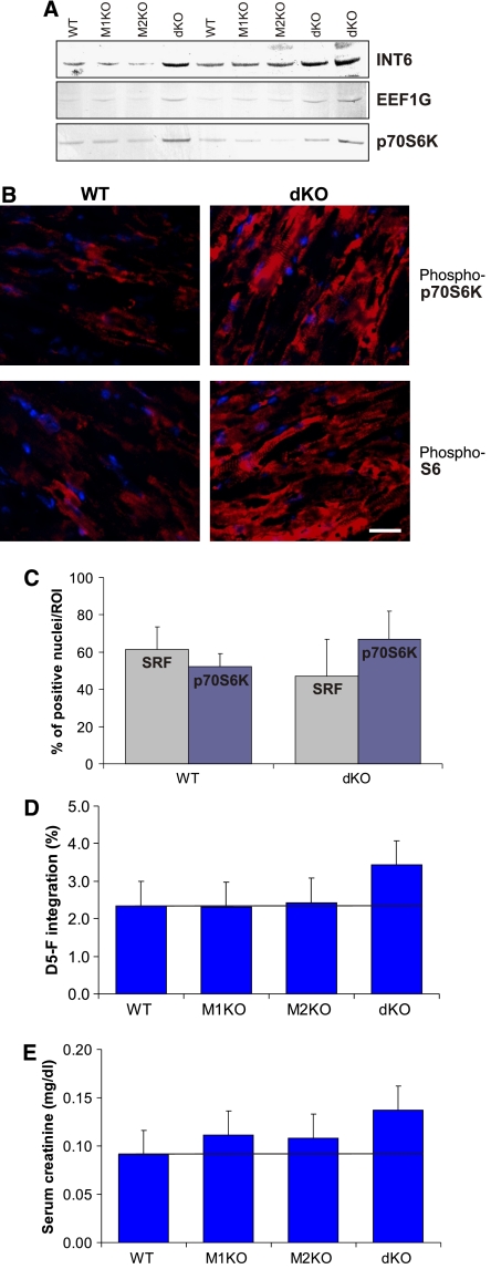
The muscle-specific RING finger proteins MuRF1 and MuRF2 have been proposed to regulate protein degradation and gene expression in muscle tissues. We have tested the in vivo roles of MuRF1 and MuRF2 for muscle metabolism by using knockout (KO) mouse models. Single MuRF1 and MuRF2 KO mice are healthy and have normal muscles. Double knockout (dKO) mice obtained by the inactivation of all four MuRF1 and MuRF2 alleles developed extreme cardiac and milder skeletal muscle hypertrophy. Muscle hypertrophy in dKO mice was maintained throughout the murine life span and was associated with chronically activated muscle protein synthesis. During ageing (months 4-18), skeletal muscle mass remained stable, whereas body fat content did not increase in dKO mice as compared with wild-type controls. Other catabolic factors such as MAFbox/atrogin1 were expressed at normal levels and did not respond to or prevent muscle hypertrophy in dKO mice. Thus, combined inhibition of MuRF1/MuRF2 could provide a potent strategy to stimulate striated muscles anabolically and to protect muscles from sarcopenia during ageing.


Figures

Figure 1
Figure 1
Synergistic control of heart muscle mass and cardiac myocyte size by MuRF1 and MuRF2. (A) Dissection of 13-day-old MuRF1 and MuRF2-dKO mice revealed grossly enlarged hearts (h), causing caudally lung compression (lu); the liver appears hyperaemic (li). (B) Effect of MuRF1/MuRF2 genotypes on heart weights. Left: hearts isolated from two matched pairs of WT and dKO mice (13 days old). Right: dKO mouse hearts (ventricles only) had 231% (P=0.001) increased HW/BW ratios, whereas weights of MuRF1, MuRF2 KO, and WT hearts did not differ significantly (young mice between d8 and d24; MuRF1: 25.5% increase, P=0.1, MuRF2: 27%, P=0.2, heart ventricles weights, respectively; dKO n=18; MuRF1 KO n=6, MuRF2 KO n=7; WT n=7). (C) Left: hematoxylin/eosin (HE) sections indicated that cardiomyocytes from young dKO hearts were hypertrophic. Scale bar, 20 μm. Right: morphometric comparison of WT and dKO sections nuclei had 59% increased length and cardiomyocytes were also 58% larger/cell density was reduced by 58% (as indicated by the number of cells in 0.2 mm2 large sections). (D) HE (top) and Masson histology (bottom) of WT and dKO hearts indicated concentric-type physiological hypertrophy at month 12, with intact inner and outer circular fiber systems and absence of fibrosis. Scale bar, 1 mm.
Figure 2
Figure 2
Phenotype of dKO mice at 18 months of age. Perinatally, dKO mice have a high mortality of 74%. The 26% of dKO mice (n=27) that survived became long-term survivors and were all alive at month 18 (unless killed, see below), thus allowing phenotypic studies in aged dKO. (A) MRI scans detected hypertrophic hearts in adult dKO mice with reduced EFs and stroke volumes (for time-resolved MRI scans, see also Supplementary Videos 4, 5, 6 and 7). LV, left ventricle; RV, right ventricle; EDV, end-diastolic volume; ESV, end-systolic volume (total number of mice scanned: two mice of each genotype. (B) Left: effect of MuRF1/2 genotypes on heart (ventricles, left) and quadriceps skeletal muscle (right) to body weight ratios. dKO mice maintain cardiac hypertrophy during ageing (dKO, 84% increase, P=0.001; MuRF1, 24.5% increase; MuRF2, 19%). In addition, dKO mice have 38.1% increased QW/BW ratios (P=0.001), whereas MuRF1 KO and MuRF2 KO genotypes have moderate effects on skeletal muscle mass (MuRF1, 16% increase, P=0.05; MuRF2, 11% increase, P=0.08). Despite increasing body mass in WT during months 4–18, skeletal muscles remain more hypertrophic in dKO mice during ageing (4–18 months of age, dKO n=27; MuRF1 KO n=52; MuRF2 KO n=81; WT n=49). For absolute weights, please refer to Supplementary Figure 8. Right: hematoxylin/eosin sections indicated that skeletal myofibers from dKO muscle show hypertrophic fibers with slightly augmented cross-section areas alternating with normal-appearing fibers (mice aged 4 months). Scale bar, 100 μm. (C) Weight gain of WT and dKO mice during ageing. Left: between months 4–18, dKO mice gain less weight (red; P<0.001) than WT. Right: dissections revealed that aged dKO mice were leaner (mice aged 18 months).
Figure 3
Figure 3
MuRF1 and MuRF2 interact with a shared set of myocellular proteins. (A) YTH screens with full-length MuRF1 and MuRF2 baits of both human cardiac (‘heart') and skeletal cDNA libraries (‘SKM') fished a total of 87 genes. The table summarizes those 35 prey clones identified independently in both MuRF1 and MuRF2 screens and thus predicted to interact with both MuRF1+2: 13 prey clone inserts code for sarcomeric proteins (4 of which are components of the Z-disk), 10 code for transcriptional regulators (2 of which are also associated with the Z-disk), 5 genes are involved in mitochondrial ATP production, and 6 genes participate in translation initiation and elongation. Numbers indicate independently identified prey clones in respective screens. M=interaction was found by mating. An SRF prey clone fished with the MuRF1 bait could not be confirmed by mating, as in our hands the 3′ UTR and not the coding sequence of SRF activated yeast growth during mating with MuRF1 and 2. (B) The interaction of selected proteins derived from the above-mentioned genes was studied in vitro by pull-downs using expressed MuRF1/MuRF2 Bcc (B-Box+coiled-coil domain) and MuRF1cc (coiled-coil domain) constructs (see also Supplementary Figure S1 and methods). MuRF1cc and MuRF1Bcc (arrows) co-eluted together with CARP, EEF1G, GFM1 MBP fusion proteins. Below: left—MuRF1cc co-eluted with myozenin-1/calsarcin-2, and MRP-L41/Pig3 MBP-fusion proteins; right—MuRF2Bcc co-eluted together with CARP, EEFG1, GFM1 MBP fusion proteins; controls—MBP plus MuRF1cc, Bcc, MuRF2Bcc, respectively, or fusion proteins only.
Figure 4
Figure 4
Altered Z-disks and mitochondrial ultrastructure in dKO myocardium. (A, B) At 18 months, we noted no ultrastructural abnormalities in myocardium from WT (A), MuRF1-KO (B), and MuRF2 KO (not shown) mice. (C, D) In dKO myocardium, sarcomere lengths and myofiber alignments are less regular (maximal variation is 2.5-fold larger than in WT sarcomeres). Z-disks have a denser appearance (arrows). Vacuoles (V) are frequently found between or embedded within mitochondria (M). Mitochondria are less regular in shape including abnormally small mitochondria and are, unlike in WT, not tightly packed together. Scale bar, 1 μm.
Figure 5
Figure 5
Characterization of altered signaling pathways in dKO myocardium by western blot studies. (A) Upregulation of the MuRF1/MuRF2 binding proteins CARP, FHL2, and SQSTM1 in dKO myocardium. Striking upregulation required the deletion of all four MuRF1/2 alleles, suggesting that both MuRF1 and MuRF2 synergistically control the transcriptional regulators CARP, FHL2, and SQSTM1. Below, cTnI and total multi-ubiquitinated protein species were not affected by the inactivation of MuRF1 and MuRF2 alleles. Among SUMO family members, we noticed for SUMO4 differential reactivity in the 8–30 kDa region. (B, C) Hyperactive stretch signaling in dKO as suggested by chronic upregulation of stretch-dependent signaling markers. (B) In myocardium, ANP is barely detectable in WT, MuRF1-KO, and MuRF2-KO hearts. ANP is strikingly upregulated in dKO myocardium (ventricles, 12 months old). Other markers for cardiomyopathy/hypertrophy remain normal or are moderately upregulated: SERCA2a (used as a marker for heart failure/calcium overload), SRF (previously implicated in stretch-dependent MuRF2 signaling), p38 Map kinase (a marker for ERK signaling and heart failure). (C) In dKO quadriceps muscles, hyperactive stretch signaling was suggested by the effect of 72 h immobilization on the stretch marker MLP: abnormally high levels of MLP/Crsp3 are maintained after 72 h immobilization in dKO quadriceps (NT=no treatment, byc=bycast immobilization).
Figure 6
Figure 6
Elevated muscle protein synthesis after inactivation of MuRF1 and MuRF2. (A) The translation elongation factor EEF1G (interacting with MuRF1 and MuRF2; see Figure 3), its binding protein elF3a subunit INT6, and the translation-promoting p70S6 kinase are upregulated in dKO myocardium. (B) Immunohistochemistry with phospho-specific antibodies demonstrated the upregulation of the activated forms of p70S6K and its substrate S6 (phospho-p70S6K and phospho-S6, respectively) in dKO myocardium, suggesting activation of the AKT/mTOR pathway. Scale bar, 20 μm. (C) Nuclear entry of phospho-p70S6K was stimulated in dKO myocardium, whereas cellular distribution of SRF did not change significantly (data represent counts of 200 nuclei on 4HPF sections). (D) Fractional synthesis rate (% per 48 h) of total cardiac muscle proteins was determined by injecting D5-F i.p. into mice of each genotype (n=6). Total cardiac muscle protein synthesis is elevated in dKO myocardium (P=0.05 in dKO versus WT, mice aged between 6 and 16 months). (E) Serum level of creatinine was increased in dKO mice (n=6 in each group, P=0.01 in dKO versus WT mice, mice aged between 6 and 16 months).

Similar articles

Cited by

References

    1. Arya R, Kedar V, Hwang JR, McDonough H, Li HH, Taylor J, Patterson C (2004) Muscle ring finger protein-1 inhibits PKC{epsilon} activation and prevents cardiomyocyte hypertrophy. J Cell Biol 167: 1147–1159 - PMC - PubMed
    1. Baar K, Torgan CE, Kraus WE, Esser K (2000) Autocrine phosphorylation of p70(S6k) in response to acute stretch in myotubes. Mol Cell Biol Res Commun 4: 76–80 - PubMed
    1. Bang ML, Mudry RE, McElhinny AS, Trombitas K, Geach AJ, Yamasaki R, Sorimachi H, Granzier H, Gregorio CC, Labeit S (2001) Myopalladin, a novel 145-kilodalton sarcomeric protein with multiple roles in Z-disc and I-band protein assemblies. J Cell Biol 153: 413–427 - PMC - PubMed
    1. Bassel-Duby R, Olson EN (2006) Signaling pathways in skeletal muscle remodeling. Annu Rev Biochem 75: 19–37 - PubMed
    1. Belle R, Minella O, Cormier P, Morales J, Poulhe R, Mulner-Lorillon O (1995) Phosphorylation of elongation factor-1 (EF-1) by cdc2 kinase. Prog Cell Cycle Res 1: 265–270 - PubMed

Publication types

MeSH terms