. 2008 Feb;153(4):693-709.
doi: 10.1038/sj.bjp.0707624. Epub 2007 Dec 24.

Testosterone protects rat hearts against ischaemic insults by enhancing the effects of alpha(1)-adrenoceptor stimulation

Affiliations

Testosterone protects rat hearts against ischaemic insults by enhancing the effects of alpha(1)-adrenoceptor stimulation

S Tsang et al. Br J Pharmacol. 2008 Feb.

Abstract

Background and purpose: Testosterone alleviates symptoms in patients with ischaemic heart disease. Androgen receptors are present in the heart, and testosterone upregulates gene expression of cardiac beta(1)-adrenoceptors. We hypothesize that testosterone may confer cardioprotection by interacting with adrenoceptors.

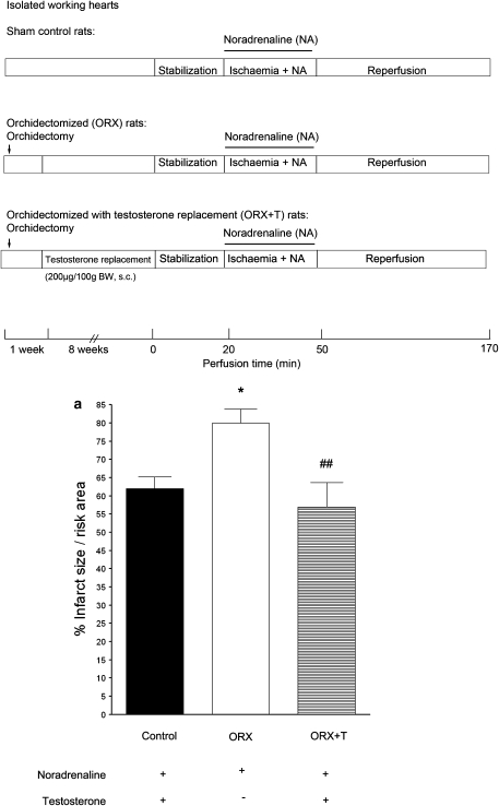
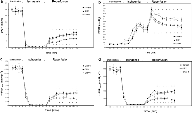
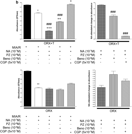
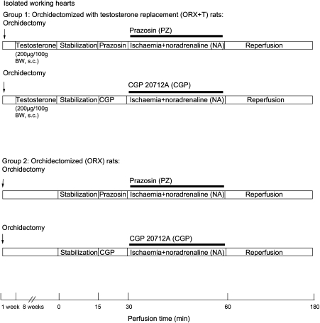
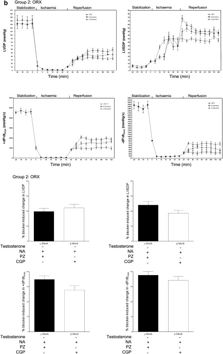
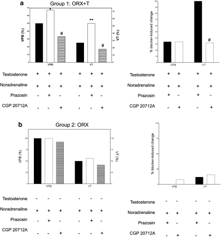
Experimental approach: In isolated perfused hearts and ventricular myocytes from orchidectomized rats without or with testosterone (200 microg/100 g) replacement, we first determined the effect of ischaemia/reperfusion in the presence of noradrenaline (10(-7) M). Then we determined the contribution of interactions between testosterone and alpha(1)- or beta(1)-adrenoceptors in cardiac injury/protection (infarct size, release of lactate dehydrogenase, viability of myocytes, recovery of contractile function and incidence of arrhythmias) upon ischaemia/reperfusion by pharmacological manipulation using selective adrenoceptor agonists (alpha(1)-adrenoceptor agonist: phenylephrine 10(-6) M; non-selective beta-adrenoceptor agonist: isoprenaline 10(-7) M) and antagonists (alpha(1): prazosin or benoxathian 10(-6) M; beta(1): CGP 20712A 5 x 10(-7) M). We also determined the expression of alpha(1) and beta(1)-adrenoceptor in the hearts from rats with and without testosterone.

Key results: Testosterone reduced injury induced by ischaemia/reperfusion and noradrenaline. This was achieved by enhancing the beneficial effect of alpha(1)-adrenoceptor stimulation, which was greater than the deleterious effect of beta(1)-adrenoceptor stimulation (also enhanced by testosterone). The effects of testosterone were abolished or attenuated by blockade of androgen receptors. Testosterone also enhanced the expression of alpha(1A) and beta(1)-adrenoceptor.

Conclusions and implications: Testosterone conferred cardioprotection by upregulating the cardiac alpha(1)-adrenoceptor and enhancing the effects of stimulation of this adrenoceptor. The effect of testosterone was at least partly mediated by androgen receptors.


Figures

Figure 1
Figure 1
Effects of ischaemic insults and administration of noradrenaline (NA) on (a) infarct size in isolated hearts, (b) LDH release and (c) viability of isolated ventricular myocytes from sham, ORX and ORX+T rats. Isolated perfused hearts were subjected to regional ischaemia by occlusion of the coronary artery for 30 min followed by release of the occlusion for 2 h. This produced reperfusion shown in the protocol (a). Infarct size was determined at the end of reperfusion. Noradrenaline (NA; 10−7M) was administered during ischaemia. As shown in (b) and (c), left ventricular myocytes were superfused for 10 min with a medium containing 10 mM 2-deoxy-D-glucose, an inhibitor of glycolysis, to induce metabolic inhibition, and 10 mM sodium dithionite, an oxygen scavenger, to produce anoxia. This simulated ischaemic insult and was followed by superfusion with the normal Krebs buffer for 10 min to simulate reperfusion. Values are mean±s.e.mean from 12 hearts in each group. *P<0.05, **P<0.01, ***P<0.001 vs sham control rats; # P<0.05, ## P<0.01, ### P<0.001 vs ORX rats. ORX, orchidectomized male rats; ORX+T, orchidectomized male rats with testosterone replacement (200 μg per 100 g).
Figure 1
Figure 1
Effects of ischaemic insults and administration of noradrenaline (NA) on (a) infarct size in isolated hearts, (b) LDH release and (c) viability of isolated ventricular myocytes from sham, ORX and ORX+T rats. Isolated perfused hearts were subjected to regional ischaemia by occlusion of the coronary artery for 30 min followed by release of the occlusion for 2 h. This produced reperfusion shown in the protocol (a). Infarct size was determined at the end of reperfusion. Noradrenaline (NA; 10−7M) was administered during ischaemia. As shown in (b) and (c), left ventricular myocytes were superfused for 10 min with a medium containing 10 mM 2-deoxy-D-glucose, an inhibitor of glycolysis, to induce metabolic inhibition, and 10 mM sodium dithionite, an oxygen scavenger, to produce anoxia. This simulated ischaemic insult and was followed by superfusion with the normal Krebs buffer for 10 min to simulate reperfusion. Values are mean±s.e.mean from 12 hearts in each group. *P<0.05, **P<0.01, ***P<0.001 vs sham control rats; # P<0.05, ## P<0.01, ### P<0.001 vs ORX rats. ORX, orchidectomized male rats; ORX+T, orchidectomized male rats with testosterone replacement (200 μg per 100 g).
Figure 2
Figure 2
Effects of ischaemic insults and administration of noradrenaline (NA) on contractile variables in isolated hearts (ad) from sham, ORX and ORX+T rats. (a) LVDP, (b) LVEDP, (c) +dP/dtmax and (d) –dP/dtmax obtained from perfused hearts in pre-ischaemic conditions (stabilization), during ischaemia and then during reperfusion. Values are mean±s.e.mean from 12 hearts from sham control, 12 hearts from ORX and 9 hearts from ORX+T group. # P<0.05, ## P<0.01 vs sham control rats; *P<0.05, **P<0.01 vs ORX rats. ORX, orchidectomized male rats; ORX+T, orchidectomized male rats with testosterone replacement (200 μg per 100 g).
Figure 3
Figure 3
Effects of MIA/R in the presence of α1 (a, b) or β1-adrenoceptors (c, d) stimulation on LDH release and viability of left ventricular myocytes from sham, ORX and ORX+T groups. For stimulation of α1-adrenoceptors, 10−6M phenylephrine (PE) in the presence of 10−6M propranolol, a β-adrenoceptor blocker, and 5 × 10⁻⁷M yohimbine, an α2-adrenoceptor blocker, was administered during MIA. Cyproterone acetate (Cyp), 10−6M, prazosin (PZ), 10−6M, was used for blockade of the androgen receptor and α1, adrenoceptor, respectively. (a) % LDH release resulting from stimulation of α1-adrenoceptors. (b) Viability resulting from stimulation of α1-adrenoceptors. Values represent mean±s.e.mean of triplicate determinations from six hearts in each group. *, **, *** P<0.05, 0.01, 0.001 vs corresponding sham control, respectively. #, ##, ### P<0.05, 0.01, 0.001 vs ORX rats, respectively. +, ++, +++ P<0.05, 0.01, 0.001 vs ORX+T group with phenylephrine, respectively. For stimulation of β1-adrenoceptors, 10−7M isoprenaline (ISO) in the presence of 10−6M phentolamine, an α-adrenoceptor antagonist, and 5 × 10⁻⁷M ICI, a β2-adrenoceptor antagonist, were administered during MIA. (c) % LDH release resulting from stimulation of β1-adrenoceptors. (d) Viability resulting from stimulation of β1-adrenoceptors. Values are mean±s.e.mean of triplicate determinations from six hearts in each group, respectively. *, **, *** P<0.05, 0.01, 0.001 vs corresponding sham control, respectively.#, ##, ### P<0.05, 0.01, 0.001 vs ORX group, respectively. +, ++, +++ P<0.05, 0.01, 0.001 vs ORX+T group with isoprenaline, respectively. ICI, ICI 118, 551; MIA/R, metabolic inhibition and anoxia/reperfusion; ORX, orchidectomized male rats; ORX+T, orchidectomized male rats with testosterone replacement (200 μg per 100 g).
Figure 3
Figure 3
Effects of MIA/R in the presence of α1 (a, b) or β1-adrenoceptors (c, d) stimulation on LDH release and viability of left ventricular myocytes from sham, ORX and ORX+T groups. For stimulation of α1-adrenoceptors, 10−6M phenylephrine (PE) in the presence of 10−6M propranolol, a β-adrenoceptor blocker, and 5 × 10⁻⁷M yohimbine, an α2-adrenoceptor blocker, was administered during MIA. Cyproterone acetate (Cyp), 10−6M, prazosin (PZ), 10−6M, was used for blockade of the androgen receptor and α1, adrenoceptor, respectively. (a) % LDH release resulting from stimulation of α1-adrenoceptors. (b) Viability resulting from stimulation of α1-adrenoceptors. Values represent mean±s.e.mean of triplicate determinations from six hearts in each group. *, **, *** P<0.05, 0.01, 0.001 vs corresponding sham control, respectively. #, ##, ### P<0.05, 0.01, 0.001 vs ORX rats, respectively. +, ++, +++ P<0.05, 0.01, 0.001 vs ORX+T group with phenylephrine, respectively. For stimulation of β1-adrenoceptors, 10−7M isoprenaline (ISO) in the presence of 10−6M phentolamine, an α-adrenoceptor antagonist, and 5 × 10⁻⁷M ICI, a β2-adrenoceptor antagonist, were administered during MIA. (c) % LDH release resulting from stimulation of β1-adrenoceptors. (d) Viability resulting from stimulation of β1-adrenoceptors. Values are mean±s.e.mean of triplicate determinations from six hearts in each group, respectively. *, **, *** P<0.05, 0.01, 0.001 vs corresponding sham control, respectively.#, ##, ### P<0.05, 0.01, 0.001 vs ORX group, respectively. +, ++, +++ P<0.05, 0.01, 0.001 vs ORX+T group with isoprenaline, respectively. ICI, ICI 118, 551; MIA/R, metabolic inhibition and anoxia/reperfusion; ORX, orchidectomized male rats; ORX+T, orchidectomized male rats with testosterone replacement (200 μg per 100 g).
Figure 4
Figure 4
Effects of MIA/R in the presence of noradrenaline (NA) on the release of LDH and viability of left ventricular myocytes from ORX rats upon blockade of either α1- and β2-adrenoceptors or β1- and β2-adrenoceptors. For blockade of α1-adrenoceptors, two selective α1-adrenoceptor antagonists, 10−6M prazosin (PZ) or 2 × 10⁻⁶M benoxathian (Beno), were administered. The β1-adrenoceptor-selective antagonist CGP at 5 × 10⁻⁷M in the presence of ICI at 5 × 10⁻⁷M was used to block both β-adrenoceptor subtypes. NA (10−7M) was administered. (a) LDH release from myocytes with or without testosterone replacement. Left panels indicate % release after different treatments and right panels indicate net changes in LDH release after blockade of either α1- and β2-adrenoceptors or β1- and β2-adrenoceptors. (b) Viability of myocytes with or without testosterone replacement. Left panels indicate viability after different treatments and right panels indicate net changes in viability after blockade of either α1- and β2-adrenoceptors or β1- and β2-adrenoceptors. The results are expressed as percentage of noradrenaline and antagonist-stimulated change in LDH release/viability. Where appropriate, data are expressed as percentage of baseline (i.e., 100% × induced value/value before stimulation. Data represent mean±s.e.mean of triplicate determinations in eight independent experiments using cell preparations from different rats. Differences between multiple groups were determined by one-way ANOVA followed by post hoc Neuman–Keuls multiple comparison tests. *P<0.05, **P<0.01 vs MIA/R. +, ++, +++ P<0.05, 0.01, 0.001 vs MIA/R with NA, respectively.### P<0.001 vs CGP. CGP, CGP 20712A; ICI, ICI 118, 551; MIA/R, metabolic inhibition and anoxia/reperfusion; ORX, orchidectomized male rats.
Figure 4
Figure 4
Effects of MIA/R in the presence of noradrenaline (NA) on the release of LDH and viability of left ventricular myocytes from ORX rats upon blockade of either α1- and β2-adrenoceptors or β1- and β2-adrenoceptors. For blockade of α1-adrenoceptors, two selective α1-adrenoceptor antagonists, 10−6M prazosin (PZ) or 2 × 10⁻⁶M benoxathian (Beno), were administered. The β1-adrenoceptor-selective antagonist CGP at 5 × 10⁻⁷M in the presence of ICI at 5 × 10⁻⁷M was used to block both β-adrenoceptor subtypes. NA (10−7M) was administered. (a) LDH release from myocytes with or without testosterone replacement. Left panels indicate % release after different treatments and right panels indicate net changes in LDH release after blockade of either α1- and β2-adrenoceptors or β1- and β2-adrenoceptors. (b) Viability of myocytes with or without testosterone replacement. Left panels indicate viability after different treatments and right panels indicate net changes in viability after blockade of either α1- and β2-adrenoceptors or β1- and β2-adrenoceptors. The results are expressed as percentage of noradrenaline and antagonist-stimulated change in LDH release/viability. Where appropriate, data are expressed as percentage of baseline (i.e., 100% × induced value/value before stimulation. Data represent mean±s.e.mean of triplicate determinations in eight independent experiments using cell preparations from different rats. Differences between multiple groups were determined by one-way ANOVA followed by post hoc Neuman–Keuls multiple comparison tests. *P<0.05, **P<0.01 vs MIA/R. +, ++, +++ P<0.05, 0.01, 0.001 vs MIA/R with NA, respectively.### P<0.001 vs CGP. CGP, CGP 20712A; ICI, ICI 118, 551; MIA/R, metabolic inhibition and anoxia/reperfusion; ORX, orchidectomized male rats.
Figure 5
Figure 5
Effects of ischaemic insult in the presence of noradrenaline (NA) on cardiac contractile variables in the isolated hearts from ORX and ORX+T rats upon blockade of either α1- and β2-adrenoceptors or β1- and β2-adrenoceptors. For blockade of α1-adrenoceptors, the selective α1-adrenoceptor antagonist 10−6M prazosin (PZ) was administered. The β1-adrenoceptor-selective antagonist CGP at 5 × 10⁻⁷M in the presence of ICI at 5 × 10⁻⁷M was used to block both β-adrenoceptor subtypes. Noradrenaline (10−7M) was administered. Analyses were made during stabilization (baseline), ischaemia and reperfusion in (a) ORX+T and (b) ORX rats. LVDP, left ventricular developed pressure; LVEDP, left ventricular end-diastolic pressure; ±dP/dtmax, velocity of contraction and relaxation, respectively. Lower panel: net changes in contractile variables after blockade of either α1- and β2-adrenoceptors or β1- and β2-adrenoceptors. Values are mean±s.e.mean of 9–12 rats. *P<0.05, **P<0.01 vs ORX+T; # P<0.05, ## P<0.01, ### P<0.001 vs CGP. CGP, CGP 20712A; ICI, ICI 118, 551; ORX, orchidectomized male rats; ORX+T, orchidectomized male rats with testosterone replacement (200 μg per 100 g).
Figure 5
Figure 5
Effects of ischaemic insult in the presence of noradrenaline (NA) on cardiac contractile variables in the isolated hearts from ORX and ORX+T rats upon blockade of either α1- and β2-adrenoceptors or β1- and β2-adrenoceptors. For blockade of α1-adrenoceptors, the selective α1-adrenoceptor antagonist 10−6M prazosin (PZ) was administered. The β1-adrenoceptor-selective antagonist CGP at 5 × 10⁻⁷M in the presence of ICI at 5 × 10⁻⁷M was used to block both β-adrenoceptor subtypes. Noradrenaline (10−7M) was administered. Analyses were made during stabilization (baseline), ischaemia and reperfusion in (a) ORX+T and (b) ORX rats. LVDP, left ventricular developed pressure; LVEDP, left ventricular end-diastolic pressure; ±dP/dtmax, velocity of contraction and relaxation, respectively. Lower panel: net changes in contractile variables after blockade of either α1- and β2-adrenoceptors or β1- and β2-adrenoceptors. Values are mean±s.e.mean of 9–12 rats. *P<0.05, **P<0.01 vs ORX+T; # P<0.05, ## P<0.01, ### P<0.001 vs CGP. CGP, CGP 20712A; ICI, ICI 118, 551; ORX, orchidectomized male rats; ORX+T, orchidectomized male rats with testosterone replacement (200 μg per 100 g).
Figure 6
Figure 6
Effects of ischaemic insult in the presence of noradrenaline on reperfusion arrhythmias in the isolated hearts from ORX and ORX+T rats upon blockade of either α1- and β2-adrenoceptors or β1- and β2-adrenoceptors. The protocol shown in Figure 5 was adopted. Values shown are the percentage of hearts exhibiting VPBs and VT in (a) ORX+T and (b) ORX rats over the first 30 min reperfusion period. Values are mean±s.e.mean rounded to the nearest whole number. Right panels: net changes in VPBs and VT after blockade of either α1- and β2-adrenoceptors or β1- and β2-adrenoceptors. Values are mean±s.e.mean of 9–12 rats. *P<0.05, ** P<0.01 relative to vehicle control and#P<0.05 relative to prazosin by χ2 test for percentages. ORX, orchidectomized male rats; ORX+T, orchidectomized male rats with testosterone replacement (200 μg per 100 g); VPBs, ventricular premature beats; VT, ventricular tachycardia.
Figure 7
Figure 7
Effect of orchidectomy (ORX) on the expression of α1A- and β1-adrenoceptors in the rat heart. The expression of subtypes of α1- and β1-adrenoceptors was evaluated by western blotting from sham control, ORX and ORX+T as described in Methods. The relative arbitrary unit for sham control group was assigned as 1. Data are normalized to average value of sham control obtained from the same blot. Values are mean±s.e.mean of 5–6 rats. *P<0.05 vs control; **P<0.01 vs control; #P<0.05 vs ORX. ORX, orchidectomized male rats; ORX+T, orchidectomized male rats with testosterone replacement (200 μg per 100 g).

Similar articles

Cited by

References

    1. Banu KS, Govindarajulu P, Aruldhas MM. Testosterone and estradiol modulate TSH-binding in the thyrocytes of Wistar rats: influence of age and sex. J Steroid Biochem Mol Biol. 2001;78:329–342. - PubMed
    1. Callies F, Stromer H, Schwinger RH, Bolck B, Hu K, Frantz S, et al. Administration of testosterone is associated with a reduced susceptibility to myocardial ischemia. Endocrinology. 2003;144:4478–4483. - PubMed
    1. Communal C, Singh K, Pimentel DR, Colucci WS. Norepinephrine stimulates apoptosis in adult rat ventricular myocytes by activation of the β-adrenergic pathway. Circulation. 1998;98:1329–1334. - PubMed
    1. Communal C, Singh K, Sawyer DB, Colucci WS. Opposing effects of β1- and β2-adrenergic receptors on cardiac myocyte apoptosis: role of a pertussis toxin-sensitive G protein. Circulation. 1999;100:2210–2212. - PubMed
    1. Du XJ, Gao XM, Kiriazis H, Moore XL, Ming Z, Su Y, et al. Transgenic α1A-adrenergic activation limits post-infarct ventricular remodeling and dysfunction and improves survival. Cardiovasc Res. 2006;71:735–743. - PubMed

Publication types

MeSH terms