. 2008 Mar 1;507(1):1141-50.
doi: 10.1002/cne.21588.

Repeated stress alters dendritic spine morphology in the rat medial prefrontal cortex

Affiliations

Repeated stress alters dendritic spine morphology in the rat medial prefrontal cortex

Jason J Radley et al. J Comp Neurol. .

Abstract

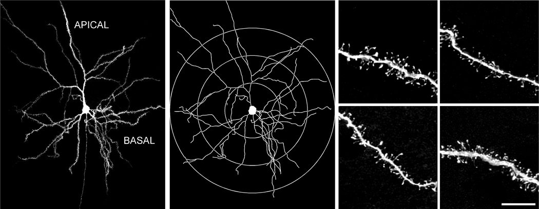
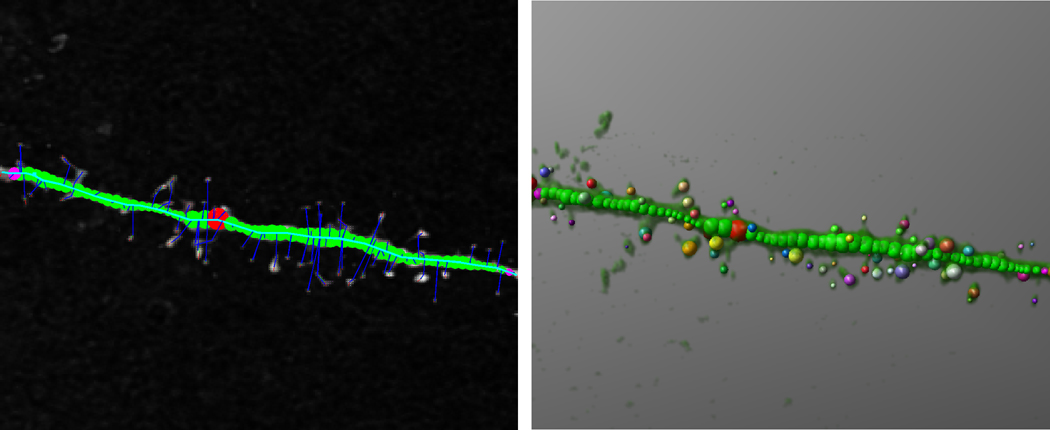
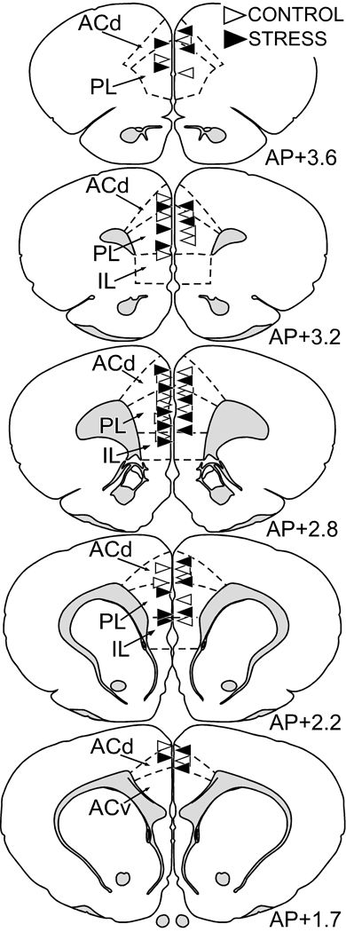
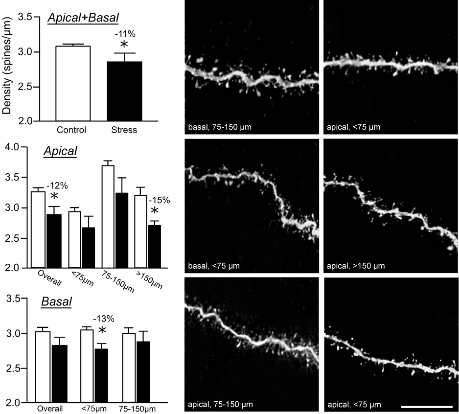
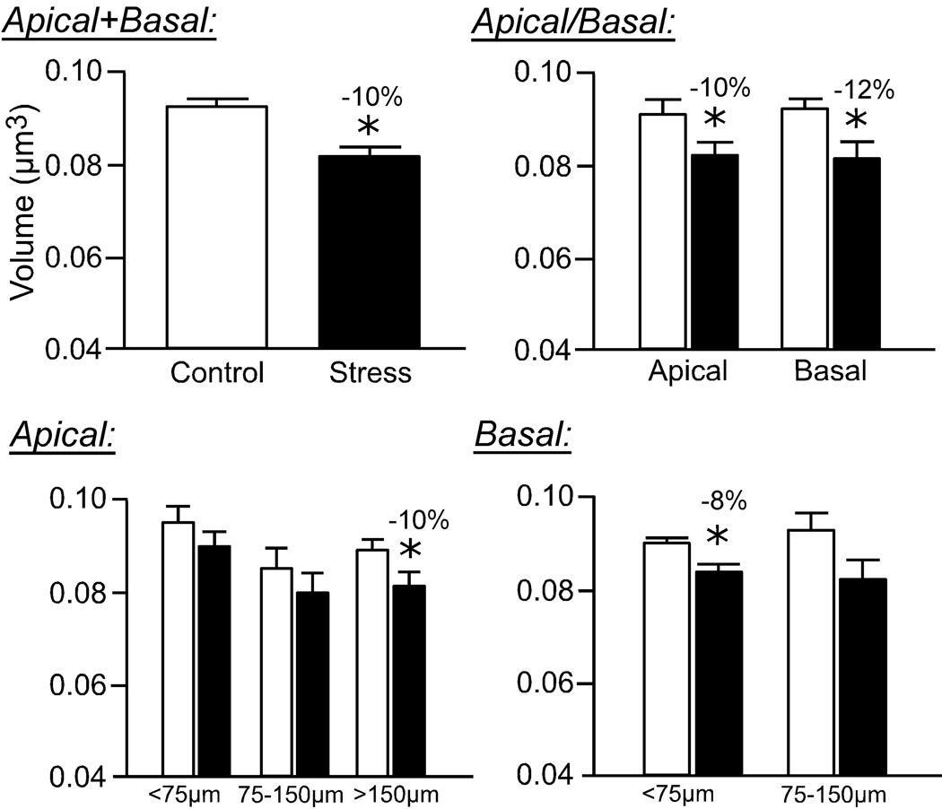
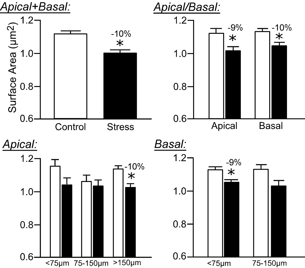
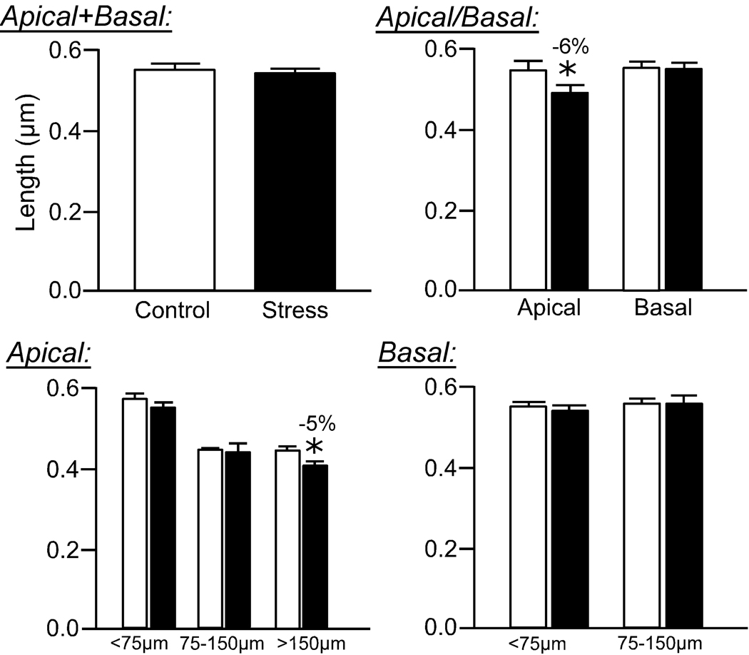
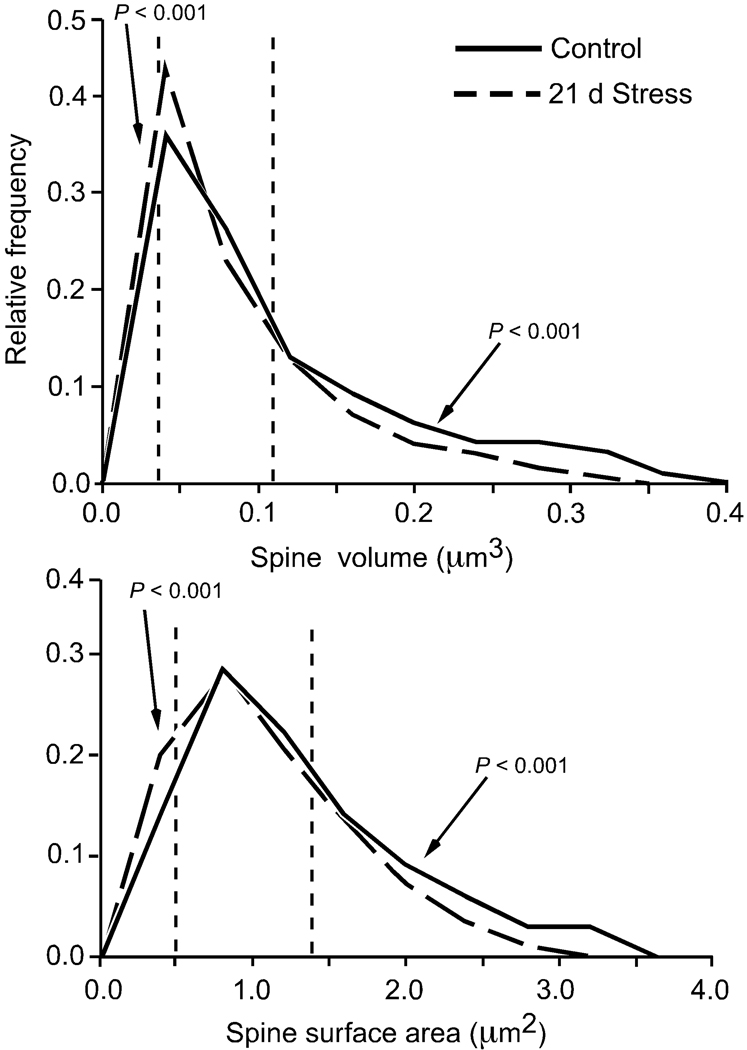
Anatomical alterations in the medial prefrontal cortex (mPFC) are associated with hypothalamopituitary adrenal (HPA) axis dysregulation, altered stress hormone levels, and psychiatric symptoms of stress-related mental illnesses. Functional imaging studies reveal impairment and shrinkage of the mPFC in such conditions, and these findings are paralleled by experimental studies showing dendritic retraction and spine loss following repeated stress in rodents. Here we extend this characterization to how repeated stress affects dendritic spine morphology in mPFC through the utilization of an automated approach that rapidly digitizes, reconstructs three dimensionally, and calculates geometric features of neurons. Rats were perfused after being subjected to 3 weeks of daily restraint stress (6 hours/day), and intracellular injections of Lucifer Yellow were made in layer II/III pyramidal neurons in the dorsal mPFC. To reveal spines in all angles of orientation, deconvolved high-resolution confocal laser scanning microscopy image stacks of dendritic segments were reconstructed and analyzed for spine volume, surface area, and length using a Rayburst-based automated approach (8,091 and 8,987 spines for control and stress, respectively). We found that repeated stress results in an overall decrease in mean dendritic spine volume and surface area, which was most pronounced in the distal portion of apical dendritic fields. Moreover, we observed an overall shift in the population of spines, manifested by a reduction in large spines and an increase in small spines. These results suggest a failure of spines to mature and stabilize following repeated stress and are likely to have major repercussions on function, receptor expression, and synaptic efficacy.


Figures

Figure 1
Figure 1. Methodology for comprehensive spine morphologic analysis of pyramidal neurons in layers II/III of dorsal mPFC
Left: Digital reconstruction of a Lucifer Yellow-filled neuron in the rat dorsal mPFC at 160x magnification on a Zeiss 410 laser-scanning microscope from 5 separate confocal digital stacks (z-step = 0.5 µm), that were deconvolved and aligned in the x-y-z planes using Vias software (Rodriguez et al., 2003; Radley et al., 2006b). Middle: 2-dimensional map for the same neuron reconstructed with Neurolucida (MBF Biosciences). The different colored branches indicate increasing branch orders within single dendritic arbors. Radial distances are shown to depict the different zones within which segments were selected for dendritic reconstructions (< 75 µm, 75–150 µm, > 150 µm) at high magnification. Right: Examples of dendritic segments, imaged at 5,000X magnification (z-step = 0.1 µm). Scale bar = 150 µm (left); 10 µm (right).
Figure 2
Figure 2. Analysis of spine morphometric parameters with the Rayburst-based automated spine analysis system
The Rayburst-based approach was used for the calculation of spine morphological parameters (Wearne et al., 2005; Rodriguez et al., 2006), involving 3D reconstruction and geometric analysis on deconvolved optical stacks of dendritic segments in two principal steps. First, the medial axis of the dendritic segment is extracted using a dynamically adjusting local segmentation technique (left). Next, a Rayburst Sampling algorithm extracts dendrite and spine geometric features (right), yielding spine volume, length, and surface area for each spine on the segment, for all angles of orientation.
Figure 3
Figure 3. Anatomical localization of Lucifer Yellow-filled layers II/III pyramidal neurons in mPFC for subregional analysis
Atlas plates (modified from Swanson, 1992) of coronal sections through a similar level of the mPFC from animals that received intracellular Lucifer Yellow injections. Neurons from layers II/III in the dorsal mPFC were identified using a fluorescent nucleic acid stain, followed by iontophoretic injections with Lucifer Yellow. The location for each filled neuron is designated for each treatment with a triangle (control, blue; stress, red). ACd, anterior cingulate cortex, dorsal subdivision; ACv anterior cingulate cortex, ventral subdivision; PL, prelimbic area; IL, infralimbic area.
Figure 4
Figure 4. Repeated restraint stress reduces dendritic spine density in layer II/III pyramidal neurons
Repeated restraint stress (21 days, 6 hours/day) induces decreases in overall (upper left) and apical dendritic spine density (middle left). This effect is most prominent in more distal portions (>150 µm) of apical dendrites. While there was no overall effect of stress on basal dendrites, a significant reduction of spine density was present at <75 µm from the soma (lower left). The spine density values for each dendritic segment were quantified using the Rayburst-based automated approach and was similar to previous estimates that involved manually counting spines from optical stacks by controlling the plane of focus for z-step increments and marking spines as they appear on the dendritic segment (Radley et al., 2006b). Examples of dendritic segments are shown in the middle (control) and right (21 days stress) columns. *, differs significantly from unstressed controls; P < 0.05. Scale bar = 10 µm.
Figure 5
Figure 5. Effects of repeated restraint stress on Rayburst volumes of dendritic spines in layers II/III pyramidal neurons in the dorsal mPFC
Values from individual spines on each dendritic segment were averaged, and then averaged for each animal (N = 5 neurons/animal), and values per animal were used for comparison of means (N = 5/group). Values represent mean + SEM. *, P < 0.05.
Figure 6
Figure 6. Effects of repeated restraint stress on Rayburst surface areas of dendritic spines in layers II/III pyramidal neurons in the dorsal mPFC
Values from individual spines on each dendritic segment were averaged, and then averaged for each animal (N = 5 neurons/animal), and values per animal were used for comparison of means (N = 5/group). Values represent mean + SEM. *, P < 0.05.
Figure 7
Figure 7. Effects of repeated restraint stress on dendritic spine lengths in layers II/III pyramidal neurons in the dorsal mPFC
Values from individual spines on each dendritic segment were averaged, and then averaged for each animal (N = 5 neurons/animal), and values per animal were used for comparison of means (N = 5/group). Values represent mean + SEM. *, P < 0.05.
Figure 8
Figure 8. Probability distributions for spine volume and surface area for distal apical dendritic segments
The distributions were derived from high-resolution imaging of spines on apical dendritic segments at radial distances greater than 150 µm relative to the soma, and analyzed using the Rayburst-based automated approach (Control = 1,629 spines; 21 days stress = 1,438 spines). The dashed vertical lines in each histogram shows the 25th and 75th percentiles of the entire spine population (i.e., 3,067 spines). These frequency distributions illustrate how repeated stress results in an overall downward shift in spine morphometric parameters.

Similar articles

Cited by

References

    1. Brown SM, Henning S, Wellman CL. Mild, short-term stress alters dendritic morphology in rat medial prefrontal cortex. Cereb Cortex. 2005;15:1714–1722. - PubMed
    1. Caspi A, Sugden K, Moffitt TE, Taylor A, Craig IW, Harrington H, McClay J, Mill J, Martin J, Braithwaite A, Poulton R. Influence of life stress on depression: moderation by a polymorphism in the 5-HTT gene. Science. 2003;301:386–389. - PubMed
    1. Cerqueira JJ, Mailliet F, Almeida OF, Jay TM, Sousa N. The prefrontal cortex as a key target of the maladaptive response to stress. J Neurosci. 2007;27:2781–2787. - PMC - PubMed
    1. Cerqueira JJ, Pego JM, Taipa R, Bessa JM, Almeida OF, Sousa N. Morphological correlates of corticosteroid-induced changes in prefrontal cortex-dependent behaviors. J Neurosci. 2005;25:7792–7800. - PMC - PubMed
    1. Cerqueira JJ, Taipa R, Uylings HB, Almeida OF, Sousa N. Specific configuration of dendritic degeneration in pyramidal neurons of the medial prefrontal cortex induced by differing corticosteroid regimens. Cereb Cortex. 2006 epub 2006 Nov 2. - PubMed

Publication types