

Neuropathological basis of magnetic resonance images in aging and dementia
- PMID: 18157909
- PMCID: PMC2624571
- DOI: 10.1002/ana.21296
Neuropathological basis of magnetic resonance images in aging and dementia
Abstract
Objective: Magnetic resonance (MR) imaging is used widely for assessment of patients with cognitive impairment, but the pathological correlates are unclear, especially when multiple pathologies are present.
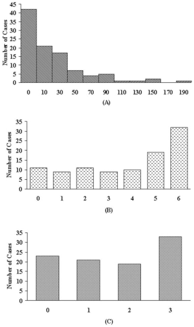
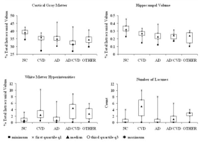
Methods: This report includes 93 subjects from a longitudinally followed cohort recruited for the study of Alzheimer's disease (AD) and subcortical cerebrovascular disease (CVD). MR images were analyzed to quantify cortical gray matter volume, hippocampal volume, white matter hyperintensities, and lacunes. Neuropathological examination quantified CVD parenchymal pathology, AD pathology (defined as Consortium to Establish a Registry for Alzheimer's Disease scores and Braak and Braak stage), and hippocampal sclerosis. Subjects were pathologically classified as 12 healthy control subjects, 46 AD, 14 CVD, 9 mixed AD/CVD, and 12 cognitively impaired patients without significant AD/CVD pathology. Multivariate models tested associations between magnetic resonance and pathological findings across the entire sample.
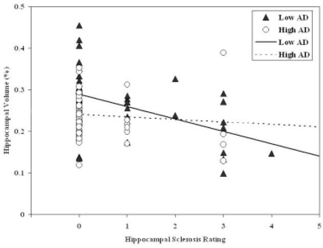
Results: Pathological correlates of cortical gray matter volume were AD, subcortical vascular pathology, and arteriosclerosis. Hippocampal volume was related to AD pathology and hippocampal sclerosis, and the effects of hippocampal sclerosis were greater for subjects with low levels of AD pathology. White matter hyperintensities were related to age and to white matter pathology. Number of MRI lacunes was related to subcortical vascular pathology.
Interpretation: In this clinical setting, the presence of lacunes and white matter changes provide a good signal for vascular disease. The neuropathological basis of MR defined cerebral cortical and hippocampal atrophy in aging and dementia is complex, with several pathological processes converging on similar brain structures that mediate cognitive decline.
Figures
References
-
- Knopman DS, DeKosky ST, Cummings JL, et al. Practice parameter: diagnosis of dementia (an evidence-based review). Report of the Quality Standards Subcommittee of the American Academy of Neurology. Neurology. 2001;56:1143–1153. - PubMed
-
- Scab JP, Jagust WJ, Wong STS, et al. Quantitative NMR measurements of hippocampal atrophy in Alzheimer’s disease. Magn Reson Med. 1988;8:200–208. - PubMed
-
- Jack CR, Petersen RC, O’Brien PC, Tangalos EG. MR-based hippocampal volumetry in the diagnosis of Alzheimer’ disease. Neurology. 1992;42:183–188. - PubMed
Publication types
MeSH terms
Grants and funding
LinkOut - more resources
Full Text Sources
Medical
