. 2007 Dec 24:7:142.
doi: 10.1186/1471-213X-7-142.

Diverse roles of actin in C. elegans early embryogenesis

Affiliations

Diverse roles of actin in C. elegans early embryogenesis

Nathalie Velarde et al. BMC Dev Biol. .

Abstract

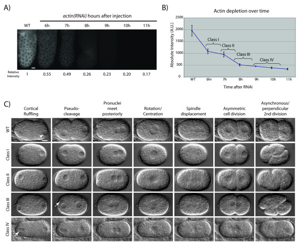
Background: The actin cytoskeleton plays critical roles in early development in Caenorhabditis elegans. To further understand the complex roles of actin in early embryogenesis we use RNAi and in vivo imaging of filamentous actin (F-actin) dynamics.

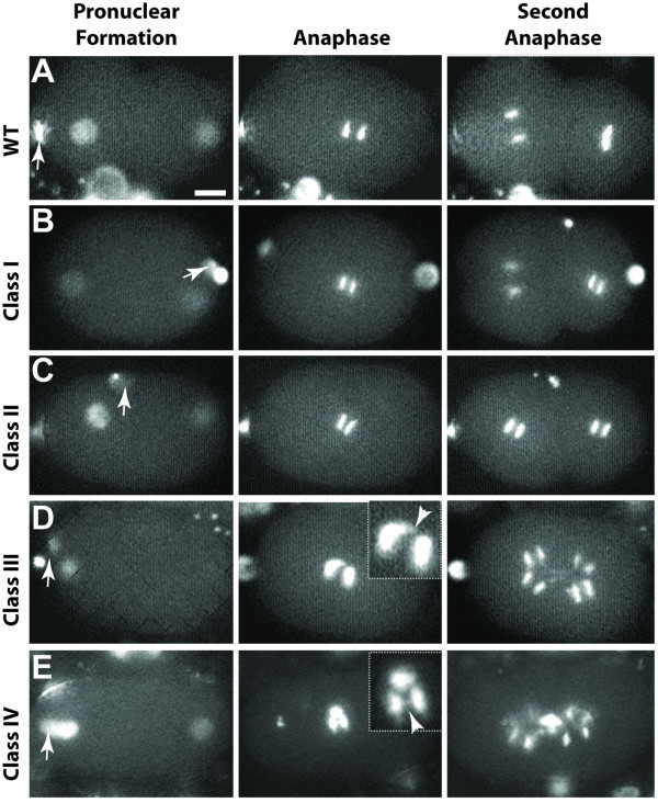
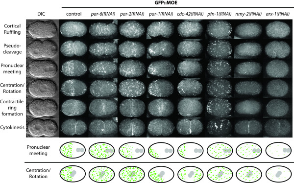
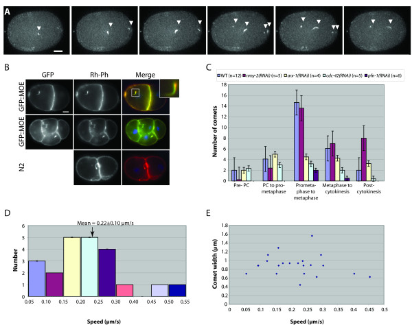
Results: Using RNAi, we found processes that are differentially sensitive to levels of actin during early embryogenesis. Mild actin depletion shows defects in cortical ruffling, pseudocleavage, and establishment of polarity, while more severe depletion shows defects in polar body extrusion, cytokinesis, chromosome segregation, and eventually, egg production. These defects indicate that actin is required for proper oocyte development, fertilization, and a wide range of important events during early embryogenesis, including proper chromosome segregation. In vivo visualization of the cortical actin cytoskeleton shows dynamics that parallel but are distinct from the previously described myosin dynamics. Two distinct types of actin organization are observed at the cortex. During asymmetric polarization to the anterior, or the establishment phase (Phase I), actin forms a meshwork of microfilaments and focal accumulations throughout the cortex, while during the anterior maintenance phase (Phase II) it undergoes a morphological transition to asymmetrically localized puncta. The proper asymmetric redistribution is dependent on the PAR proteins, while both asymmetric redistribution and morphological transitions are dependent upon PFN-1 and NMY-2. Just before cytokinesis, actin disappears from most of the cortex and is only found around the presumptive cytokinetic furrow. Finally, we describe dynamic actin-enriched comets in the early embryo.

Conclusion: During early C. elegans embryogenesis actin plays more roles and its organization is more dynamic than previously described. Morphological transitions of F-actin, from meshwork to puncta, as well as asymmetric redistribution, are regulated by the PAR proteins. Results from this study indicate new insights into the cellular and developmental roles of the actin cytoskeleton.


Figures

Figure 1
Figure 1
Progressive depletion of actin over time in C. elegans gonads and embryos. (A) Fixed gonads stained with anti-actin antibody in wild-type and actin(RNAi) treated animals at successive timepoints after injection of dsRNA, visualized with FITC-conjugated secondary antibody. The distal end of the gonad is shown. For each timepoint, the maximum signal intensity was quantified in each of three sampled areas per gonad, and the average of these was then compared to WT to obtain a measure of relative intensity. Scale bar is 10 μm. (B) Graph of actin depletion over time in actin(RNAi) gonads based on data shown in (A). Brackets indicate approximate time intervals during which the four classes of actin(RNAi) phenotypes were observed in the early embryo for this batch of dsRNA. Average maximum intensities and standard deviations are based on cumulative data from 3–5 gonads and three sampled areas per gonad (i.e. 9–15 data points) for each timepoint. Numbers on the vertical axis represent units of intensity as measured by Openlab 3.1.7 software. (C) Time-course depletion of actin and requirements for early embryogenesis. Each row, labeled by class, shows DIC images from a time series analysis spanning from just after the completion of meiosis to the second mitotic division. For each timepoint, hallmark events in wild-type (top row) are listed above each column. actin(RNAi) Class I phenotypes (n = 11 embryos): Loss of cortical contractions, no pseudocleavage, and synchronous second cell division. actin(RNAi) Class II phenotypes (n = 12): Pronuclei meet more centrally, loss of spindle displacement, and parallel and synchronous second cell division. Note the increase in distance between the paternal nucleus and the posterior cortex. actin(RNAi) Class III phenotypes (n = 12): First cell division is incomplete with nuclear reformation close to contractile ring, and multiple nuclei in subsequent divisions. actin(RNAi) Class IV phenotypes (n = 14): Failure to extrude polar bodies, loss of cytokinesis and multiple nuclei. Arrows point to polar bodies (where observed). Scale bar is 10 μm.
Figure 2
Figure 2
actin(RNAi) affects extrusion of polar bodies, chromosome segregation, and relative timing and rotation of mitotic spindles. (A) In wild type, both polar bodies are extruded at the anterior end of the embryo (arrow indicates second polar body). Anaphase during the second round of mitosis occurs asynchronously, with the larger AB cell dividing first. The spindle in the P1 cell rotates and divides later and perpendicularly to the spindle in AB. (B) Class I (n = 5) embryos depleted of actin show synchronous second anaphase spindles. This is a unique case in which the polar bodies appear at the posterior pole of the embryo, indicating a reversal of polarity or that they have migrated with the secretion of the eggshell. (C) Class II (n = 5) embryos show anaphase spindles in the second division that are both synchronous and oriented parallel to each other. (D) Class III (n = 4) embryos show chromosome segregation and spindle elongation defects at anaphase in the first and second divisions. (E) Class IV (n = 4) embryos fail to extrude both polar bodies, which remain attached to the maternal pronucleus. Chromosome segregation and spindle elongation defects can also be seen (insets in D and E). Arrows in A-E point to polar bodies. Arrowheads in insets (D and E) point to chromatin bridges. Scale bar is 10 μm.
Figure 3
Figure 3
Polarity determinants and F-actin regulators affect F-actin dynamics during early embryogenesis. Columns show successive images from time-series analyses, from pronuclear formation to first mitotic division. Labels for each row of micrographs indicate hallmark events in WT at corresponding timepoints. Wild-type: F-actin becomes enriched in the anterior half of the embryo during the first cell division; maximum enrichment occurs after pronuclear meeting. Note the change from F-actin meshwork to discrete foci. par-6(RNAi) (n = 8): Initial clearing of the posterior end is lost by the time of pronuclear centration and rotation. The cell undergoes an equal cell division. par-2(RNAi) (n = 5): An initial displacement of F-actin occurs as in WT, but after pronuclear meeting F-actin begins to accumulate at the posterior. par-1(RNAi) (n = 11): F-actin becomes hyper-asymmetrically enriched, occupying approximately one-quarter the length of the embryo. No F-actin accumulates at the posterior. cdc-42(RNAi) (n = 6): F-actin progressively becomes asymmetrically distributed until pronuclear meeting, at which time F-actin begins to accumulate at the posterior. F-actin forms a weak filamentous network at the cortex and few foci are observed. pfn-1(RNAi) (n = 6): Only discrete foci are observed at the cortical surface of the embryo. These foci appear to move passively toward the anterior until pronuclear meeting, at which point foci begin to agglomerate into larger foci that disperse throughout the entire embryo cortex. Cytokinesis does not occur. nmy-2(RNAi) (n = 4): Weak depletion of NMY-2 disrupts the actomyosin network at the cortex. Very weak filamentous structures are observed with few thick foci forming at pronuclear meeting. The contractile ring is slow to form and eventually disintegrates (data not shown). arx-1(RNAi) (n = 4): Almost no detectable F-actin structures are observed at the cortical surface. F-actin was recruited to the contractile ring and cytokinesis occurred as in WT. The last two rows depict actin morphology at the cortex (in green) during pronuclear meeting and pronuclear centration and rotation (pronuclei in gray). Scale bar is 10 μm.
Figure 4
Figure 4
Actin comets are dynamic and cell cycle-dependent. (A) Comets vary in speed, length, width, and direction of movement. Six consecutive frames show the 3D-Max Projection of 8 sections (1 μm Z-step at 6 s intervals) through the top half of an embryo at pronuclear meeting. (B) Rh-phalloidin labels actin comets and colocalizes with GFP::MOE. Fixed 2- and 4-cell embryos from wild-type N2 and GFP::MOE transgenic animals (inset, magnified image of one comet tail). No DAPI staining is observed at the tips of comet tails. (C) The number of actin comets peaks just prior to prometaphase of the first cell cycle in WT and is influenced by RNAi of several actin-binding proteins. Actin comets were counted in the top half of the embryo during five consecutive time intervals of 3–4 minutes each, bounded by the following landmarks: 3 minutes before pseudocleavage (PC), pseudocleavage, prometaphase, metaphase, completion of cytokinesis, cytokinesis + 3 min. RNAi of Arp2/3 (ARX-1), CDC-42, and profilin (PFN-1), but not non-muscle myosin (NMY-2), significantly reduces the maximum number of comets. (D) Distribution and mean speed (0.22 ± 0.10 μm/s) of comet tails in the one-cell embryo. Measured speeds of individual comets (n = 21) were binned into 0.05 μm intervals. (E) A scatterplot of tail width and speed shows that tail width and speed are not correlated. Scale bars in A and B are 10 μm.

References

    1. Alberts B. Molecular biology of the cell. 4th. New York , Garland Science; 2002. p. xxxiv, 1463, [86] p..
    1. Pollard TD, Blanchoin L, Mullins RD. Molecular mechanisms controlling actin filament dynamics in nonmuscle cells. Annu Rev Biophys Biomol Struct. 2000;29:545–576. doi: 10.1146/annurev.biophys.29.1.545. - DOI - PubMed
    1. Wedlich-Soldner R, Li R. Closing the loops: new insights into the role and regulation of actin during cell polarization. Exp Cell Res. 2004;301:8–15. doi: 10.1016/j.yexcr.2004.08.011. - DOI - PubMed
    1. Willis JH, Munro E, Lyczak R, Bowerman B. Conditional dominant mutations in the Caenorhabditis elegans gene act-2 identify cytoplasmic and muscle roles for a redundant actin isoform. Mol Biol Cell. 2006;17:1051–1064. doi: 10.1091/mbc.E05-09-0886. - DOI - PMC - PubMed
    1. Fehrenbacher K, Huckaba T, Yang HC, Boldogh I, Pon L. Actin comet tails, endosomes and endosymbionts. J Exp Biol. 2003;206:1977–1984. doi: 10.1242/jeb.00240. - DOI - PubMed

Publication types

MeSH terms

LinkOut - more resources