Anthocyanin inhibits propidium iodide DNA fluorescence in Euphorbia pulcherrima: implications for genome size variation and flow cytometry
- PMID: 18158306
- PMCID: PMC2710214
- DOI: 10.1093/aob/mcm303
Anthocyanin inhibits propidium iodide DNA fluorescence in Euphorbia pulcherrima: implications for genome size variation and flow cytometry
Abstract
Background: Measuring genome size by flow cytometry assumes direct proportionality between nuclear DNA staining and DNA amount. By 1997 it was recognized that secondary metabolites may affect DNA staining, thereby causing inaccuracy. Here experiments are reported with poinsettia (Euphorbia pulcherrima) with green leaves and red bracts rich in phenolics.
Methods: DNA content was estimated as fluorescence of propidium iodide (PI)-stained nuclei of poinsettia and/or pea (Pisum sativum) using flow cytometry. Tissue was chopped, or two tissues co-chopped, in Galbraith buffer alone or with six concentrations of cyanidin-3-rutinoside (a cyanidin-3-rhamnoglucoside contributing to red coloration in poinsettia).
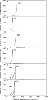
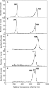
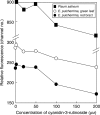
Key results: There were large differences in PI staining (35-70 %) between 2C nuclei from green leaf and red bract tissue in poinsettia. These largely disappeared when pea leaflets were co-chopped with poinsettia tissue as an internal standard. However, smaller (2.8-6.9 %) differences remained, and red bracts gave significantly lower 1C genome size estimates (1.69-1.76 pg) than green leaves (1.81 pg). Chopping pea or poinsettia tissue in buffer with 0-200 microm cyanidin-3-rutinoside showed that the effects of natural inhibitors in red bracts of poinsettia on PI staining were largely reproduced in a dose-dependent way by this anthocyanin.
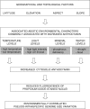
Conclusions: Given their near-ubiquitous distribution, many suspected roles and known affects on DNA staining, anthocyanins are a potent, potential cause of significant error variation in genome size estimations for many plant tissues and taxa. This has important implications of wide practical and theoretical significance. When choosing genome size calibration standards it seems prudent to select materials producing little or no anthocyanin. Reviewing the literature identifies clear examples in which claims of intraspecific variation in genome size are probably artefacts caused by natural variation in anthocyanin levels or correlated with environmental factors known to induce variation in pigmentation.
Figures


Similar articles
-
The rare orange-red colored Euphorbia pulcherrima cultivar 'Harvest Orange' shows a nonsense mutation in a flavonoid 3'-hydroxylase allele expressed in the bracts.BMC Plant Biol. 2018 Oct 3;18(1):216. doi: 10.1186/s12870-018-1424-0. BMC Plant Biol. 2018. PMID: 30285622 Free PMC article.
-
Hybrid de novo transcriptome assembly of poinsettia (Euphorbia pulcherrima Willd. Ex Klotsch) bracts.BMC Genomics. 2019 Nov 27;20(1):900. doi: 10.1186/s12864-019-6247-3. BMC Genomics. 2019. PMID: 31775622 Free PMC article.
-
Population changes of Bemisia tabaci (Hemiptera: Aleyrodidae) on different colored poinsettia leaves with different trichome densities and chemical compositions.J Econ Entomol. 2023 Aug 10;116(4):1276-1285. doi: 10.1093/jee/toad100. J Econ Entomol. 2023. PMID: 37279557
-
Anthocyanin metabolic engineering of Euphorbia pulcherrima: advances and perspectives.Front Plant Sci. 2023 May 15;14:1176701. doi: 10.3389/fpls.2023.1176701. eCollection 2023. Front Plant Sci. 2023. PMID: 37255565 Free PMC article. Review.
-
Plant DNA flow cytometry and estimation of nuclear genome size.Ann Bot. 2005 Jan;95(1):99-110. doi: 10.1093/aob/mci005. Ann Bot. 2005. PMID: 15596459 Free PMC article. Review.
Cited by
-
Karyology and Genome Size Analyses of Iranian Endemic Pimpinella (Apiaceae) Species.Front Plant Sci. 2022 Jun 15;13:898881. doi: 10.3389/fpls.2022.898881. eCollection 2022. Front Plant Sci. 2022. PMID: 35783941 Free PMC article.
-
A guided tour of large genome size in animals: what we know and where we are heading.Chromosome Res. 2011 Oct;19(7):925-38. doi: 10.1007/s10577-011-9248-x. Chromosome Res. 2011. PMID: 22042526 Review.
-
Genome Size Variation and Evolution Driven by Transposable Elements in the Genus Oryza.Front Plant Sci. 2022 Jul 7;13:921937. doi: 10.3389/fpls.2022.921937. eCollection 2022. Front Plant Sci. 2022. PMID: 35874017 Free PMC article.
-
Genome size variation and evolution in allotetraploid Arabidopsis kamchatica and its parents, Arabidopsis lyrata and Arabidopsis halleri.AoB Plants. 2014 May 26;6:plu025. doi: 10.1093/aobpla/plu025. AoB Plants. 2014. PMID: 24887004 Free PMC article.
-
The Application of Flow Cytometry for Estimating Genome Size, Ploidy Level Endopolyploidy, and Reproductive Modes in Plants.Methods Mol Biol. 2021;2222:325-361. doi: 10.1007/978-1-0716-0997-2_17. Methods Mol Biol. 2021. PMID: 33301101
References
-
- Arabidopsis Genome Initiative. Analysis of the genome sequence of the flowering plant. Arabidopsis thaliana. Nature. 2000;408:796–815. - PubMed
-
- Arumuganathan KE, Earle ED. Nuclear DNA content of some important plant species. Plant Molecular Biology Reporter. 1991;9:208–219.
-
- Bassi P. Quantitative variations of nuclear DNA during plant development: a critical analysis. Biological Reviews of the Cambridge Philosophical Society. 1990;65:185–225.
-
- Bennett MD. Intraspecific variation in DNA amount and the nucleotypic dimension in plant genetics. In: Freeling M, editor. UCLA Symposium on molecular and cellular biology, Plant genetics. Vol. 35. New York: Alan Liss; 1985. pp. 283–302.
Publication types
MeSH terms
Substances
LinkOut - more resources
Full Text Sources
Research Materials
Miscellaneous

