. 2008 Jan 21;205(1):105-15.
doi: 10.1084/jem.20071367. Epub 2007 Dec 24.

Initiation of the adaptive immune response to Mycobacterium tuberculosis depends on antigen production in the local lymph node, not the lungs

Affiliations

Initiation of the adaptive immune response to Mycobacterium tuberculosis depends on antigen production in the local lymph node, not the lungs

Andrea J Wolf et al. J Exp Med. .

Abstract

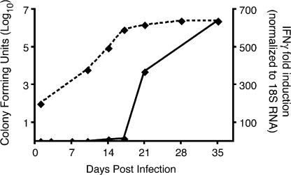
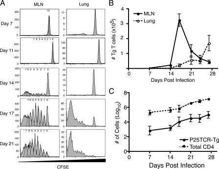
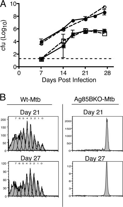
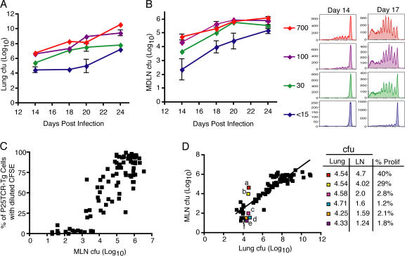
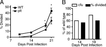
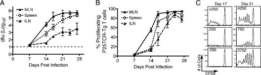
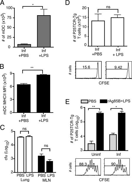
The onset of the adaptive immune response to Mycobacterium tuberculosis is delayed compared with that of other infections or immunization, and allows the bacterial population in the lungs to expand markedly during the preimmune phase of infection. We used adoptive transfer of M. tuberculosis Ag85B-specific CD4(+) T cells to determine that the delayed adaptive response is caused by a delay in initial activation of CD4(+) T cells, which occurs earliest in the local lung-draining mediastinal lymph node. We also found that initial activation of Ag85B-specific T cells depends on production of antigen by bacteria in the lymph node, despite the presence of 100-fold more bacteria in the lungs. Although dendritic cells have been found to transport M. tuberculosis from the lungs to the local lymph node, airway administration of LPS did not accelerate transport of bacteria to the lymph node and did not accelerate activation of Ag85B-specific T cells. These results indicate that delayed initial activation of CD4(+) T cells in tuberculosis is caused by the presence of the bacteria in a compartment that cannot be mobilized from the lungs to the lymph node, where initial T cell activation occurs.


Figures

Figure 1.
Figure 1.
Timing of the appearance of adaptive immunity compared with bacterial growth in the lungs of M. tuberculosis–infected C57BL/6 mice. C57BL/6 mice were infected by the aerosol route with 50 cfu of M. tuberculosis (H37Rv). At designated time points, the lungs were removed and assayed for bacteria by plating and for IFNγ gene expression by real-time RT-qPCR. Four mice were assayed at each time point. Error bars represent the SEM.
Figure 2.
Figure 2.
Proliferation of P25TCR-Tg CD4+ T cells occurs in the mediastinal lymph node 11 d after infection. (A) CFSE proliferation profile of P25TCR-Tg CD4+ T cells in the mediastinal lymph node and lungs over the course of the infection with M. tuberculosis. Plots are representative of five mice at each time point. Data from days 7, 14, 17, and 21 are from one experiment; day 11 results are from a separate experiment, using the same bacterial inoculum and number of adoptively transferred cells. (B) Total number of P25TCR-Tg CD4+ T cells in the mediastinal lymph node (solid line) and lung (dashed line). Replicates were averaged, and the error bars represent the SEM of five mice per time point. (C) Comparison of the number of total CD4+ T cells and the P25TCR-Tg CD4+ T cells in the lungs with time. Error bars represent the mean ± the SD.
Figure 3.
Figure 3.
P25TCR-Tg CD4+ T cell responses are specific for M. tuberculosis Antigen 85B. (A) CFSE-labeled CD4+ P25TCR-Tg CD4+ T cells were adoptively transferred into C57BL/6J mice, and after 24 h, mice were infected with either wild-type M. tuberculosis (Wt-Mtb, solid lines) or Antigen 85BKO M. tuberculosis (Ag85BKO-Mtb, dashed lines with open symbols) by the aerosol route. The initial inoculum for both strains was ∼100 bacteria/mouse. Total bacterial load was assessed over the first 28 d of infection in the lungs (circles) and the mediastinal lymph node (squares). At each time point, two Ag85BKO-Mtb–infected mice were assessed with a difference of <0.5 log10. Error bars for WT-Mtb represent the mean ± the SD of five mice. The dashed line represents the limit of detection for the cfu assay (18.75 cfu/mouse). The mediastinal lymph node cfu of all the mice at day 7 were below the limit of detection. (B) CFSE proliferation profile of P25TCR-Tg CD4+ T cells in the mediastinal lymph node.
Figure 4.
Figure 4.
Dependence of initiation of proliferation on the inoculum of M. tuberculosis. Bacterial cfu in the lung (A) and mediastinal lymph node (B) over the course of the infection after inocula of <15, 30, 100, or 700 bacteria per mouse. (B) Histograms are representative of P25TCR-Tg CD4+ T cell proliferation in the mediastinal lymph node 14 (left column) or 17 (right column) d after infection in mice that received the designated inocula. (C) Correlation of mediastinal lymph node bacterial burden and proliferation of P25TCR-Tg CD4+ T cells. Data are from three independent experiments, and include data from mice that received distinct inocula and that were harvested on various days after infection. (D) Correlation of lung and mediastinal lymph node cfu and P25TCR-Tg CD4+ T cell proliferation. Six mice with similar lung cfu and dissimilar mediastinal lymph node cfu are highlighted. Inset table shows the log10 lung cfu (Lung), log10 mediastinal lymph node cfu (LN), and the percentage of P25TCR-Tg CD4+ T cells that had undergone one or more cycles of proliferation in the mediastinal lymph node for each of the highlighted mice.
Figure 5.
Figure 5.
Delayed dissemination of M. tuberculosis in plt mice results in further delayed T cell activation. (A) Proliferation of CFSE-labeled P25TCR-Tg CD4+ T cells after adoptive transfer to M. tuberculosis–infected wild-type C57BL/6J or plt mice. Results shown are the percentage of P25TCR-Tg CD4+ T cells that had started to divide, to correct for the partial defect in the number of adoptively transferred cells that trafficked to the mediastinal lymph node of plt mice. To detect P25TCR-Tg CD4+ T cells on days 10 and 14, it was necessary to pool lymph nodes from 5 mice, therefore statistical analysis was not done on those samples. Error bars on days 19 and 21 represent the mean ± the SD of 3 C57BL/6J mice and 5 plt mice. *, P < 0.05 by Student's t test. The percentage of CD69+ P25TCR-Tg CD4+ T cells was also fourfold lower in plt mice compared with wild-type at day 14 (not depicted). (B) Bar graph displaying the percentage of P25TCR-Tg CD4+ T cells that had divided and the cfu at days 14 and 19 for the plt mice as the percentage of wild-type levels.
Figure 6.
Figure 6.
M. tuberculosis disseminates and activates P25TCR-Tg CD4+ T cells in lymphoid tissues that do not drain the lungs. (A) Bacterial load in the mediastinal lymph node (MLN), spleen, and inguinal lymph node (ILN) with time. The dashed line represents the limit of detection (18.75 cfu/mouse). Error bars represent the SD. (B) The percentage of P25TCR-Tg CD4+ T cells that have undergone at least 1 cycle of proliferation in the mediastinal lymph node (MLN), spleen, and inguinal lymph node (ILN). Error bars represent the SEM. (C) CFSE histograms form the inguinal lymph node at days 17 and 21 after infection; each histogram represents one mouse. Values in the top left corner represent the number of cfu found in the inguinal lymph node of that mouse.
Figure 7.
Figure 7.
Airway administration of LPS does not increase translocation of M. tuberculosis from the lungs to the mediastinal lymph node, and does not accelerate P25TCR-Tg CD4+ T cell responses. Mice that were either uninfected or infected 10 d earlier with wild-type M. tuberculosis received 5 × 10⁶ CFSE-labeled P25TCR-Tg CD4+ T cells 4 d before harvest. 24 h later, i.e., 3 d before harvest, mice were given sterile PBS or 1 μg LPS with or without 50 ng of recombinant Antigen 85B (rAg85B) by the intranasal route. (A) The number of myeloid dendritic cell in the mediastinal lymph node. (B) Surface MHC class II mean fluorescence intensity (MFI) on mediastinal lymph node myeloid dendritic cell. (C) Cfu in the lung and mediastinal lymph node (MLN) in mice infected with wild-type M. tuberculosis that either received PBS or LPS i.n. (D) Change in the number of P25TCR-Tg CD4+ T cells caused by proliferation as assessed in the mediastinal lymph node at day 14 after infection (4 d after LPS or control). Histograms show day 14 CFSE proliferation profiles of representative mice given either PBS (D, left) or LPS (D, right) i.n. (E) Comparison of the number of P25TCR-Tg CD4+ T cells in the mediastinal lymph node of uninfected or M. tuberculosis–infected mice that received either PBS or rAg85B + LPS i.n. Histograms represent the proliferation profile of the CFSE-labeled P25TCR-Tg cells in uninfected + rAg85B + LPS (left) and infected + rAg85B + LPS (right). The number of cells was determined by multiplying the percentage as determined by flow cytometry by the total number of cells in the lymph node as assessed by trypan exclusion. Three mice were examined per group, and the error bars represent the SD. Statistics were done using Student t test. *, P < 0.05; **, P < 0.01. ns, not significant.

References

    1. Mogues, T., M.E. Goodrich, L. Ryan, R. LaCourse, and R.J. North. 2001. The relative importance of T cell subsets in immunity and immunopathology of airborne Mycobacterium tuberculosis infection in mice. J. Exp. Med. 193:271–280. - PMC - PubMed
    1. Perlman, D.C., W.M. El-Sadr, E.T. Nelson, J.P. Matts, E.E. Telzak, N. Salomon, K. Chirgwin, and R. Hafner. 1997. Variation of chest radiographic patterns in pulmonary tuberculosis by degree of human immunodeficiency virus-related immunosuppression. Clin. Infect. Dis. 25:242–246. - PubMed
    1. Poulsen, A. 1950. Some clinical features of tuberculosis I. Incubation period. Acta Tuberc. Pneumol. Scand. 24:311–346. - PubMed
    1. Wallgren, A. 1948. The time-table of tuberculosis. Tubercle. 29:245–251. - PubMed
    1. Chackerian, A.A., J.M. Alt, T.V. Perera, C.C. Dascher, and S.M. Behar. 2002. Dissemination of Mycobacterium tuberculosis is influenced by host factors and precedes the initiation of T-cell immunity. Infect. Immun. 70:4501–4509. - PMC - PubMed

Publication types