. 2007 Dec 31;179(7):1355-63.
doi: 10.1083/jcb.200702143. Epub 2007 Dec 24.

A novel insertion pathway of mitochondrial outer membrane proteins with multiple transmembrane segments

Affiliations

A novel insertion pathway of mitochondrial outer membrane proteins with multiple transmembrane segments

Hidenori Otera et al. J Cell Biol. .

Abstract

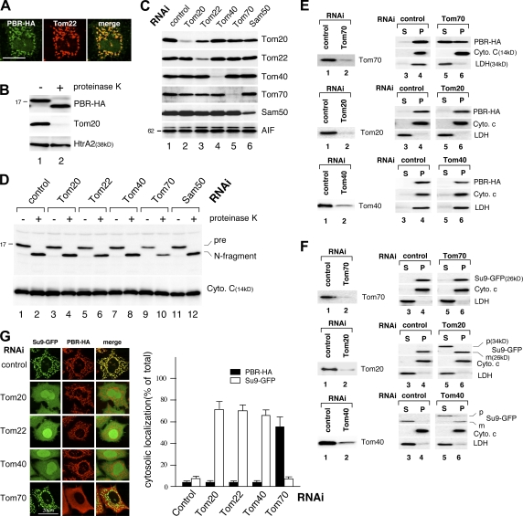
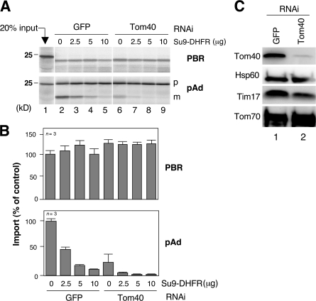
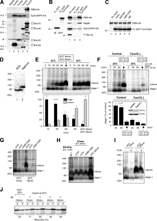
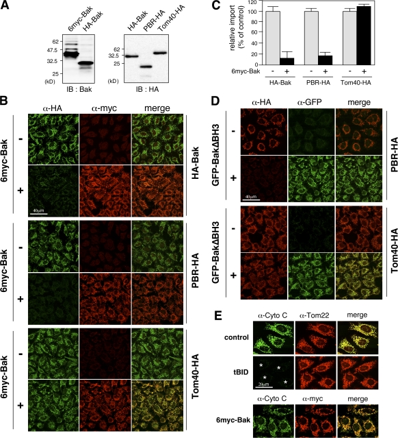
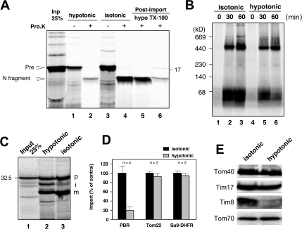
The central channel Tom40 of the preprotein translocase of outer membrane (TOM) complex is thought to be responsible for the import of virtually all preproteins synthesized outside the mitochondria. In this study, we analyze the topogenesis of the peripheral benzodiazepine receptor (PBR), which integrates into the mitochondrial outer membrane (MOM) through five hydrophobic transmembrane segments (TMSs) and functions in cholesterol import into the inner membrane. Analyses of in vitro and in vivo import into TOM component-depleted mitochondria reveal that PBR import (1) depends on the import receptor Tom70 but requires neither the Tom20 and Tom22 import receptors nor the import channel Tom40, (2) shares the post-Tom70 pathway with the C-tail-anchored proteins, and (3) requires factors of the mitochondrial intermembrane space. Furthermore, membrane integration of mitofusins and mitochondrial ubiquitin ligase, the MOM proteins with two and four TMSs, respectively, proceeds through the same initial pathway. These findings reveal a previously unidentified pathway of the membrane integration of MOM proteins with multiple TMSs.


Figures

Figure 1.
Figure 1.
MOM integration of PBR in TOM component–depleted semi-intact and intact cells. (A) HeLa cells were semipermeabilized with digitonin and incubated with reticulocyte lysate–synthesized PBR-HA. After fixation and permeabilization, the cells were processed for immunofluorescence microscopy using anti-HA and anti-Tom22 antibodies. (B) Reticulocyte lysate–synthesized biotin-labeled PBR-HA was incubated with semi-intact cells as in A. The cells were treated with 50 μg/ml proteinase K at 26°C for 3 min and analyzed by SDS-PAGE and immunoblotting using HRP-conjugated streptavidin (PBR-HA), anti-Tom20, or anti-HtrA2 antibodies. (C) HeLa cells subjected to RNAi for the indicated proteins were analyzed by SDS-PAGE and subsequent immunoblotting using the indicated antibodies. AIF, loading control. (D) TOM component knockdown semi-intact cells were subjected to import assay as in B. Cytochrome c, loading control. (E and F) HeLa cells subjected to RNAi were transfected with the expression vector for Su9-GFP or PBR-HA. The cells were treated with digitonin to permeabilize the plasma membrane and centrifuged to separate the supernatant (S) and membrane (P) fractions (Otera et al., 2005), which were analyzed by SDS-PAGE and immunoblotting using the antibodies against HA or the indicated proteins. (G) Su9-GFP and PBR-HA were coexpressed in TOM component knockdown HeLa cells. The cells were analyzed by immunofluorescence microscopy. The cells (100 cells in three distinct fields) exhibiting cytosolic localization of Su9-DHFR or PBR-HA in each RNAi experiment were counted. Error bars represent SD. Bars, 20 μm.
Figure 2.
Figure 2.
Effect of Tom40 channel block on MOM integration of PBR-HA. (A) Reticulocyte lysate–synthesized 35S–PBR-HA or 35S-pAd was imported into the control or Tom40 knockdown mitochondria in the presence of recombinant Su9-DHFR. The reaction mixtures were treated with (for PBR) or without (for pAd) proteinase K and analyzed by SDS-PAGE and subsequent digital autoradiography. (B) The band intensities (proteinase K–resistant band for PBR and mature band for pAd) were quantified setting those in the absence of Su9-DHFR at 100%. Results obtained from three independent experiments are shown. (C) The mitochondria used in A were analyzed by SDS-PAGE and immunoblotting using antibodies against the indicated proteins. This procedure depleted Tom40 by ∼95%. Error bars represent SD.
Figure 3.
Figure 3.
Analysis of import steps of PBR. (A and B) Reticulocyte lysate–synthesized PBR-HA, Su9-DHFR-HA, or T7–Bcl-XL was incubated with FLAG-tagged Tom proteins or FLAG-GFP-Tom70ΔN purified from HeLa cells. They were subjected to pull-down reaction and subsequent immunoblotting using the indicated antibodies. (C) Reticulocyte lysate–synthesized PBR-HA was incubated with 100 U/ml apyrase, 10 mM AMP-PNP, or 1 mM novobiocin (NB). The reaction mixtures were subjected to pull-down assay as in B. (D) Rat liver mitochondria were subjected to BN-PAGE followed by immunoblotting using anti-PBR antibodies. (E and F) Reticulocyte lysate–synthesized 35S–PBR-HA was incubated with control or Tom70-knockdown mitochondria under the indicated conditions. The reaction mixtures were analyzed by BN-PAGE and subsequent digital autoradiography. Band intensities of stage I and the mature form are shown, setting those of stage I (25°C for 15 min) and the mature form (25°C for 90 min plus 33°C for 60 min; E) or that of control (30 min) at 100% (F). Results obtained from three independent experiments are shown. Error bars represent SD. (G) Reticulocyte lysate–synthesized 35S–PBR-HA was passed through a spin column and subjected to import in the absence or presence of 1 mM ATP or AMP-PNP under the indicated conditions. Other conditions were set as in E. (H) Reticulocyte lysate–synthesized 35S–PBR-HA was subjected to mitochondrial import at 25°C for 15 min (binding). The mitochondria were reisolated and incubated at 33°C for 60 min (chase) in the import buffer with or without 1 mM ATP or AMP-PNP. Other conditions were set as in E. (I) Reticulocyte lysate–synthesized 35S–PBR-HA was imported into mitochondria under the indicated conditions. The reaction mixtures were treated with or without proteinase K and analyzed by BN-PAGE and digital autoradiography. (J) Reticulocyte lysate–synthesized 35S–PBR-HA was subjected to mitochondrial import. The mitochondria were treated with 100 mM Na2CO3, pH 11.5, and centrifuged to separate the supernatant (S) and membrane (P) fractions, which were analyzed by SDS-PAGE and subsequent digital autoradiography.
Figure 4.
Figure 4.
Inhibition of MOM integration of PBR by an excess amount of Bak. (A) The reticulocyte lysate–synthesized proteins used (4 μl each) were analyzed by SDS-PAGE and immunoblotting (IB) using the indicated antibodies. (B) The indicated proteins were subjected to mitochondrial import in semi-intact cells in the presence or absence of an excess amount of 6myc-Bak (70 μl; import substrates, 20 μl each) and subsequent immunofluorescence microscopy. Imported proteins and 6myc-Bak are shown in green and red, respectively. (C) The extent of import was quantified using ImageJ (National Institutes of Health). Each graph indicates the mean ± SD (error bars) of three independent experiments of at least 100 cells. The fluorescence intensities of 6myc-Bak (−) cells were set at 100%. (D) Mitochondrial import of PBR-HA and Tom40-HA was performed as in B in the presence or absence of GFP-BakΔBH3. (E) Semi-intact cells were treated with tBid or 6myc-Bak, and the cells were analyzed by immunofluorescence microscopy using the antibodies against cytochrome c (green), Tom22 (red), or myc-tag (red). Asterisks indicate cytochrome c–released cells.
Figure 5.
Figure 5.
Depletion of IMS proteins inhibits PBR import. (A) Reticulocyte lysate–synthesized [35S]PBR-HA was imported into intact or hypotonic buffer–treated mitochondria. The reaction mixtures were aliquoted and incubated with or without 100 μg/ml proteinase K. The other aliquots (post-import) were treated with proteinase K under hypotonic conditions or in the presence of 1% Triton X-100. Samples were analyzed by SDS-PAGE and subsequent digital autoradiography. (B and C) Reticulocyte lysate–synthesized 35S-Tom22 (B) and 35S–Su9-DHFR (C) were imported into mitochondria and analyzed by BN-PAGE (for Tom22) or SDS-PAGE (for Su9-DHFR). p, precursor; i, intermediate; m, mature form. (D) Import efficiencies of PBR, Tom22, and Su9-DHFR were calculated setting each activity (PBR, proteinase K–resistant bands [percentage of input]; Tom22, ∼400-kD band; Su9-DHFR, m/(p + I + m]) of untreated mitochondria at 100%. Results obtained from two or four independent experiments are shown. Error bars represent SD. (E) Intact and hypotonic buffer–treated mitochondria were analyzed by SDS-PAGE and subsequent immunoblotting using the indicated antibodies.

References

    1. Ahting, U., T. Weizenegger, W. Neupert, and D. Rapaport. 2005. Signal-anchor proteins follow a unique insertion pathway into the outer membrane of mitochondria. J. Biol. Chem. 280:48–53. - PubMed
    1. Bohnert, M., N. Pfanner, and M. van der Laan. 2007. A dynamic machinery for import of mitochondrial precursor proteins. FEBS Lett. 581:2802–2810. - PubMed
    1. Eura, Y., N. Ishihara, S. Yokota, and K. Mihara. 2003. Two mitofusin proteins, mammalian homologues of FZO, with distinct functions are both required for mitochondrial fusion. J. Biochem. (Tokyo). 134:333–344. - PubMed
    1. Fan, A.C.Y., M.K. Bhangoo, and J.C. Yound. 2006. Hsp90 functions in the targeting and outer membrane translocation steps of Tom70-mediated mitochondrial import. J. Biol. Chem. 281:33313–33324. - PubMed
    1. Fritz, S., D. Rapaport, E. Klanner, W. Neupert, and B. Westermann. 2001. Connection of the mitochondrial outer and inner membranes by Fzo1 is critical for organeller function. J. Cell Biol. 152:683–692. - PMC - PubMed

Publication types

MeSH terms