. 2007 Dec 31;179(7):1569-82.
doi: 10.1083/jcb.200704010. Epub 2007 Dec 24.

Sequential roles for myosin-X in BMP6-dependent filopodial extension, migration, and activation of BMP receptors

Affiliations

Sequential roles for myosin-X in BMP6-dependent filopodial extension, migration, and activation of BMP receptors

Xinchun Pi et al. J Cell Biol. .

Erratum in

  • J Cell Biol. 2009 Feb 23;184(4):611

Abstract

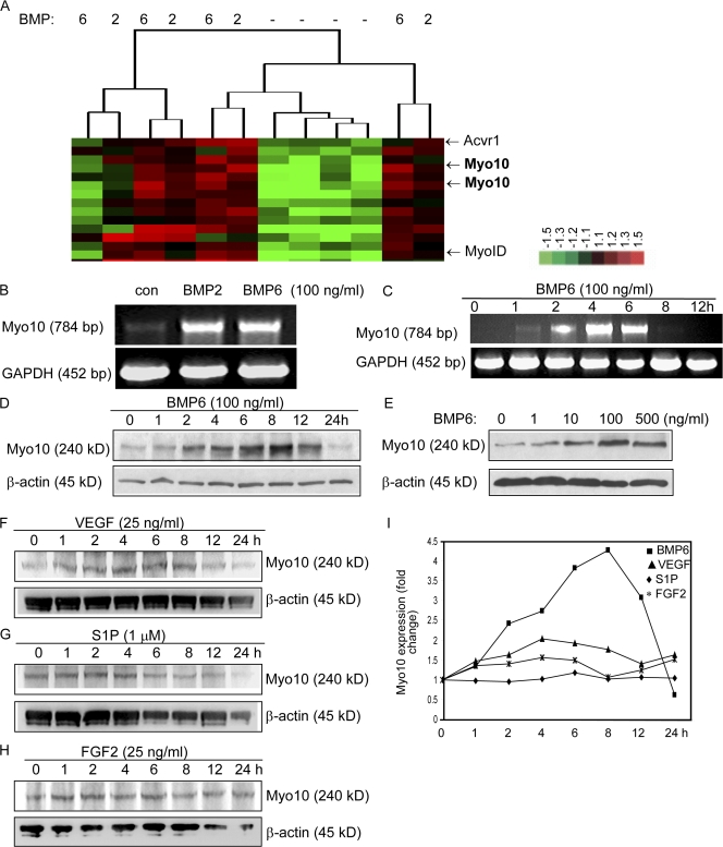
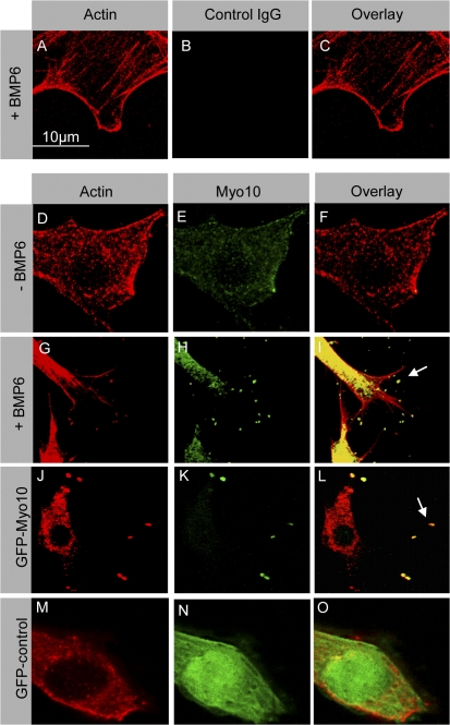
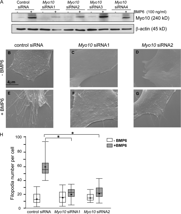
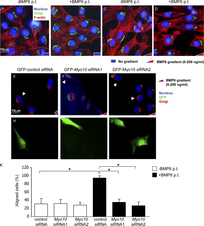
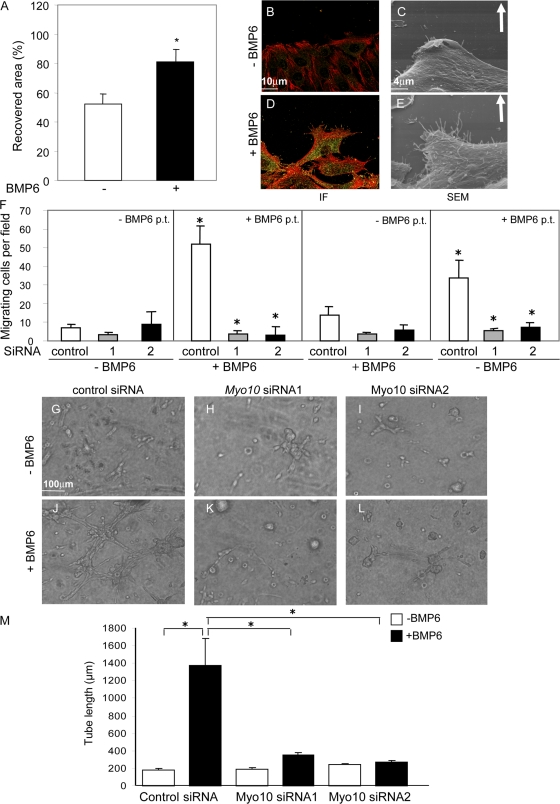
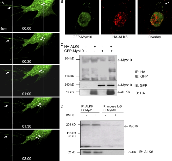
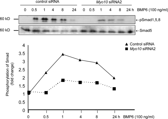
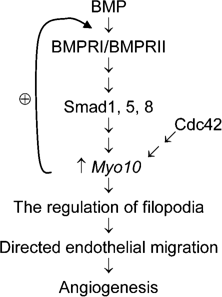
Endothelial cell migration is an important step during angiogenesis, and its dysregulation contributes to aberrant neovascularization. The bone morphogenetic proteins (BMPs) are potent stimulators of cell migration and angiogenesis. Using microarray analyses, we find that myosin-X (Myo10) is a BMP target gene. In endothelial cells, BMP6-induced Myo10 localizes in filopodia, and BMP-dependent filopodial assembly decreases when Myo10 expression is reduced. Likewise, cellular alignment and directional migration induced by BMP6 are Myo10 dependent. Surprisingly, we find that Myo10 and BMP6 receptor ALK6 colocalize in a BMP6-dependent fashion. ALK6 translocates into filopodia after BMP6 stimulation, and both ALK6 and Myo10 possess intrafilopodial motility. Additionally, Myo10 is required for BMP6-dependent Smad activation, indicating that in addition to its function in filopodial assembly, Myo10 also participates in a requisite amplification loop for BMP signaling. Our data indicate that Myo10 is required to guide endothelial migration toward BMP6 gradients via the regulation of filopodial function and amplification of BMP signals.


Figures

Figure 1.
Figure 1.
Myo10 is up-regulated by BMP2 and BMP6 treatment. (A) Microarray analysis was performed with RNA samples purified from MECs treated with BMP2, BMP6, or DMSO for 4 h (Ren et al., 2007). Differentially expressed genes were selected with a p-value ≤0.05 and ratio fold change ≥±1.5 and were subjected to hierarchical cluster analysis. Median-centered clusters were viewed with JavaTreeView. Fold change relative to common reference is indicated by red (+1.5; full scale) and green (−1.5; full scale) intensity. This image is a group of clustered genes with selected genes labeled. (B) MECs were treated with BMP2 and BMP6, and the total RNA was used for RT-PCR analysis with Myo10-specific primers. Glyceraldehyde-3-phosphate dehydrogenase (GAPDH) was used as an internal control. (C) RT-PCR analysis of Myo10 transcripts after BMP6 treatment for the indicated times. (D) Western blotting analysis of Myo10 protein expression after BMP6 treatment for the indicated times. (E) Western blotting analysis of Myo10 protein expression after BMP6 treatment for 8 h with the indicated dosages. (F–I) Myo10 is up-regulated by BMP6 and VEGF but not by S1P and FGF-2 treatment. (F–H) MECs were treated with VEGF (F), S1P (G), and FGF2 (H) for the indicated times. The cell lysates were subjected to Western blotting analysis of Myo10 protein expression and actin as the internal control. (I) Comparison of Myo10 expression level after BMP6, VEGF, S1P, and FGF-2 treatment. The band intensity was measured and quantitated by ImageJ.
Figure 2.
Figure 2.
Myo10 induced by BMP6 is localized in filopodia. MECs were treated with BMP6 for 8 h (A–C and G–I) or DMSO (D–F) and were fixed with PFA. Cells were stained with phalloidin (A, D, G, J, and M) and chicken anti–mouse Myo10 pAb (E and H) or control IgG (B) as a negative control. Exogenous Myo10 (GFP tagged) was expressed by transiently transfecting MECs with a plasmid encoding GFP-Myo10 (J–L). GFP control plasmids (M–O) were transfected in MECs as the negative controls for GFP-Myo10. Actin was visualized using Texas red–phalloidin (red). Myo10 was visualized using chicken anti-Myo10 pAb (green; E and H) or GFP (green; K and N). The arrows represent filopodia.
Figure 3.
Figure 3.
Myo10 siRNAs inhibit BMP6-induced filopodia. (A) MECs transfected with Myo10 siRNAs were lysed and subjected to Western blotting analysis with anti-Myo10 pAb (top). A β-actin blot of the same membrane controlled for sample loadings (bottom). (B–H) Knockdown of endogenous Myo10 in MECs by Myo10 siRNAs decreased filopodial number in the BMP6-treated conditions. MECs were transfected with GFP-tagged Myo10 siRNA1 (C and F) or siRNA2 (D and G) or by the GFP-tagged control vector containing irrelevant siRNA sequence (B and E). Cells were treated with 100 ng/ml BMP6 (E–G) or DMSO (B–D) as a control, and GFP-positive cells were subjected for SEM analysis. (H) The bar graphs show quantification of filopodial numbers in MECs transfected with GFP-tagged control siRNA, GFP-Myo10 siRNA1, or GFP-Myo10 siRNA2. *, P < 0.05; n = 3. n = 20 cells per sample.
Figure 4.
Figure 4.
Cell alignment is dependent on Myo10. (A–D) MECs aligned toward the BMP6 gradient. Cells cultured on the coverslips were pretreated with BMP6 (B and D) or DMSO (A and C) for 4 h, were transferred to the Dunn chamber with the media containing 0–200 ng/ml BMP6 gradient (C and D) or not containing gradient (A and B), and were maintained for 4 h. Cells were fixed and stained with phalloidin, DAPI, and GM130. Actin (Texas red–phalloidin), red; Golgi (anti-GM130 mAb), green; nucleus (DAPI), blue. (E–K) Knockdown of endogenous Myo10 in MECs by Myo10 siRNAs inhibited cell alignment. (E–J) Cells were transfected with GFP-Myo10 siRNA1 or GFP-Myo10 siRNA2 and controlled by transfecting cells with GFP-tagged control vector containing irrelevant siRNA sequence. The cells were pretreated with BMP6 for 4 h and were transferred into the Dunn chamber for another 4 h with 0–200 ng/ml of the media containing BMP6 gradient. The cells were fixed, and immunofluorescent images were taken. GFP, green; Golgi (anti-GM130 mAb), red; nucleus (DAPI), blue. White arrowheads represent the transfected cells with siRNAs (GFP positive). (K) Quantitative analysis of the aligned cells toward the higher BMP6 gradient. Error bars represent SD. n = 3; *, P < 0.05. n = at least 57 cells per sample. p.t., pretreatment.
Figure 5.
Figure 5.
Myo10 knockdown inhibits directed cell migration and angiogenesis induced by BMP6. (A–E) BMP6 increased Myo10 proteins in filopodia, filopodial number, and directed migration. Wound-healing assay was performed with MECs grown on 35-mm wells. (A) The bar graph shows the recovered area after cells were stimulated with BMP6 for 16 h. (B and D) The immunofluorescent staining of cells. Actin (Texas red–phalloidin), red; Myo10 (anti-Myo10 pAb), green; overlay of the red and green, yellow. The increased yellow staining demonstrated the increased localization of Myo10 protein at the filopodial tips of the cells on the leading edge, where the filopodial number also increased. (C and E) The increase of filopodial number in SEM images of cells. Arrows represent the migration direction of the cells. IF, immunofluorescence; SEM, scanning EM. (F) Myo10 knockdown inhibited directed cell migration induced by BMP6. MECs were transfected with GFP-Myo10 siRNA1, GFP-Myo10 siRNA2, or control siRNA, were pretreated with BMP6 for 4 h, and were trypsinized for the Boyden chamber assay. The cells migrating through the filter were counted. p.t., pretreatment. (G–M) Myo10 knockdown inhibited tube formation induced by BMP6 using 3D collagen assay. MECs were transfected with GFP-Myo10 siRNA1, GFP-Myo10 siRNA2, or control siRNA and were lysed for the 3D collagen angiogenesis assay. (G–L) The brightfield images were taken after 24 h of incubation. (M) Quantitation of the tube formation. Error bars represent SD. n = 3; *, P < 0.05.
Figure 6.
Figure 6.
Myo10 interacts with ALK6. (A) Selected sequence from a time-lapse video focusing on the movements of puncta containing GFP-ALK6 proteins. Arrows represent the puncta containing GFP-ALK6. The images were taken every 30 s for 120 s after MECs were treated with BMP6 for 4 h. The mean rate of the forward movements for the puncta was ∼42 ± 16 nm s−1 (n = 10 puncta). (B) Colocalization of exogenous GFP-tagged Myo10 and HA-tagged ALK6 at tips of filopodia in HeLa cells. Myo10 (GFP), green; ALK6 (anti-HA mAb), red. The arrow indicates colocalization. (C) Exogenous Myo10 associated with exogenous ALK6. GFP-Myo10 and HA-ALK6 were expressed in 293T cells, and immunoprecipitation was performed with anti-HA mAb followed by Western blotting against GFP. (D) Endogenous Myo10 associated with ALK6. MEC lysates were immunoprecipitated by anti-ALK6 mAb, with mouse IgG as a control, followed by Western blotting against Myo10.
Figure 7.
Figure 7.
Myo10 knockdown inhibits BMP6-induced Smad1, 5, and 8 activation. MECs were transfected with Myo10 siRNA2 or control siRNA and were stimulated with BMP6 for the indicated times for the activation of Smads. Cells were lysed for Western blotting analysis of the antiphospho-Smad1, 5, and 8 pAb, anti-Smad5 pAb, and anti-Myo10 pAb. This is a representative image of three independent experiments.
Figure 8.
Figure 8.
Schematic model for Myo10 as a sensor for filopodia to sense the BMP6 gradient for directed cell migration and angiogenesis.

References

    1. Bar, T., and J.R. Wolff. 1972. The formation of capillary basement membranes during internal vascularization of the rat's cerebral cortex. Z. Zellforsch. Mikrosk. Anat. 133:231–248. - PubMed
    1. Berg, J.S., and R.E. Cheney. 2002. Myosin-X is an unconventional myosin that undergoes intrafilopodial motility. Nat. Cell Biol. 4:246–250. - PubMed
    1. Bohil, A.B., B.W. Robertson, and R.E. Cheney. 2006. Myosin-X is a molecular motor that functions in filopodia formation. Proc. Natl. Acad. Sci. USA. 103:12411–12416. - PMC - PubMed
    1. Deckers, M.M., R.L. van Bezooijen, G. van der Horst, J. Hoogendam, C. van Der Bent, S.E. Papapoulos, and C.W. Lowik. 2002. Bone morphogenetic proteins stimulate angiogenesis through osteoblast-derived vascular endothelial growth factor A. Endocrinology. 143:1545–1553. - PubMed
    1. Folkman, J., and M. Klagsbrun. 1987. Vascular physiology. A family of angiogenic peptides. Nature. 329:671–672. - PubMed

Publication types

MeSH terms