. 2007 Dec 31;179(7):1481-96.
doi: 10.1083/jcb.200710061. Epub 2007 Dec 24.

Huntingtin-interacting protein 14, a palmitoyl transferase required for exocytosis and targeting of CSP to synaptic vesicles

Affiliations

Huntingtin-interacting protein 14, a palmitoyl transferase required for exocytosis and targeting of CSP to synaptic vesicles

Tomoko Ohyama et al. J Cell Biol. .

Abstract

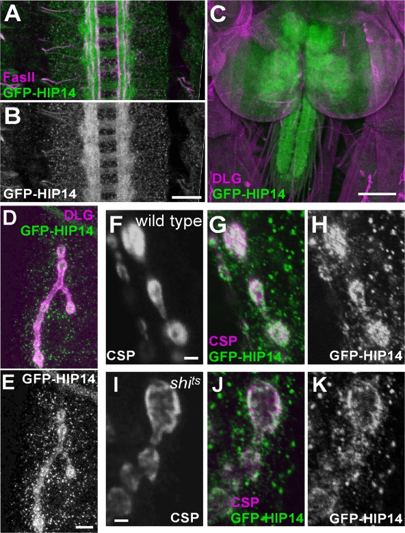
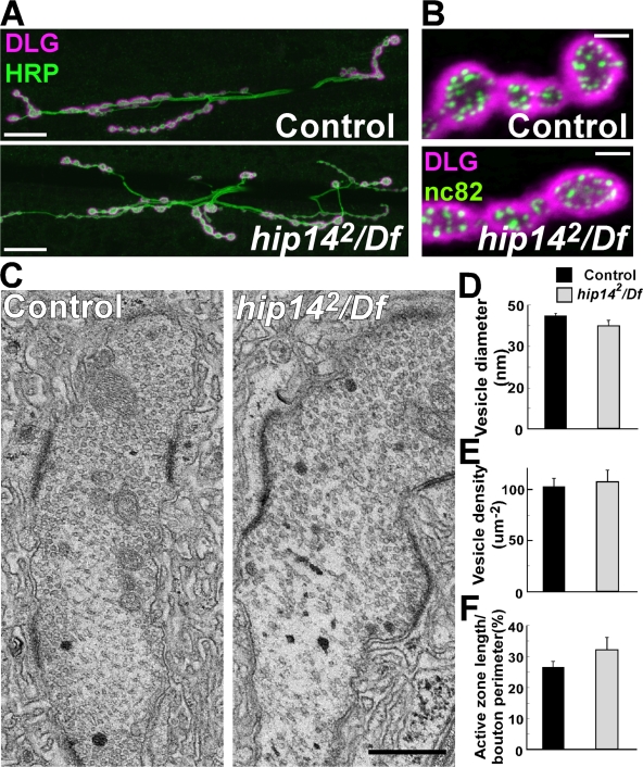
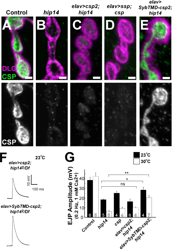
Posttranslational modification through palmitoylation regulates protein localization and function. In this study, we identify a role for the Drosophila melanogaster palmitoyl transferase Huntingtin-interacting protein 14 (HIP14) in neurotransmitter release. hip14 mutants show exocytic defects at low frequency stimulation and a nearly complete loss of synaptic transmission at higher temperature. Interestingly, two exocytic components known to be palmitoylated, cysteine string protein (CSP) and SNAP25, are severely mislocalized at hip14 mutant synapses. Complementary DNA rescue and localization experiments indicate that HIP14 is required solely in the nervous system and is essential for presynaptic function. Biochemical studies indicate that HIP14 palmitoylates CSP and that CSP is not palmitoylated in hip14 mutants. Furthermore, the hip14 exocytic defects can be suppressed by targeting CSP to synaptic vesicles using a chimeric protein approach. Our data indicate that HIP14 controls neurotransmitter release by regulating the trafficking of CSP to synapses.


Figures

Figure 1.
Figure 1.
Characterization of the ey-FLP screen phenotypes of 3L1 mutants. (A) ERGs of controls (y w ey-FLP GMR-lacZ; FRT80B/RpS174 P{w + } FRT80B) and 3L1 mutants (y w ey-FLP GMR-lacZ; 3L11, 3L12, or 3L13 FRT80B/RpS174 P{w + } FRT80B). The positions of on and off transients are indicated by gray arrowheads. (B and C) Confocal images showing control (y w ey-FLP GMR-lacZ; FRT80B/RpS174 P{w + } FRT80B) and 3L1 mutant (y w ey-FLP GMR-lacZ; 3L12 FRT80B/RpS174 P{w + } FRT80B) adult brains stained with mAb 24B10. (D and E) EM of control (D) and 3L12 mutant (E) PR terminals in the lamina (for genotypes, see A). Some of the capitate projections (cp) and mitochondria (m) are indicated. (F) Quantification of capitate projection distribution in controls and 3L11, 3L12, and 3L13 mutants. Error bars represent SEM. Bars: (B and C) 50 μm; (D and E) 1 μm.
Figure 2.
Figure 2.
Identification of 3L1. (A) P-element fine mapping. Numbers separated by a backslash indicate recombinants out of total flies scored. Recombination distance in centiMorgans for the two closest P elements is indicated. A deficiency that complements (green) and one that fails to complement (red) are shown. The calculated mapping location based on P-element mapping of 3L1 is shown as a red star, and the coding regions of nearby genes (black boxes) are shown. The region used for rescue is indicated by the blue bar. (B) Intron-exon structure of CG6017 and protein structure of HIP14. The start codon is marked in green, and the stop codon in indicated in red. The position of the N- or C-termEGFP of the genomic rescue constructs are indicated by the green inserts. HIP14 contains five ankyrin repeats (green boxes), five TMDs (yellow boxes), and a DHHC-CRD domain (blue box). The molecular nature of the three hip14 alleles is indicated. (C) Lethal stage of hip14 mutant combinations. (D) Lethality rescue of hip14 mutant combinations by genomic and cDNA constructs expressed using elav-GAL4. Columns are divided such that the yellow portion indicates the result from the genomic construct, and white indicates the result from the cDNA. (C and D) L3, animals do not survive beyond the third instar larval stage; P, death during late pupal stage; ND, not tested; R, rescued lethality.
Figure 3.
Figure 3.
Localization of HIP14. Confocal images from genomic GFP-tagged hip14 transgenic animals. (A and B) Stage 14 embryo (y w; P{w + NtermGFP-hip14 + }) with GFP-HIP14 (green) and Fasciclin II (magenta). The GFP-HIP14 (green) channel is shown separately in B. (C) Third instar larval (y w; P{w + NtermGFP-hip14 + }) CNS with GFP-HIP14 (green) and DLG (magenta). (D and E) Third instar larvae NMJ boutons in genomic GFP-tagged hip14 transgenic animals (y w; P{w + NtermGFP-hip14 + }) with GFP-HIP14 (green) and DLG (magenta) to label the synaptic areas. The green channel is shown separately in E. (F–H) Third instar larval NMJ boutons in wild type (y w, shibirets/+; P{w + NtermGFP-hip14 + }/+) with GFP-HIP14 (green) and CSP (magenta) at ambient temperature. Both channels are separately shown in F and H. Images are single confocal sections. (I–K) Third instar larval NMJ boutons in shibirets mutant (y w, shibirets/Y; P{w + NtermGFP-hip14 + }/+) at restrictive temperature or 32°C with GFP-HIP14 (green) and CSP (magenta). Both channels are separately shown in I and K. Images are single confocal sections. Bars: (A and B) 20 μm; (C) 100 μm; (D and E) 10 μm; (F–K) 2 μm.
Figure 4.
Figure 4.
NMJ morphology is normal in hip14 mutants. (A) Confocal images of the third instar larval NMJ on muscles 6 and 7 labeled with anti-HRP (green) to mark all neuronal membranes and DLG (magenta) to label the pre- and postsynaptic regions in control (y w; FRT80B) and hip142/Df mutants (y w ey-FLP GMR-lacZ; hip142 FRT80B/Df(3L)brm11). (B) mAb nc82 (Bruchpilot) and DLG labeling of boutons of control (y w; FRT80B) and hip142/Df mutants (y w ey-FLP GMR-lacZ; hip142 FRT80B/Df(3L)brm11). (C) Ultrastructure of NMJ boutons in control (y w; FRT80B) and hip142/Df mutants (y w ey-FLP GMR-lacZ; hip142 FRT80B/Df(3L)brm11). (D–F) Quantification of synaptic features. Error bars represent SD. Bars: (A) 20 μm; (B) 2 μm; (C) 0.5 μm.
Figure 5.
Figure 5.
Evoked neurotransmitter release is impaired in hip14 mutants. (A and B) FM 1-43 dye uptake on controls (y w; FRT80B), hip141/Df mutants (y w ey-FLP GMR-lacZ; hip141 FRT80B/Df(3L)brm11), and hip142/Df mutants (y w ey-FLP GMR-lacZ; hip142 FRT80B/Df(3L)brm11). (A) Preparations were stimulated for 1 min in the presence of 4 μM of dye, 90 mM KCl, and 1.5 mM Ca2+ to label the exo-endo cycling pool. (B) Quantification of the labeling intensity of FM 1-43 shown in A. (C and E) Sample EJPs recorded in 1 mM of external Ca2+ at 0.2 Hz in controls and hip142/Df. Bath temperature was kept at 23 (C) or 30°C (E). (D and F) Quantification of EJP amplitudes recorded at 23°C are shown in D for all genotypes, including csp mutants (cspu1/cspx1) and cDNA rescue by presynaptic expression (elav-GAL4/+; UAS-cDNAhip14/+; hip142/Df(3L)brm11). Quantification for controls, hip142/Df, and csp mutants at 30°C is shown in F. Recordings were performed for 1 min, and 12 EJP amplitudes were averaged per recording. (G) Frequency and amplitude of miniature synaptic currents in hip14 mutants. Miniature EJPs were recorded in the presence of 5 μM tetrodotoxin in 0.5 mM Ca2+. (H) GluRIII immunostaining at NMJs of control and hip142/Df animals. (B, D, F, and G) The number of animals tested is indicated in the bars. Error bars represent SEM. **, P < 0.01 (t test). Bars: (A) 5 μm; (H) 2 μm.
Figure 6.
Figure 6.
CSP and SNAP25 are mislocalized in hip14 mutants. (A–D) Confocal images showing labeling of control (y w; FRT80B; left) and hip142/Df mutant (y w ey-FLP GMR-lacZ; hip142 FRT80B/Df(3L)brm11; right) boutons on muscle 4 segment A4 for Syt I (A), n-Syb (B), CSP (C), and SNAP25 (D; green). (E) Quantification of labeling intensity for synaptic markers shown in A–D. **, P < 0.01 (t test). Error bars represent SEM. The number of animals tested is indicated in the bars. (F) Western blots of larval brain extracts of controls, hip141/Df, and hip142/Df using antibodies against CSP and SNAP25. Protein loading normalized using anti-actin labeling. (G and H) Confocal images showing labeling of control (G) and hip142/Df mutant (H) VNC with CSP (green) and DLG (magenta). The green channel is shown separately in the bottom panel. n, neuropil; c, cell body. (I) Hydroxylamine treatment of hip14 mutant brains. Larval brain extracts of controls (lanes 1 and 2) and hip142/Df (lanes 3 and 4) were treated with hydroxylamine (lanes 2 and 4) or Tris (lanes 1 and 3). After treatment, proteins were subjected to SDS-PAGE and immunoblotted with CSP antibodies. Bars: (A–D) 10 μm; (G and H) 20 μm.
Figure 7.
Figure 7.
Chimeric but not wild-type CSP rescues the localization of CSP and the exocytic defects in hip14 mutants. (A–E) Confocal images showing labeling of larval filets with CSP (green) and DLG (magenta) to indicate the synaptic areas. Genotypes: control (y w; FRT80B; A), hip142/Df mutant (y w ey-FLP GMR-lacZ; hip142 FRT80B/Df(3L)brm11; B), elav>csp2; hip14 (elav-GAL4/+; P{w + UAS-csp2}/+; hip142 FRT80B/Df (3L)brm11; C), elav>ssp; csp (elav-GAL4/+;; P{w + UAS-csp-11c/s}, cspu1/cspx1; D), and elav>SybTMD-csp2; hip14 (elav-GAL4/+; P{w + UAS-SybTMD-csp}/+ hip142 FRT80B/Df(3L)brm11; E). CSP labeling is separately shown on the bottom panels. (F) Sample EJPs recorded in 1 mM Ca2+ at 0.2-Hz stimulation at 23°C when wild-type CSP2 or chimeric CSP is overexpressed in hip142/Df neurons. (G) Quantification of EJP amplitudes are recorded in 1 mM Ca2+ at 23°C (black bars) and at 30°C (white bars) in controls, hip14 mutant, csp mutants (w, cspu1/cspx1), CSP2 overexpressed neuronally in hip14 mutant background, and SybTMD-CSP2 overexpressed neuronally in hip14 mutant background. For recordings at 23°C (black bars), hip142/Df was used, whereas at 30°C (white bars), the hip142/hip141 allelic combination was used. Recordings were performed for 1 min at 0.2 Hz, and 12 EJP amplitudes were averaged per recording. *, P < 0.05; **, P < 0.01 (t test). Error bars represent SEM. The number of animals tested is indicated in the bars. Bars, 2 μm.

References

    1. Arnold, C., N. Reisch, C. Leibold, S. Becker, K. Prufert, K. Sautter, D. Palm, S. Jatzke, S. Buchner, and E. Buchner. 2004. Structure-function analysis of the cysteine string protein in Drosophila: cysteine string, linker and C terminus. J. Exp. Biol. 207:1323–1334. - PubMed
    1. Babu, P., R.J. Deschenes, and L.C. Robinson. 2004. Akr1p-dependent palmitoylation of Yck2p yeast casein kinase 1 is necessary and sufficient for plasma membrane targeting. J. Biol. Chem. 279:27138–27147. - PubMed
    1. Bellen, H.J., R.W. Levis, G. Liao, Y. He, J.W. Carlson, G. Tsang, M. Evans-Holm, P.R. Hiesinger, K.L. Schulze, G.M. Rubin, et al. 2004. The BDGP gene disruption project: single transposon insertions associated with 40% of Drosophila genes. Genetics. 167:761–781. - PMC - PubMed
    1. Bronk, P., J.J. Wenniger, K. Dawson-Scully, X. Guo, S. Hong, H.L. Atwood, and K.E. Zinsmaier. 2001. Drosophila Hsc70-4 is critical for neurotransmitter exocytosis in vivo. Neuron. 30:475–488. - PubMed
    1. Bronk, P., Z. Nie, M.K. Klose, K. Dawson-Scully, J. Zhang, R.M. Robertson, H.L. Atwood, and K.E. Zinsmaier. 2005. The multiple functions of cysteine-string protein analyzed at Drosophila nerve terminals. J. Neurosci. 25:2204–2214. - PMC - PubMed

Publication types

MeSH terms