. 2008 Jan 15;117(2):144-54.
doi: 10.1161/CIRCULATIONAHA.107.726752. Epub 2007 Dec 24.

Reversibility of PRKAG2 glycogen-storage cardiomyopathy and electrophysiological manifestations

Affiliations

Reversibility of PRKAG2 glycogen-storage cardiomyopathy and electrophysiological manifestations

Cordula M Wolf et al. Circulation. .

Abstract

Background: PRKAG2 mutations cause glycogen-storage cardiomyopathy, ventricular preexcitation, and conduction system degeneration. A genetic approach that utilizes a binary inducible transgenic system was used to investigate the disease mechanism and to assess preventability and reversibility of disease features in a mouse model of glycogen-storage cardiomyopathy.

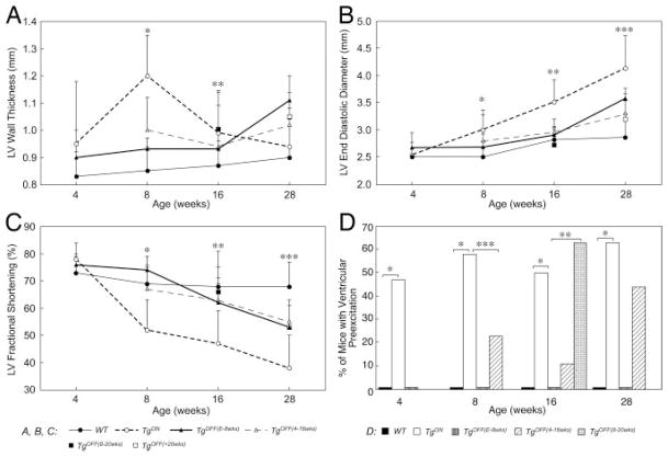
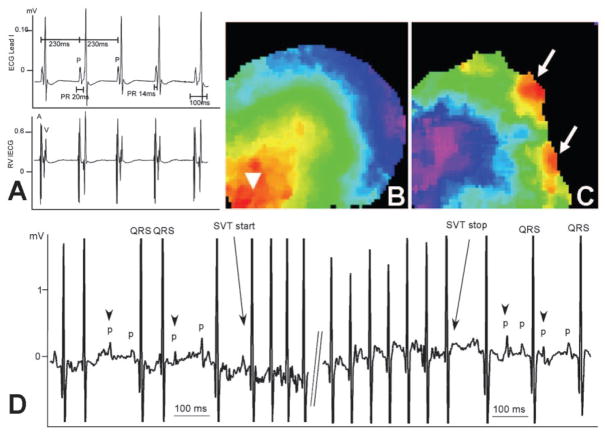
Methods and results: Transgenic (Tg) mice expressing a human N488I PRKAG2 cDNA under control of the tetracycline-repressible alpha-myosin heavy chain promoter underwent echocardiography, ECG, and in vivo electrophysiology studies. Transgene suppression by tetracycline administration caused a reduction in cardiac glycogen content and was initiated either prenatally (Tg(OFF(E-8 weeks))) or at different time points during life (Tg(OFF(4-16 weeks)), Tg(OFF(8-20 weeks)), and Tg(OFF(>20 weeks))). One group never received tetracycline, expressing transgene throughout life (Tg(ON)). Tg(ON) mice developed cardiac hypertrophy followed by dilatation, ventricular preexcitation involving multiple accessory pathways, and conduction system disease, including sinus and atrioventricular node dysfunction.

Conclusions: Using an externally modifiable transgenic system, cardiomyopathy, cardiac dysfunction, and electrophysiological disorders were demonstrated to be reversible processes in PRKAG2 disease. Transgene suppression during early postnatal development prevented the development of accessory electrical pathways but not cardiomyopathy or conduction system degeneration. Taken together, these data provide insight into mechanisms of cardiac PRKAG2 disease and suggest that glycogen-storage cardiomyopathy can be modulated by lowering glycogen content in the heart.


Figures

Figure 1
Figure 1
Modulation of transgene expression. A, Tetracycline-controlled transcriptional activator (tTA) system under the control of a cardiac-specific myosin heavy chain promoter (α-MHC) was used to regulate expression of the PRKAG2 transgene, which carried the disease-causing Asn488Ile mutation (PRKAG2N488I). Disease-causing transgene expression is suppressed in the presence of tetracycline (or doxycycline), which binds to the tTA protein. Pro indicates promoter; PCMV, human cytomegalovirus promoter IE; VP16, viral protein 16; tetR, tetracycline repressor; and tetO, tetracycline operator. B through E, Histological heart sections from a WT mouse (B) and transgenic (C through E) mice, stained with periodic acid Schiff for glycogen detection in purple, show decreased glycogen content in TgOFF (D: TgOFF(E-8 weeks); E: TgOFF(4–16 weeks)) compared with TgON (C) hearts. Bar=50 μm.
Figure 2
Figure 2
Echocardiographic and ECG parameters. A, Cardiac hypertrophy seen in TgON mice starting at 8 weeks of age (*,** WT vs TgON: P<0.001) is delayed by prenatal (*TgON vs TgOFF(E-8 weeks), P<0.001) and early (*TgON vs TgOFF(4–16 weeks), P<0.001) transgene suppression and reversed by transgene suppression later in life (TgON at 8 weeks vs TgOFF(8–20 weeks) at 16 weeks, P=0.022, paired t test). B, Left ventricular dilation starting at 8 weeks of age in TgON mice with enlarged left ventricular end-diastolic diameters (*WT vs TgON, P<0.001; **WT vs TgON, P=0.025; ***WT vs TgON, P=0.006) is delayed by early transgene suppression (*TgON vs TgOFF(E-8 weeks), P=0.032; TgON vs TgOFF(4–16 weeks), P=0.021; TgON vs TgOFF(8–20 weeks), P<0.001) and reversed by transgene suppression at 20 weeks of age (***TgON vs TgOFF(>20 weeks), P=0.014). C, Cardiac dysfunction as shown in percentage of fractional shortening begins at 8 weeks of age in TgON mice (*WT vs TgON, P<0.001; **WT vs TgON, P<0.001; ***WT vs TgON, P=0.004). Transgene suppression early in life preserves (*TgON vs TgOFF(E-8 weeks), P<0.001; TgON vs TgOFF(4–16 weeks), P=0.002) and later in life restores (*TgON vs TgOFF(8–20 weeks), P=0.002) cardiac function. *,**,***Independent t tests; data shown as mean and SD. D, Half of TgON but no WT mice develop ventricular preexcitation as seen on surface ECG starting at 4 weeks of age (*WT vs TgON, P<0.001). No TgOFF(E-8 weeks) mice ever develop ventricular preexcitation. Transgene suppression starting at 4 weeks of life causes a loss of ventricular preexcitation (***TgON at 4 weeks vs TgOFF(4–16 weeks) at 8 weeks, P<0.001, paired t test; **TgON vs TgOFF(4–16 weeks), P<0.001), whereas transgene suppression after 8 weeks of age does not reverse signs of ventricular preexcitation on surface ECG.
Figure 3
Figure 3
CCS disease. A, TgON mice display ventricular preexcitation on ECG. The right ventricular intracardiac recording (RV IECG) demonstrates short and varying distances between the atrial (A) and ventricular (V) activation signal, which suggests multiple electrical accessory pathways. B and C, Optical mapping of the left ventricle in WT mice shows normal electrical activation initiating at the apex (B), whereas there are multiple areas of preexcitation evident at the base of the heart (arrows) in TgON mice (C). D, Later in life (>20 weeks old), TgON mice develop severe conduction system disease with sinus bradycardia, sinus block, and varying degrees of AV block (arrowheads). Fifty percent of these mice exhibit recurrent long runs of supraventricular tachycardia (SVT).
Figure 4
Figure 4
Histopathology. A, WT mice demonstrate an intact annulus fibrosis separating the interatrial (IAS) from the interventricular (IVS) septum on histopathology with Masson trichrome–stained sections in a 4-chamber view. B, The same anatomic region in TgON mice appears interrupted (arrows) and is enriched with glycogen-containing vacuolated cells (arrowheads). C, TgOFF(E-8 weeks) mice develop a normal annulus fibrosis without disorganization or interruption, even after transgene reexpression. D, Transgene suppression starting at 4 weeks of life in TgOFF(4–16 weeks) diminishes the amount of vacuole-containing myocytes within the annulus fibrosis. Bar=100 μm.
Figure 5
Figure 5
Prevention of ventricular preexcitation but not conduction system disease by transgene suppression during prenatal and early postnatal development. A, Right ventricular (RV) intracardiac recordings of TgOFF(E-8 weeks) mice show clear His signals with normal conduction times. B, Optical mapping of the left ventricle shows normal elliptical “anisotropic” pattern (arrowhead). C, Adenosine administration with atrial pacing provokes AV block without evidence for accessory AV nodal connections. D, At 28 weeks of age, TgOFF(E-8 weeks) mice show conduction system disease that includes higher-degree AV block (arrows).
Figure 6
Figure 6
Reversibility of ventricular preexcitation with transgene suppression. A and B, Surface ECG lead I recordings of 2 different TgOFF(4–16 weeks) mice at 3 time points. There was a loss of preexcitation on ECG after transgene suppression. C, Intracardiac electrophysiological testing revealed conduction via accessory pathways on atrial pacing in some of these TgOFF(4–16 weeks) mice. Transgene reexpression caused reoccurrence of preexcitation in some (A) and no WPW recurrence in others (B). RV indicates right ventricular.

Similar articles

Cited by

References

    1. Arad M, Benson DW, Perez-Atayde AR, McKenna WJ, Sparks EA, Kanter RJ, McGarry K, Seidman JG, Seidman CE. Constitutively active AMP kinase mutations cause glycogen storage disease mimicking hypertrophic cardiomyopathy. J Clin Invest. 2002;109:357–362. - PMC - PubMed
    1. Blair E, Redwood C, Ashrafian H, Oliveira M, Broxholme J, Kerr B, Salmon A, Ostman-Smith I, Watkins H. Mutations in the gamma(2) subunit of AMP-activated protein kinase cause familial hypertrophic cardiomyopathy: evidence for the central role of energy compromise in disease pathogenesis. Hum Mol Genet. 2001;10:1215–1220. - PubMed
    1. Arad M, Seidman CE, Seidman JG. AMP-activated protein kinase in the heart: role during health and disease. Circ Res. 2007;100:474–488. - PubMed
    1. Hardie DG, Sakamoto K. AMPK: a key sensor of fuel and energy status in skeletal muscle. Physiology. 2006;21:48–60. - PubMed
    1. Ingwall JS. Transgenesis and cardiac energetics: new insights into cardiac metabolism. J Mol Cell Cardiol. 2004;37:613–623. - PubMed

Publication types

MeSH terms