. 2007 Dec 28;28(6):1002-14.
doi: 10.1016/j.molcel.2007.12.002.

Interplay of chromatin modifiers on a short basic patch of histone H4 tail defines the boundary of telomeric heterochromatin

Affiliations

Interplay of chromatin modifiers on a short basic patch of histone H4 tail defines the boundary of telomeric heterochromatin

Mohammed Altaf et al. Mol Cell. .

Abstract

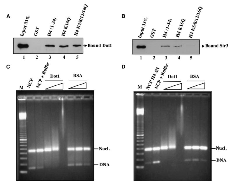
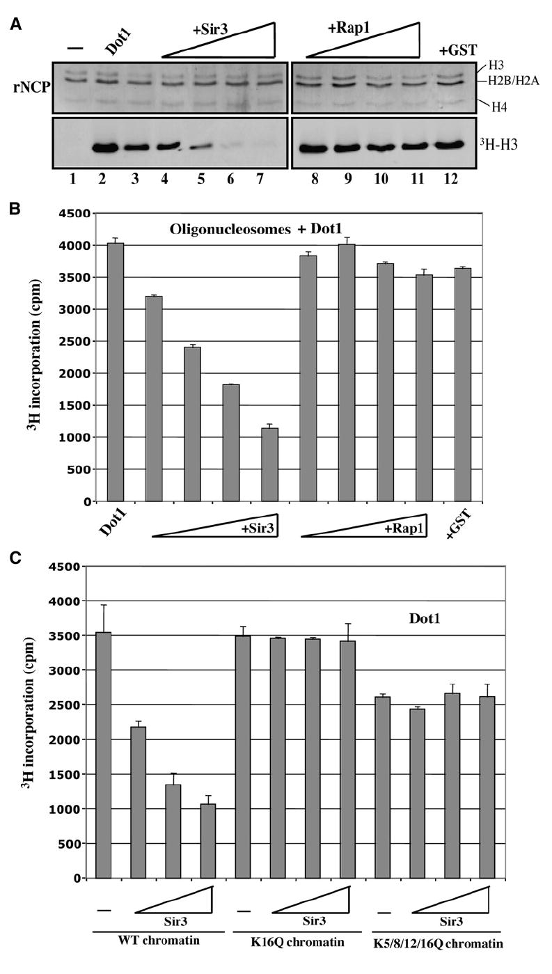
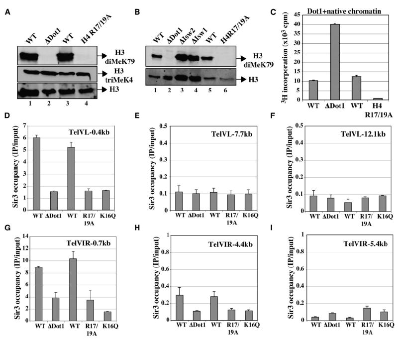
Dot1 (Disruptor of telomeric silencing-1) is a histone H3 lysine 79 methyltransferase that contributes to the establishment of heterochromatin boundary and has been linked to transcription elongation. We found that histone H4 N-terminal domain, unlike other histone tails, interacts with Dot1 and is essential for H3 K79 methylation. Furthermore, we show that the heterochromatin protein Sir3 inhibits Dot1-mediated methylation and that this inhibition is dependent on lysine 16 of H4. Sir3 and Dot1 bind the same short basic patch of histone H4 tail, and Sir3 also associates with the residues surrounding H3 K79 in a methylation-sensitive manner. Thus, Sir3 and Dot1 compete for the same molecular target on chromatin. ChIP analyses support a model in which acetylation of H4 lysine 16 displaces Sir3, allowing Dot1 to bind and methylate H3 lysine 79, which in turn further blocks Sir3 binding/spreading. This draws a detailed picture of the succession of molecular events occurring during the establishment of telomeric heterochromatin boundaries.


Figures

Figure 1
Figure 1. Histone H4 N-Terminal Tail Is Essential for Dot1-Dependent Methylation of Nucleosomal H3 at Lysine 79
(A) Histone H4 tail is required for H3 K79 methylation in vivo. Western blot of RIPA extracts from wild-type, H4ΔN (Δ4–19), H3ΔN (Δ3–29), H2AΔN (Δ5–21), and H2AΔC (Δ121–132) yeast cells using anti-H3 diMeK79 antibody. (B) Histone H4 lysine residues targeted for acetylation are not required for H3 K79 methylation in vivo. Western analysis of RIPA extracts was performed as in (A) including cells carrying K to Q substitutions for all four lysine residues of H4 tail domain. Anti-total H3 signals are included to monitor loading amounts. (C) Histone H3 methylation at lysine 4 is not affected by H4 tail deletion. Western analysis of RIPA extracts was as in (A) using anti-H3 diMeK4 and anti-total H4 antibodies. (D and E) Dot1 requires histone H4 tail to methylate H3 K79 on nucleosomes in vitro. HMT assays were performed using recombinant Dot1 and recombinant nucleosome core particles (rNCP) with or without H4 N-terminal tail (Δ2–20). Reactions were analyzed by gel/fluorography in (D) and filter binding/liquid scintillation counting in (E) (average of duplicate reactions with standard deviations).
Figure 2
Figure 2. Dot1 Directly Binds the Histone H4 Tail, but This Interaction Is Not Required for Binding to Nucleosomes
(A) Dot1 binds histone H4 tail region irrespective of acetylatable lysine residues. GST pull-down assays were performed using recombinant His-Dot1 and beads harboring GST-H4 fusion proteins (wild-type or K-to-Q substitution mutants at the indicated positions). GST is used as negative control. After washes, input (33%) and bound (100%) material were run on a 12% SDS-PAGE and revealed by western analysis using anti-His antibody. (B) Sir3 binds histone H4 tail region in a lysine-dependent manner. GST pull-down assays were as in (A) but using recombinant His-Sir3 (620–978). (C and D) Dot1 binds nucleosome core particles irrespective of histone H4 tail. Gel mobility shift assays were performed using recombinant nucleosome core particles (1.5 μg) with or without histone H4 tail (Δ2–20) and increasing amounts of Dot1 (0.1, 0.3, and 1 μg). Binding reactions were run on a 1.8% agarose gel followed by ethidium bromide staining. Mononucleosome and naked DNA bands are indicated. BSA was used as control. Marker is 100 bp ladder.
Figure 3
Figure 3. Heterochromatin Protein Sir3 Specifically Blocks Nucleosomal H3 Methylation by Dot1 in an H4 Lysine 16-Dependent Manner
(A) Sir3 inhibits nucleosomal H3 methylation by Dot1. HMT assays are as in Figure 1D using recombinant nucleosome particles, Dot1, and increasing amounts of Sir3 or Rap1 proteins (0.5, 1.0, 1.5, and 2.0 μg). (B) HMT assays were performed as in Figure 1E using purified native oligonucleosomes from human, Dot1, and increasing amounts of Sir3 or Rap1 (0.5–2.0 μg). (C) Sir3 requires histone H4 lysine 16 to inhibit Dot1-dependent methylation in vitro. HMT assays as in (B) using purified native chromatin from yeast cells expressing wild-type histone H4 or substitution mutants for the indicated positions of lysine residues. Increasing amounts of Sir3 (1, 2, and 4 μg) were added to the reactions.
Figure 4
Figure 4. Histone H4 Basic Patch Residues Are Essential for Chromatin Methylation by Dot1 In Vivo and In Vitro
(A) Point mutants of histone H4 arginine residues at positions 17 and 19 lead to loss of Dot1-dependent methylation of H3 in vivo. Western analysis of RIPA extracts (as in Figures 1A–1C) from DOT1 deletion or histone H4 R17/19A substitution mutant cells and their isogenic wild-types was performed using anti-H3 diMeK79, anti-H3 triMeK4, and anti-H3 (total). (B) Abundant Isw1/2 ATP-dependent chromatin remodelers that interact with histone H4 basic patch do not regulate bulk levels of Dot1-dependent H3 methylation in vivo. Western analysis was performed as in (A), including RIPA extracts from isw1 and isw2 mutant cells. (C) Dot1 requires arginine residues of histone H4 tail basic patch to methylate H3 K79 in chromatin in vitro. HMT assays were performed as in Figure 1E using Dot1 and yeast native chromatin purified from DOT1 deletion or histone H4 R17/19A substitution mutant cells and their isogenic wild-types. Since the majority of H3 K79 is methylated in wild-type cells in vivo, ΔDot1 mutant chromatin was used as control for the H4 mutant chromatin (neither contain MeK79, as shown in [A]). (D–I) In vivo association of Sir3 protein with telomeric regions is greatly decreased in dot1, H4 K16Q, and H4 R17/19A mutant cells. Sir3 binding to the telomeres of chromosome V left arm (D–F) and VI right arm (G–I) was analyzed by ChIP in the indicated strains. Occupancy represents IP/input signal (%) using a strain carrying chromosomal SIR3 tagged with 13 Myc epitopes. Signals are determined by real-time PCR with primers corresponding to the indicated positions relative to the end of the chromosomes. These values are based on two independent experiments with standard errors.
Figure 5
Figure 5. Dot1 and H3 K79 Methylation Directly Affect the Ability of Sir3 to Bind Chromatin
(A) Overexpression of Dot1 increases methylation of H3 K79 near telomeres with a corresponding decrease in Sir3 occupancy while not affecting acetylation of H4 K16. ChIP analysis as in Figure 4E was performed in a strain carrying a galactose-inducible DOT1 gene on a high-copy plasmid (over a wild-type DOT1 background). IPs were done on chromatin prepared from mid-log phase cells grown from 2 hr in glucose or in galactose. IPs were done with anti-H3 triMeK79 and anti-total H3 (left), anti-Myc (middle), or anti-H4 AcK16 and anti-total H3 (right). Data are presented as the change of IP/input signals when Dot1 is overexpressed (standard errors are based on two independent experiments). Total H3 signal is used to correct for potential changes/variations in nucleosome occupancy (for MeK79 and AcK16). (B) Sir3 binds to peptides corresponding to histone H3 K79 region in a methylation-sensitive manner. Sir3 pull-down assays were performed with the indicated biotinylated peptides. Mock reaction is Sir3 incubated without peptides. (C) Even monomethylation of K79 disrupts Sir3 interaction with H3 peptides. Pull downs were done as in (B) with Sir3 and the indicated peptides. The membrane was also probed with anti-biotin to monitor relative peptide amounts bound to the beads. (D) Sir3 interaction with H3 N-terminal peptides is also regulated by site-specific methylation but seems less sensitive. Pull-down assays were done as in (C) with Sir3 and the indicated peptides (reactions done at the same time but run on two different gels).
Figure 6
Figure 6. Increased Sas2-Dependent Acetylation of H4 K16 Greatly Enhances H3 K79 Methylation and Htz1 Incorporation near Telomeres
Overexpression of Sas2 H4 K16 acetyltransferase greatly stimulates methylation of H3 K79 near a telomere in vivo. ChIP analysis as in Figure 5A was performed in a strain carrying a galactose-inducible SAS2 gene on a high-copy plasmid (over a wild-type SAS2 background). IPs were done on chromatin prepared from mid-log phase cells grown from 2 hr in glucose or in galactose. IPs were done with anti-H4 AcK16 (A), anti-H3 triMeK79 (B), Anti-Sir2 (C), anti-Htz1 (D), and anti-total H3. Data are presented as the change occupancy when Sas2 is overexpressed. Histone marks/variants are corrected for nucleosome occupancy using total H3 signal.
Figure 7
Figure 7. Model of Succession of Events during the Establishment of Telomeric Heterochromatin Boundaries in Yeast
Histone H4 tail and its K16 acetylation site is depicted as coming out the nucleosome cores. Methylation of H3 K79 is depicted by the “M”-marked red circle in the cores, and “Z”-marked ovals represent histone variant Htz1.

References

    1. Bird AW, Yu DY, Pray-Grant MG, Qiu Q, Harmon KE, Megee PC, Grant PA, Smith MM, Christman MF. Acetylation of histone H4 by Esa1 is required for DNA double-strand break repair. Nature. 2002;419:411–415. - PubMed
    1. Carmen AA, Milne L, Grunstein M. Acetylation of the yeast histone H4 N terminus regulates its binding to heterochromatin protein SIR3. J Biol Chem. 2002;277:4778–4781. - PubMed
    1. Clapier CR, Nightingale KP, Becker PB. A critical epitope for substrate recognition by the nucleosome remodeling ATPase ISWI. Nucleic Acids Res. 2002;30:649–655. - PMC - PubMed
    1. Dhillon N, Kamakaka RT. A histone variant, Htz1p, and a Sir1p-like protein, Esc2p, mediate silencing at HMR. Mol Cell. 2000;6:769–780. - PubMed
    1. Fazzio TG, Gelbart ME, Tsukiyama T. Two distinct mechanisms of chromatin interaction by the Isw2 chromatin remodeling complex in vivo. Mol Cell Biol. 2005;25:9165–9174. - PMC - PubMed

Publication types

MeSH terms