. 2008 Mar 1;44(5):893-906.
doi: 10.1016/j.freeradbiomed.2007.11.015. Epub 2007 Dec 5.

Identifying the site of spin trapping in proteins by a combination of liquid chromatography, ELISA, and off-line tandem mass spectrometry

Affiliations

Identifying the site of spin trapping in proteins by a combination of liquid chromatography, ELISA, and off-line tandem mass spectrometry

Olivier M Lardinois et al. Free Radic Biol Med. .

Abstract

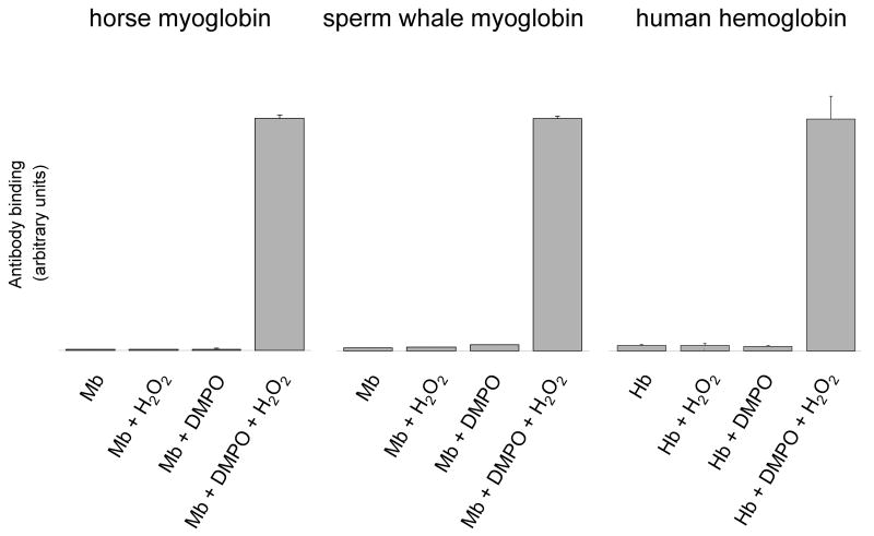
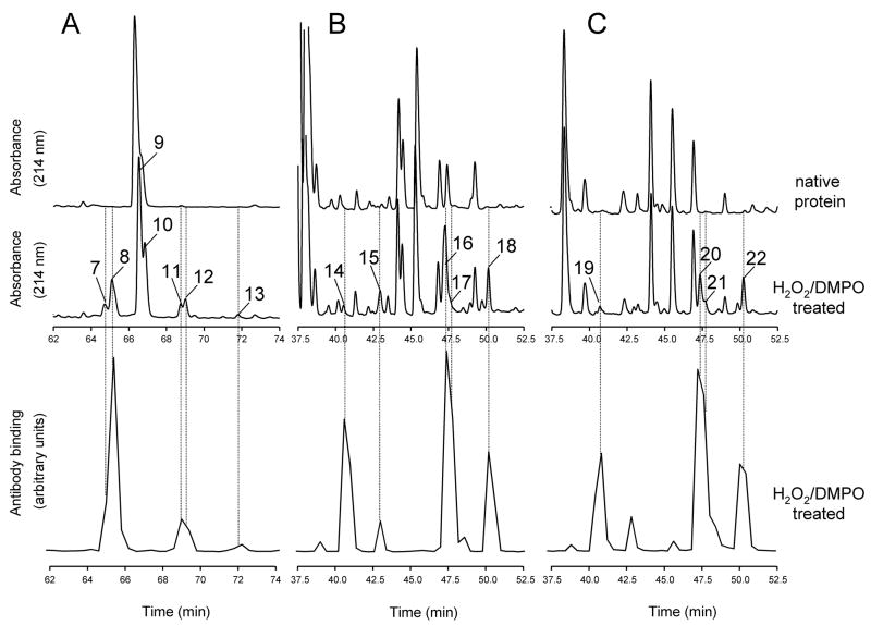
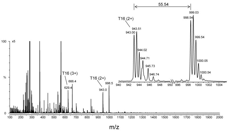
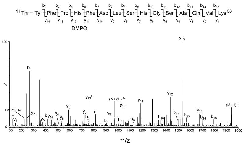
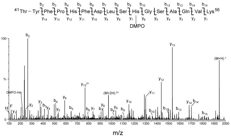
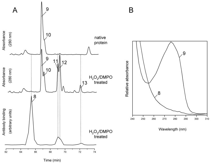
An off-line mass spectrometry method that combines immuno-spin trapping and chromatographic procedures has been developed for selective detection of the nitrone spin trap 5,5-dimethyl-1-pyrroline-N-oxide (DMPO) covalently attached to proteins, an attachment which occurs only subsequent to DMPO trapping of free radicals. In this technique, the protein-DMPO nitrone adducts are digested to peptides with proteolytic agents, peptides from the enzymatic digest are separated by HPLC, and enzyme-linked immunosorbent assays (ELISA) using polyclonal anti-DMPO nitrone antiserum are used to detect the eluted HPLC fractions that contain DMPO nitrone adducts. The fractions showing positive ELISA signals are then concentrated and characterized by tandem mass spectrometry (MS/MS). This method, which constitutes the first liquid chromatography-ELISA-mass spectrometry (LC-ELISA-MS)-based strategy for selective identification of DMPO-trapped protein residues in complex peptide mixtures, facilitates location and preparative fractionation of DMPO nitrone adducts for further structural characterization. The strategy is demonstrated for human hemoglobin, horse heart myoglobin, and sperm whale myoglobin, three globin proteins known to form DMPO-trappable protein radicals on treatment with H(2)O(2). The results demonstrate the power of the new experimental strategy to select DMPO-labeled peptides and identify sites of DMPO covalent attachments.


Figures

Figure 1
Figure 1
Detection of myoglobin and hemoglobin radical-derived nitrone adducts by immuno-spin trapping. Reaction mixtures contained globin (500 μM), DMPO (100 mM) and H2O2 (500 μM), or subsets of these as indicated. The reactions were initiated by adding H2O2 to the mixture of globin and the spin trap. After 10 min incubation at 25 °C, reactions were diluted with deionized H2O and the globin radical-derived nitrone adducts were detected by ELISA. Values are mean ± S.D (n = 3).
Figure 2
Figure 2
Effects of chaotropes, organic solvents and solvent modifiers on nitrone adduct stability. Reaction mixtures containing the Mb-derived nitrone adducts with and without the indicated concentration of chaotropes, organic solvents or organic modifiers were incubated for 14 hours at 37 ºC. Results obtained with HCl are for a 30 s incubation at 25 ºC. At the conclusion of the experiments, reactions were diluted with coating buffer for ELISA analysis as described under Materials and Methods. Gu.HCl, guanidine hydrochloride; FA, formic acid; TFA, trifluoroacetic acid; AA, acetic acid. Values are mean ± S.D (n = 3).
Figure 3
Figure 3
(A), detection of horse heart myoglobin radical-derived nitrone adducts by immuno-spin trapping: effect of acid-solvent extraction. Reaction mixtures contained horse heart myoglobin (500 μM), DMPO (100 mM) and H2O2 (500 μM), or subsets of these as indicated. The reactions were initiated by adding H2O2 to the mixture of myoglobin and the spin trap. After 10 min incubation at 25 °C, the heme was removed from the globin by acid-solvent extraction as described under Materials and Methods. At the conclusion of the solvent extraction process, reactions were diluted with coating buffer for subsequent ELISA analysis. Values are mean ± S.D (n = 3). (B), effect of trifluoroacetic acid on nitrone adduct stability: time course of adduct decay. DMPO-modified peptides were purified from the tryptic digest of hemoglobin, lyophilized and resuspended in H2O + 0.5% TFA. Excess NH4HCO3 was then added at various times after initiation of the reaction to stop further reaction. The zero time estimates refer to control experiments in which excess NH4HCO3 was added before TFA. At the conclusion of the experiments, reactions were diluted with deionized H2O for ELISA analysis. ▲, DMPO- cysteinyl adduct (peak 1 in Fig. 4); ■, DMPO-tyrosyl adduct (peak 4 in Fig. 4); ●, DMPO-histidinyl adduct (peak 6 in Fig. 4). Values are mean ± S.D (n = 3).
Figure 4
Figure 4
Comparison of peptide maps of human hemoglobin. (A), reversed-phase HPLC of the tryptic peptides derived from intact native Hb and from Hb reacted with H2O2 in the presence of DMPO; (B), expanded view of panel A. Absorbance was monitored at 214 nm. Fractions were collected every 0.4 min in Eppendorf tubes containing excess NH4HCO3, and an aliquot of each fraction was analyzed by ELISA. The fractions showing a strong positive ELISA signal or corresponding to chromatographic peaks showing marked decreases in intensity relative to controls were further characterized by tandem mass spectrometry. Peak assignment and mass analysis are summarized in Table 1.
Figure 5
Figure 5
Comparison of peptide maps from horse heart and sperm whale myoglobin. Reversed-phase HPLC of peptides derived from intact Mb and from Mb reacted with H2O2 in the presence of DMPO. Absorbance was monitored at 214 nm. Fractions were collected every 0.4 min in Eppendorf tubes containing excess NH4HCO3, and an aliquot of each fraction was analyzed by ELISA. Chromatograms show only the time window in which some peptides exhibiting a strong positive ELISA signal eluted. (A), tryptic maps for HoMb; (B), chymotryptic maps for HoMb; (C), chymotryptic maps for SwMb. Peak assignment and mass analysis are summarized in Table 1.
Figure 6
Figure 6
Full scan mass spectrum of one HPLC fraction from the peptide map of horse heart myoglobin showing a strong ELISA signal (peak 7 in Fig. 5A eluting at approximately 64.8 min). The fraction was lyophilized, resuspended in 50:50 acetonitrile/H2O + 0.1 % formic acid, and directly infused into the electrospray source of a Micromass Q-TOF hybrid tandem mass spectrometer. An expanded view of a pair of ions having the same charge state (+2 ion) and a mass-to-charge (m/z) difference of 55.5 is shown. This mass difference corresponds to the loss of one molecule of DMPO.
Figure 7
Figure 7
Deconvoluted MS/MS spectrum of human hemoglobin DMPO-modified peptide 41–56 (peak 5 in Fig. 4B eluting at approximately 46.0 min). The MS/MS spectrum was acquired from parent at m/z 973.0 (+2 ion), which corresponds in mass to tryptic peptide αT16 plus DMPO. For clarity, not all identified fragment ions are labeled on the spectrum.
Figure 8
Figure 8
Deconvoluted MS/MS spectrum of human hemoglobin DMPO-modified peptide 41–56 (peak 6 in Fig. 4B eluting at approximately 47.4 min). The MS/MS spectrum was acquired from parent at m/z 973.0 (+2 ion), which corresponds in mass to tryptic peptide αT16 plus DMPO. For clarity reasons, not all identified fragment ions are labeled on the spectrum.
Figure 9
Figure 9
Tryptic maps and absorbance spectra from intact horse heart Mb and from Mb reacted with H2O2 in the presence of DMPO. (A), HPLC tryptic maps monitored at 280 nm. Fractions were collected every 0.4 min in Eppendorf tubes containing excess NH4HCO3, and an aliquot of each fraction was analyzed by ELISA. Chromatograms show only the time window in which some peptides exhibiting a strong positive ELISA signal eluted. Peak assignment and mass analysis are summarized in Table 1. (B), absorbance spectra recorded by the diode-array detector in the eluent of the HPLC system. The spectra are scaled arbitrarily.

References

    1. Stubbe J, van der Donk WA. Protein radicals in enzyme catalysis. Chem Rev. 1998;98:705–762. - PubMed
    1. Dalle-Donne I, Scaloni A, Giustarini D, Cavarra E, Tell G, Lungarella G, Colombo R, Rossi R, Milzani A. Proteins as biomarkers of oxidative/nitrosative stress in diseases: the contribution of redox proteomics. Mass Spec Rev. 2005;24:55–99. - PubMed
    1. Davies MJ, Fu S, Wang H, Dean RT. Stable markers of oxidant damage to proteins and their application in the study of human disease. Free Radic Biol Med. 1999;27:1151–1163. - PubMed
    1. Davies MJ, Hawkins CL. EPR spin trapping of protein radicals. Free Radic BiolMed. 2004;36:1072–1086. - PubMed
    1. Augusto O, Vaz SM. EPR spin-trapping of protein radicals to investigate biological oxidative mechanisms. Amino Acids. 2007;32:535–542. - PubMed

Publication types