. 2008 Apr;40(2):127-41.
doi: 10.1016/j.tice.2007.11.001. Epub 2007 Dec 26.

Ultrastructure and morphology of midgut visceral muscle in early pupal Aedes aegypti mosquitoes

Affiliations

Ultrastructure and morphology of midgut visceral muscle in early pupal Aedes aegypti mosquitoes

E P Bernick et al. Tissue Cell. 2008 Apr.

Abstract

These studies focus on the pupal Aedes aegypti midgut muscularis for the first 26 h following larval-pupal transition. The midgut muscularis of Ae. aegypti pupae during this first half of the pupal stadium is a grid of both circularly and longitudinally oriented muscle bands, arranged in a manner resembling that of the larvae. While many muscle bands exhibit signs of degeneration during the time period studied, not all bands degrade, nor is this degradation simultaneous. Band deterioration involves destruction of internal elements while the muscle fiber plasma membrane remains intact. Deterioration of contractile elements may involve proteosome-like structures and associated enzymes. Many features of the larval muscularis including cruciform cells, bifurcating circular bands, and bifurcating longitudinal bands of muscle are retained during the time period investigated. Neuromuscular junctions along some muscle bands are retained through at least 16 h into the pupal stadium. The selective nature of muscle fiber degradation, coupled with the retention of larval features and neural input, may allow for limited functionality of the muscularis during metamorphosis. Evidence of sexual dimorphism in the midgut muscularis of male and female Ae. aegypti pupae was not observed during the time period studied.


Figures

Fig. 1
Fig. 1
Midguts from Ae. aegypti 4th instar larvae (A), pupae (B), and adults (C and D). The anterior midgut, labeled with a grey interior, has eight associated gastric cecae in the larvae. The cecae are lost early into the pupal stadium and are not regenerated. The posterior midgut, filled with cross-hatchings, develops into the adult stomach. The Malpighian tubules are located at the junction of the posterior midgut and the hindgut. Image C represents an adult female, and D represents an adult male.
Fig. 2
Fig. 2
(A) Midgut from a female Ae. aegypti pupa, approximately 6.75–7.25 h following larval–pupal transition. Anterior midgut is located in the upper right, and the posterior midgut in the lower left. Scale bar represents 600 µm. (B) Midgut from a female Ae. aegypti pupa, approximately 9 h following larval–pupal transition. Anterior midgut is located in the upper right, and the posterior midgut in the lower left. Scale bar represents 600 µm. (C) Midgut from a female Ae. aegypti pupa, approximately 16.5–17 h following larval–pupal transition. Anterior midgut is located in the upper right, and the posterior midgut in the lower left. Scale bar represents 600 µm. (D) Midgut from a female Ae. aegypti pupa, approximately 21.5 h following larval–pupal transition. Anterior midgut is located in the upper right, and the posterior midgut in the lower left. Scale bar represents 600 µm.
Fig. 3
Fig. 3
(A) Bifurcating circular muscle bands (BC) in the anterior midgut of a female Ae. aegypti pupa, approximately 6.75–7.25 h following larval–pupal transition. Longitudinal bands of muscle (L) can also be observed. Scale bar represents 27 µm. (B) Bifurcating longitudinal muscle bands (BL) found in the posterior midgut of a female Ae. aegypti pupa, approximately 5.5–6 h following larval–pupal transition. Circular muscle bands (C) and Malpighian tubules (MT) are present. Scale bar represents 50 µm.
Fig. 4
Fig. 4
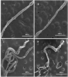
(A) Longitudinal (L) and circular (C) muscle bands in the posterior midgut of a female Ae. aegypti pupa, approximately 5.5–6 h following larval–pupal transition. Scale bar represents 50 µm. (B) Muscularis associated with the posterior midgut of a female Ae. aegypti pupa, approximately 9 h following larval–pupal transition. Defined longitudinal (L) and circular (C) bands of muscle can be seen. Malpighian tubules (MT) are located at the junction between the posterior midgut and hindgut. Scale bar represents 50 µm. (C) Muscularis associated with the posterior midgut of a male Ae. aegypti pupa, approximately 16.5–17 h following larval–pupal transition. Longitudinal (L) and circular (C) bands of muscle are present, but appear flattened and less robust than those observed in younger pupae. Scale bar represents 50 µm. (D) Cruciform cells (CC) and incomplete circular muscle bands (IC) found in the anterior midgut of a male Ae. aegypti pupa, approximately 19 h following larval–pupal transition. Longitudinal (L) and circular (C) bands of muscle are also present. Scale bar represents 75 µm. (E) Muscularis associated with the posterior midgut of a female Ae. aegypti pupa, approximately 17 h following larval–pupal transition. Circular muscle bands (C) are severely atrophied, as evidenced by decreased diameter of some muscle bands, and longitudinal bands of muscle are difficult to discern. Filamentous branches (FB) are associated with the midgut. Scale bar represents 50 µm.
Fig. 5
Fig. 5
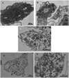
(A) Muscularis associated with the anterior midgut of a female Ae. aegypti pupa, approximately 6.75–7.25 h following larval–pupal transition. Longitudinal (L) and circular (C) bands of muscle are present. Scale bar represents 60 µm. (B) Muscularis associated with the anterior midgut of a male Ae. aegypti pupa, approximately 19 h following larval–pupal transition. Incomplete bands of circular muscle (IC) are present, but circular and longitudinal muscle bands are difficult to discern. Scale bar represents 43 µm. (C) Muscularis associated with the anterior midgut of a female Ae. aegypti pupa, approximately 21.5 h following larval–pupal transition. Longitudinal (L) and circular (C) muscle bands are present. Scale bar represents 50 µm. (D) Muscularis associated with the anterior midgut of a female Ae. aegypti pupa, approximately 21.5 h following larval–pupal transition. Higher magnification of boxed area in (C). Scale bar represents 25 µm.
Fig. 6
Fig. 6
(A) Cruciform muscle fiber containing both circularly (CF) and longitudinally (LF) oriented contractile elements from the anterior midgut of a male Ae. aegypti pupa, approximately 2.5–3 h following larval–pupal transition. A membrane invagination (MI) resembling a transverse tubule is present. Mitochondria (M) are present in the cytoplasm unoccupied by contractile elements. Scale bar represents 1 µm. (B) Cruciform muscle fiber containing both circularly (CF) and longitudinally (LF) oriented contractile filaments from the anterior midgut of a male Ae. aegypti pupa, approximately 2.5–3 h following larval–pupal transition. Mitochondria (M) are present in the cytoplasm unoccupied by contractile elements. Scale bar represents 0.5 µm. (C) Circular (C) and longitudinal (L) muscle associated with the posterior midgut of a male Ae. aegypti pupa, approximately 2.25–2.75 h following larval–pupal transition. A nucleus (N) and mitochondria (M) are present in the cytoplasm of the circular muscle. The circular muscle lies close to the basement membrane (BM). Scale bar represents 1 µm. (D) Longitudinal muscle, rich with actin and myosin filaments (AMF) from the posterior midgut of a male Ae. aegypti pupa approximately 2.25–2.75 h following larval–pupal transition. Scale bar represents 1 µm.
Fig. 7
Fig. 7
(A) Circular muscle (C) from the posterior midgut of a male Ae. aegypti pupa, approximately 8 h following larval–pupal transition. Small proteosome sized flecks are present in the muscle fiber. The basement membrane (BM) lies between the muscle and midgut epithelium (ME). Scale bar represents 1 µm. (B) Circular (C) and longitudinal (L) muscle from the posterior midgut of a male Ae. aegypti pupa, approximately 15.5–16 h following larval–pupal transition. Mitochondria (M) are present in the circular muscle. Both longitudinal and circular muscle bands contain small, dark staining structures. Scale bar represents 0.5 µm. (C) Longitudinal muscle (L) lying close to the midgut basement membrane (BM) from the anterior midgut of a female Ae. aegypti pupa, approximately 16.5–17 h following larval–pupal transition. A membrane invagination (MI) resembling a transverse tubule is present. Scale bar represents 0.5 µm. (D) Longitudinal muscle (L) lying close to the basement membrane (BM) from the anterior midgut of a female Ae. aegypti pupa, approximately 14–14.5 h following larval–pupal transition. Regions of the cytoplasm are vacant (V), unoccupied by contractile elements. Scale bar represents 0.5 µm.
Fig. 8
Fig. 8
(A) Circular (C) and longitudinal (L) muscle fibers from the posterior midgut of a female Ae. aegypti pupa, approximately 23.5–24 h following larval–pupal transition. Scale bar represents 1 µm. (B) Circular muscle (C) from the anterior midgut of a female Ae. aegypti pupa, approximately 23–23.5 h following larval–pupal transition. Cytoplasm unoccupied by contractile elements appears grainy. The midgut epithelium (ME) and basement membrane (BM) can be observed. Scale bar represents 1 µm.
Fig. 9
Fig. 9
(A) Longitudinal muscle from the posterior midgut of a male Ae. aegypti pupa, approximately 2.25–2.75 h following larval–pupal transition. The fiber contains intracellular spaces (IS) and abundant actin and myosin filaments (AMF). Scale bar represents 0.5 µm. (B) Longitudinal muscle (L) from (A) at a higher magnification. Mitochondria (M) are present. Midgut epithelium (ME) and associated basement membrane (BM) can be observed. Scale bar represents 0.5 µm. (C) Longitudinal muscle from the anterior midgut of a female Ae. aegypti pupa, approximately 23–23.5 h following larval–pupal transition. Numerous mitochondria (M) are found in the fiber, largely vacant of contractile elements. Scale bar represents 1 µm. (D) Longitudinal muscle from the anterior midgut of a female Ae. aegypti pupa approximately 23–23.5 h following larval–pupal transition. Fine, dark staining strands (FS) are present in the cytoplasm. Scale bar represents 1 µm. (E) Longitudinal muscle from the anterior midgut of a female Ae. aegypti pupa, approximately 21–21.5 h following larval–pupal transition. Mitochondria (M) and tangentially oriented filaments (TF) are present. Scale bar represents 0.5 µm.
Fig. 10
Fig. 10
(A) Longitudinal muscle, rich with actin and myosin filaments (AMF) from the posterior midgut of a male Ae. aegypti pupa, approximately 15.5–16 h following larval–pupal transition. Membrane invaginations (MI) resembling transverse tubules are present. A neuron (N) is closely associated with this muscle fiber. Scale bar represents 0.5 µm. (B) Neuromuscular junction (NMJ) seen in (A) at a higher magnification. Neurosecretory vesicles (NSV) are present in the neuron. Actin and myosin filaments (AMF) can be observed in the longitudinal muscle fiber. Scale bar represents 0.2 µm.
Fig. 11
Fig. 11
(A) Circular muscle (C) from the posterior midgut of a male Ae. aegypti pupa, approximately 2.25–2.75 h following larval–pupal transition. The muscle fiber lies closely to the midgut epithelium (ME) and basement membrane (BM). Z lines (Z) are easily observable. Mitochondria (M) are present. Scale bar represents 1 µm. (B) Circular muscle (C) from the posterior midgut of a female Ae. aegypti pupa, approximately 17–18 h following larval–pupal transition. The muscle fiber lies closely to the midgut epithelium (ME) and associated basement membrane (BM). A faint Z line (Z) is present in the upper left portion of the muscle fiber. Scale bar represents 2 µm.
Fig. 12
Fig. 12
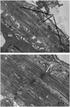
(A) Circular muscle (C) from the posterior midgut of a male Ae. aegypti pupa, approximately 15.5–16 h following larval–pupal transition. The muscle lies close to the midgut epithelium (ME). Perforated Z lines (Z) are present. Scale bar represents 1 µm. (B)A perforated Z line from (A) at a higher magnification. Some of the myosin filaments associated with the circular muscle (C) to the Z line, as opposed to penetrating the perforated regions. Scale bar represents 0.5 µm.

References

    1. Beaulaton J, Lockshin RA. Ultrastructural study of the normal degeneration of the intersegmental muscles of Antherea polyphemus and Manduca sexta (Insecta, Lepidoptera) with particular reference to cellular autophagy. J. Morphol. 1976;154:39. - PubMed
    1. Bernick EP, Moffett SB, Moffett DF. Organization, ultrastructure, and development of midgut visceral muscle in larval Aedes aegypti. Tissue Cell. 2007;39:277–292. - PMC - PubMed
    1. Christophers SR. Aedes aegypti, The Yellow Fever Mosquito: Its Life History, Binomics, and Structure. London, England: Cambridge University Press; 1960.
    1. Clark TM, Hutchinson MJ, Heugel KL, Moffett SB, Moffett DF. Additional morphological and physiological heterogeneity within the midgut of larval Aedes aegypti revealed by histology, electrophysiology, and effects of Bacillus thuringiensis endotoxin. Tissue Cell. 2005;37:457–468. - PubMed
    1. Clements AN. The Biology of Mosquitoes, vol. 1: Development, Nutrition, and Reproduction. New York: Chapman and Hall; 1992.

Publication types