

Neurotoxic effects of DSP-4 on the central noradrenergic system in male zebra finches
- PMID: 18160108
- PMCID: PMC2271146
- DOI: 10.1016/j.bbr.2007.11.004
Neurotoxic effects of DSP-4 on the central noradrenergic system in male zebra finches
Abstract
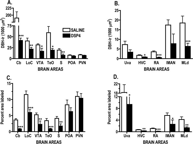
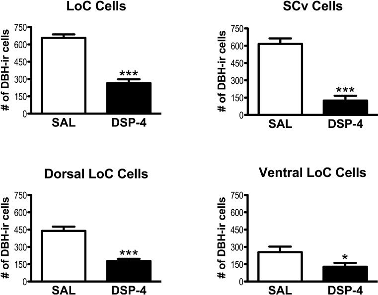
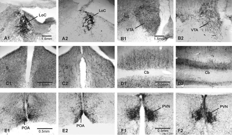
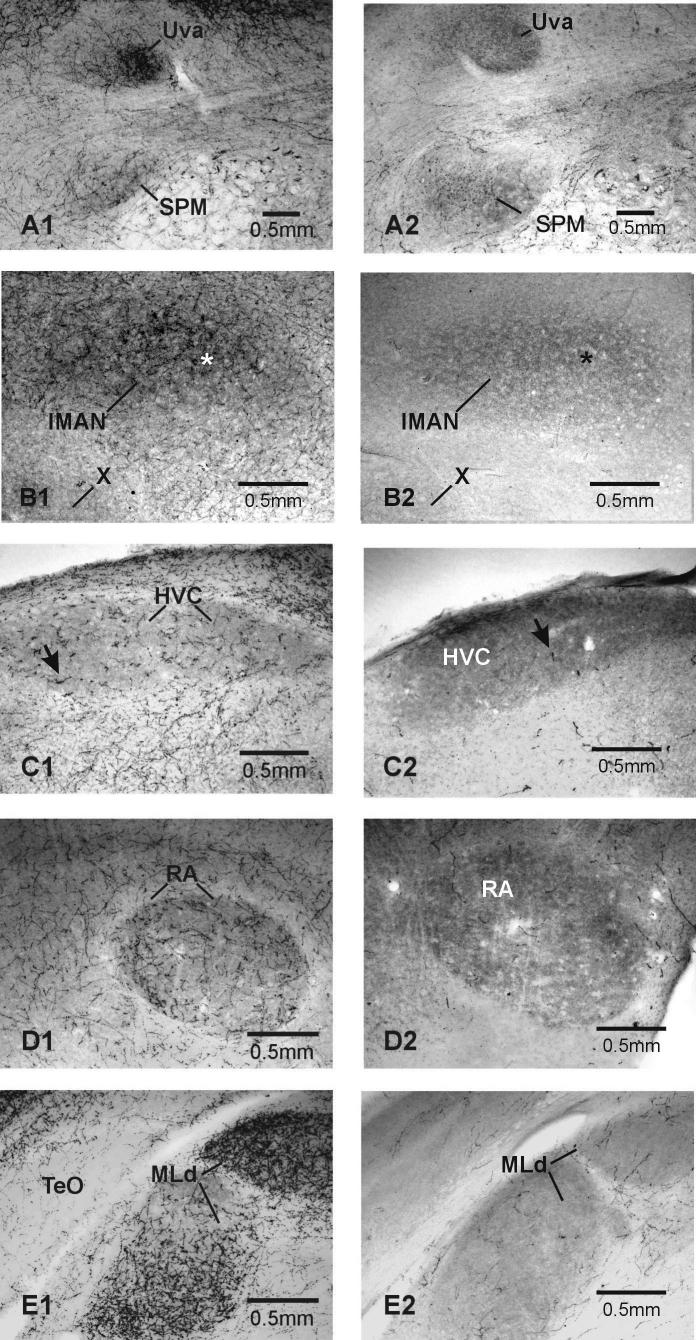
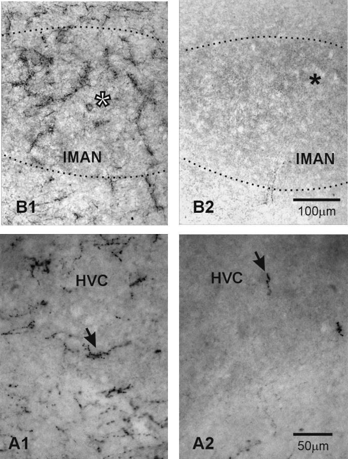
When administered systemically, the noradrenergic neurotoxin N-(2-chloroethyl)-N-ethyl-2-bromobenzylamine (DSP-4) appears to target the noradrenergic innervation originating in the locus coeruleus causing long-term decrements in noradrenergic function. In songbirds, DSP-4-treatment decreased female-directed singing by males and copulation solicitation responses of females to male songs. However, DSP-4 treatment in songbirds did not lower measures of NE function in the brain to the same extent as it does in mammals. The current study had two goals: determining if two DSP-4 treatments 10 days apart would cause significant decrements in noradrenergic function in male zebra finches and determining if, as in other species, the noradrenergic innervation of midbrain and cortical areas would be profoundly affected while hypothalamic areas were spared. Dopamine-beta-hydroxylase immunoreactivity (DBH-ir) was quantified in thirteen brain regions (five vocal control nuclei, one auditory nucleus, two hypothalamic nuclei, and five additional areas that demonstrated high DBH labeling in controls). Within 20 days, DSP-4 treatment profoundly reduced the number of DBH-ir cells in both the locus coeruleus and ventral subcoeruleus. Unlike a previous study, DBH labeling delineated four out of five vocal control nuclei and an auditory nucleus. As expected, DSP-4 treatment significantly decreased DBH labeling in all areas examined in the mesencephalon and telencephalon without significantly affecting DBH-ir in hypothalamic areas. This double treatment regime appears to be much more effective in decreasing noradrenergic function in songbirds than the single treatment typically used.
Figures
Similar articles
-
DSP-4, a noradrenergic neurotoxin, produces sex-specific effects on pairing and courtship behavior in zebra finches.Behav Brain Res. 2013 Sep 1;252:164-75. doi: 10.1016/j.bbr.2013.05.056. Epub 2013 Jun 5. Behav Brain Res. 2013. PMID: 23747610
-
Immunohistochemical analysis of the neurotoxic effects of DSP-4 identifies two populations of noradrenergic axon terminals.Neuroscience. 1989;30(1):181-97. doi: 10.1016/0306-4522(89)90364-3. Neuroscience. 1989. PMID: 2747911
-
A role for norepinephrine in the regulation of context-dependent ZENK expression in male zebra finches (Taeniopygia guttata).Eur J Neurosci. 2005 Apr;21(7):1962-72. doi: 10.1111/j.1460-9568.2005.04028.x. Eur J Neurosci. 2005. PMID: 15869489
-
Selective Lifelong Destruction of Brain Monoaminergic Nerves Through Perinatal DSP-4 Treatment.Curr Top Behav Neurosci. 2016;29:51-71. doi: 10.1007/7854_2015_398. Curr Top Behav Neurosci. 2016. PMID: 26427851 Review.
-
Selective effects of DSP-4 on locus coeruleus axons: are there pharmacologically different types of noradrenergic axons in the central nervous system?Prog Brain Res. 1991;88:257-68. doi: 10.1016/s0079-6123(08)63815-7. Prog Brain Res. 1991. PMID: 1726027 Review.
Cited by
-
Individual differences in the motivation to communicate relate to levels of midbrain and striatal catecholamine markers in male European starlings.Horm Behav. 2011 Nov;60(5):529-39. doi: 10.1016/j.yhbeh.2011.08.001. Epub 2011 Aug 31. Horm Behav. 2011. PMID: 21907203 Free PMC article.
-
What birdsong can teach us about the central noradrenergic system.J Chem Neuroanat. 2010 Mar;39(2):96-111. doi: 10.1016/j.jchemneu.2009.08.003. Epub 2009 Aug 15. J Chem Neuroanat. 2010. PMID: 19686836 Free PMC article. Review.
-
Norepinephrine inhibition in juvenile male zebra finches modulates adult song quality.Brain Res Bull. 2013 Jan;90:132-6. doi: 10.1016/j.brainresbull.2012.10.010. Epub 2012 Nov 15. Brain Res Bull. 2013. PMID: 23160069 Free PMC article.
-
Locus Coeruleus in Non-Mammalian Vertebrates.Brain Sci. 2022 Jan 20;12(2):134. doi: 10.3390/brainsci12020134. Brain Sci. 2022. PMID: 35203898 Free PMC article. Review.
-
Interference of the noradrenergic neurotoxin DSP4 with neuronal and nonneuronal monoamine transporters.Naunyn Schmiedebergs Arch Pharmacol. 2009 Dec;380(6):523-9. doi: 10.1007/s00210-009-0459-z. Epub 2009 Oct 17. Naunyn Schmiedebergs Arch Pharmacol. 2009. PMID: 19838680
References
-
- Al-Zahrani SSA, Al-Ruwaitea ASA, Ho M- Y, Bradshaw CM, Szabadi E. Destruction of central noradrenergic neurons with DSP4 impairs the acquistion of temporal discrimination but does not affect memory for duraton in a delayed conditional discrimination task. Psychopharm. 1997;130:166–73. - PubMed
-
- Appeltants D, Del Negro C, Balthazart J. Noradrenergic control of auditory information processing in female canaries. Behav Brain Res. 2002;133:221–35. - PubMed
-
- Bailhache T, Balthazart J. The catecholaminergic system of the quail brain: Immunocytochemical studies of dopamine--hydroxylase and tyrosine hydroxylase. J Comp Neurol. 1993;329:230–56. - PubMed
-
- Ball GF, Castro JM, Bernard DJ. Sex differences in the volume of avian song control nuclei: Comparative studies and the issue of brain nucleus delineation. Psychoneuroendocrinol. 1994;19:485–504. - PubMed
Publication types
MeSH terms
Substances
Grants and funding
LinkOut - more resources
Full Text Sources
Research Materials
Miscellaneous
