. 2008 Mar;158(1):22-31.
doi: 10.1016/j.molbiopara.2007.11.007. Epub 2007 Nov 19.

A Presenilin-like protease associated with Plasmodium falciparum micronemes is involved in erythrocyte invasion

Affiliations

A Presenilin-like protease associated with Plasmodium falciparum micronemes is involved in erythrocyte invasion

Xuerong Li et al. Mol Biochem Parasitol. 2008 Mar.

Abstract

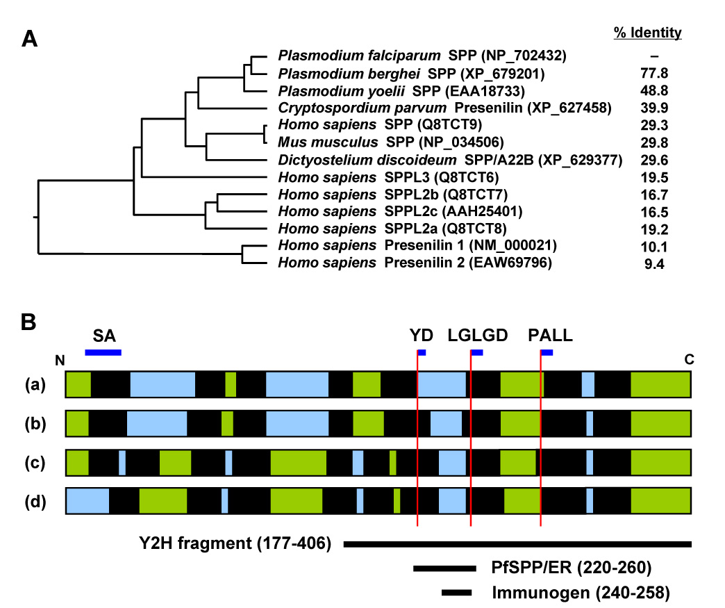
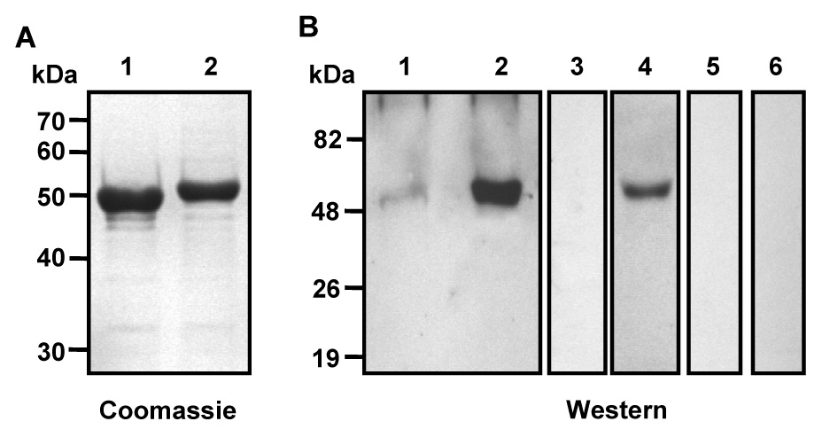
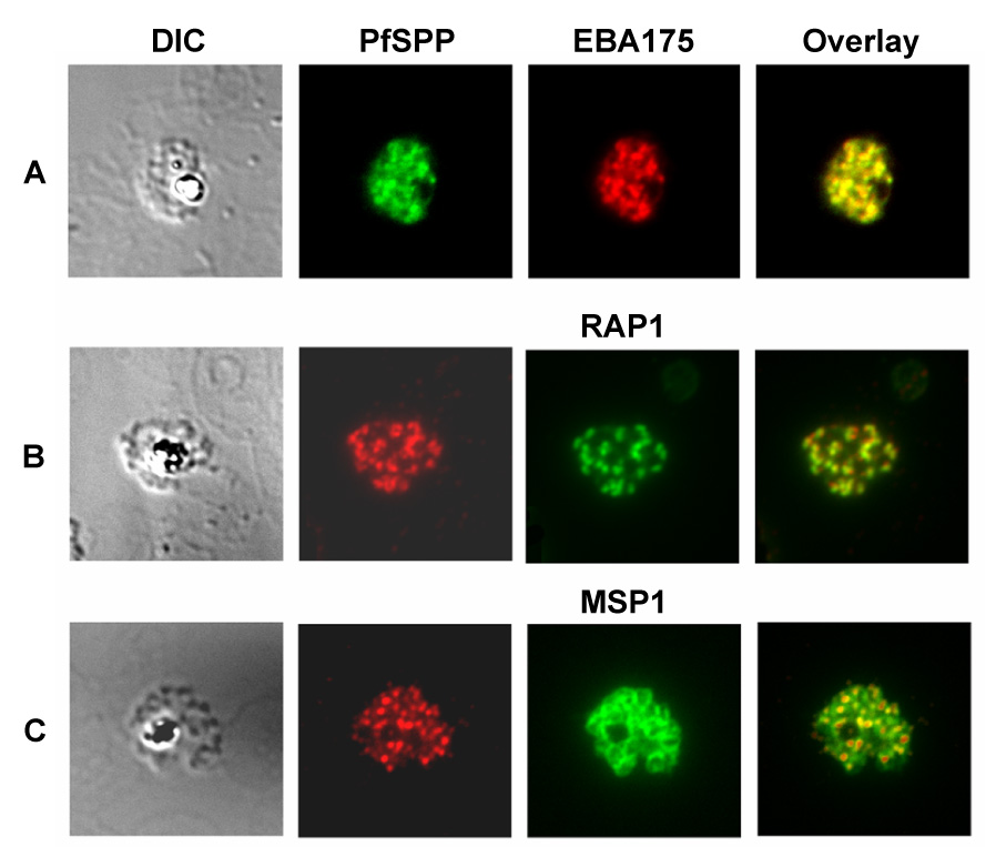
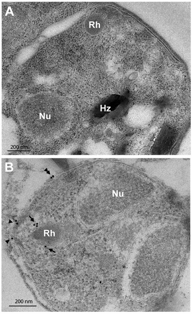
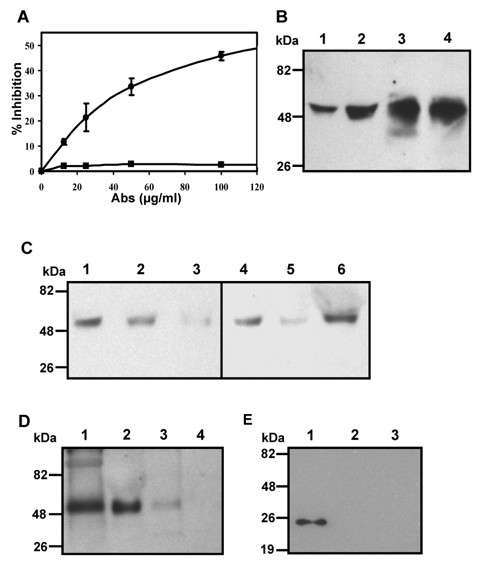
We describe identification of a Plasmodium falciparum microneme protease involved in RBC invasion. From the yeast two-hybrid screening of a P. falciparum cDNA library, we have identified a 47 kDa membrane protein that interacted with the 5ABC domain of human RBC band 3. This protein shared homology with a Presenilin-type aspartyl protease, the signal peptide peptidase (SPP). An antibody raised against a predicted exposed region of this protein reacted specifically to a single band of approximately 47 kDa in the P. falciparum protein extract. Immunofluorescence microscopy suggested that this protein co-localized with the microneme protein EBA-175 in schizonts, and immunoelectron microscopy established that it is primarily localized to micronemes in merozoites. Functional characterization of Plasmodium falciparum signal peptide peptidase (PfSPP), demonstrates that an antibody to PfSPP blocks RBC invasion by P. falciparumin vitro. Native and recombinant PfSPP bound directly to the 5ABC domain of band 3 in solution and the binding of PfSPP to RBCs was chymotrypsin-sensitive, but trypsin and neuraminidase-resistant. Together, these results suggest that host band 3 interacts with PfSPP during RBC invasion presumably following parasite microneme discharge. PfSPP is the first microneme-associated intramembrane aspartyl protease identified in the apicomplexan parasites that interacts with a major transmembrane receptor on host erythrocytes.


Figures

Fig. 1
Fig. 1. In silico analysis of P. falciparum PfSPP
(A) Phylogenetic tree of P. falciparum PfSPP was generated and percent identity was calculated by ClustalW method (MegAlign 5.08, DNASTAR) using the default settings. Protein accession numbers are given in parenthesis. (B) Topology models of PfSPP. The map shows the relative position of predicted signal-anchor sequence (SA; amino acids 19–38), two active site motifs YD (amino acids 221–222) and LGLGD (259–263), and the PALL (335–338) motif. Transmembrane (TM) regions are shown in black, cytosolic regions in green, and extracytosolic regions in light blue. (a) TMpred model: TM1, amino acids 22–47; TM2, 85–103; TM3, 110–128; TM4, 166–182; TM5, 199–221, TM6, 256–274; TM7, 309–327; TM8, 336–354. (b) TMHMM-2.0 model: TM1, 21–43; TM2, 80–102; TM3, 109–128; TM4, 165–182; TM5, 203–225, TM6, 253–275; TM7, 308–330; TM8, 335–354. (c) Phobius model: TM1, 21–37; TM2, 43–61; TM3, 82–103; TM4, 109–129; TM5, 164–181, TM6, 187–205; TM7, 221–236; TM8, 256–275; TM9, 307–330; TM10, 336–354. (d) SOSUI model: TM1, 31–53; TM2, 80–102; TM3, 108–1030; TM4, 162–184; TM5, 187–209, TM6, 214–236; TM7, 257–279; TM8, 308–329; TM9, 335–354. The region (amino acids 177–406) of PfSPP encoded by the cDNA insert in the yeast two-hybrid screen (Y2H fragment), recombinant PfSPP/ER, and a segment of ER3 used to generate anti-peptide antibodies are shown.
Fig. 2
Fig. 2. Native P. falciparum PfSPP
(A) Coomassie blue gel shows the affinity-purified recombinant proteins MBP (lane 1) and MBP-PfSPP (lane 2). (B) Characterization of anti-PfSPP Abs. Western blot shows the mono-specific anti-PfSPP Abs reacted specifically to the recombinant PfSPP/ER (lanes 1 and 2) and native P. falciparum PfSPP (lanes 4 and 6). Pre-immune controls are shown in lanes 3 and 5. Lane 1, MBP; lanes 2 and 3, MBP-PfSPP/ER; lanes 4 and 5, P. falciparum extract; and lane 6, human RBC ghosts.
Fig. 3
Fig. 3. Co-localization of PfSPP in mature P. falciparum
Indirect immunofluorescence microscopy using specific antibodies showed PfSPP is co-localized with EBA-175 (a microneme protein), but neither with RAP1 (a rhoptry marker) nor MSP1 (merozoite surface protein) in the P. falciparum (3D7) schizonts.
Fig. 4
Fig. 4. Immunoelectron microscopy
PfSPP was labeled with mono-specific PfSPP Abs using gold particles (15 nm) in P. falciparum merozoites. (A) The pre-immune control showed no specific labeling. (B) The anti-PfSPP Abs showed specific labeling of the merozoite with gold particles in the micronemes (arrows), the apical surface area (arrowheads). The parasite nucleus (Nu), rhoptries (Rh), and hemozoin (Hz) are indicated.
Fig. 5
Fig. 5. Inhibition of RBC invasion and PfSPP-RBC interactions
(A) The graph shows the rate of inhibition of P. falciparum invasion into human RBCs is dependent upon the concentration of anti-PfSPP Abs (•) but not pre-immune IgG (■) present in the culture medium at the time of invasion. The inhibition rate was determined relative to the no-antibody control taken as 100% invasion (0% inhibition). Pre-immune samples showed ~2% inhibition (>98% invasion rate) as compared to the no-antibody control (data not shown). (B) Western blotting using anti-PfSPP Abs shows that a significant amount of full-length PfSPP was found in the preparations of P. falciparum culture supernatant separated at 40,000 g for 15 min (lane 1) and 12,000 g for 20 min (lane 2). Lane 3, pellet from 40,000 g centrifugation; lane 4, pellet from 12,000 g centrifugation. An equivalent amount of two supernatant samples (lanes 1 and 2) and the two pellet samples (lanes 3 and 4) were loaded. (C) RBC binding assay in suspension using the culture supernatant prepared by 12,000 g centrifugation. Normal (untreated, lane 1), trypsin-treated (lane 4), chymotrypsin-treated (lane 5), and neuraminidase-treated (lane 6) intact human RBC samples were analyzed by Western blotting using anti-PfSPP Abs following the binding assay. Soluble GST-5ABC (lane 3) was added to the binding assay to block the binding of native PfSPP to normal RBCs. GST served as negative control (lane 2). (D) Western blotting of samples using anti-PfSPP Abs is shown in each panel. Specific-binding of native PfSPP to recombinant 5ABC domain. Lane 1, P. falciparum protein extract prepared using TX-100 (PE); lane 2, PE + GST-5ABC (beads); lane 3, PE + GST (beads); lane 4, GST-5ABC beads only. (E) Specific-binding of PfSPP and 5ABC in solution by Western blotting using anti-PfSPP Abs. Lane 1, MBP-PfSPP/ER (beads) + Trx-5ABC; lane 2, MBP (beads) + Trx-5ABC; lane 3, MBP-PfSPP/ER (beads) + Trx.
Fig. 6
Fig. 6. A proposed model for the interaction of PfSPP with band 3
The schematic diagram depicts the location of putative binding domains of P. falciparum PfSPP and human erythrocyte band 3 as identified in the yeast two-hybrid screen.

References

    1. Cowman AF, Crabb BS. Invasion of red blood cells by malaria parasites. Cell. 2006;124:755–766. - PubMed
    1. Oh SS, Chishti AH. Host receptors in malaria merozoite invasion. Curr Top Microbiol Immunol. 2005;295:203–232. - PubMed
    1. Okoyeh JN, Pillai CR, Chitnis CE. Plasmodium falciparum field isolates commonly use erythrocyte invasion pathways that are independent of sialic acid residues of glycophorin A. Infect Immun. 1999;67:5784–5791. - PMC - PubMed
    1. Baum J, Pinder M, Conway DJ. Erythrocyte invasion phenotypes of Plasmodium falciparum in The Gambia. Infect Immun. 2003;71:1856–1863. - PMC - PubMed
    1. Lobo CA, de Frazao K, Rodriguez M, et al. Invasion profiles of Brazilian field isolates of Plasmodium falciparum: phenotypic and genotypic analyses. Infect Immun. 2004;72:5886–5891. - PMC - PubMed

Publication types

Substances

LinkOut - more resources