. 2008 Apr;82(7):3530-7.
doi: 10.1128/JVI.00586-07. Epub 2007 Dec 26.

Temporal association of the herpes simplex virus genome with histone proteins during a lytic infection

Affiliations

Temporal association of the herpes simplex virus genome with histone proteins during a lytic infection

Jaewook Oh et al. J Virol. 2008 Apr.

Abstract

Previous work has determined that there are nucleosomes on the herpes simplex virus (HSV) genome during a lytic infection but that they are not arranged in an equally spaced array like in cellular DNA. However, like in cellular DNA, the promoter regions of several viral genes have been shown to be associated with nucleosomes containing modified histone proteins that are generally found associated with actively transcribed genes. Furthermore, it has been found that the association of modified histones with the HSV genome can be detected at the earliest times postinfection (1 h postinfection) and increases up to 3 h postinfection. However from 3 h to 6 h postinfection (the late phase of the replication cycle), the association decreases. In this study we have examined histone association with promoter regions of all kinetic classes of genes. This was done over the time course of an infection in Sy5y cells using sucrose gradient sedimentation, bromodeoxyuridine labeling, chromatin immunoprecipitation assays, Western blot analysis, trypsin and DNase digestion, and quantitative real-time PCR. Because no histones were detected inside HSV type 1 capsids, the viral genome probably starts to associate with histones after being transported from infecting virions into the host nucleus. Promoter regions of all gene classes (immediate early, early, and late) bind with histone proteins at the start of viral gene expression. However, after viral DNA replication initiates, histones appear not to associate with newly synthesized viral genomes.


Figures

FIG. 1.
FIG. 1.
Viral DNA and histone protein in HSV capsids treated with trypsin. Capsids from KOS strain-infected Vero cells were purified by sucrose gradient centrifugation. Crude capsids were prepared from cell lysates on a 35% sucrose cushion and treated with 5 μg/ml or 10 μg/ml trypsin as described in Materials and Methods. (A) Treated capsids were then subjected to 5 to 50% sucrose gradient centrifugation and capsids visualized as light-scattering bands. (B) DNA purified from capsid bands was used for quantitative PCR, using HSV-1 TK promoter and GAPDH primers. Error bars indicate standard deviations. (C) Purified capsid fractions were used for Western blots with histone H3, late viral protein VP16, and capsid protein VP5 antibody.
FIG. 2.
FIG. 2.
Viral DNA and histone protein in HSV capsids treated with DNase I. Capsids were prepared as described in the legend to Fig. 1, except crude capsid preparations were treated with DNase I (0.5 unit or 1.0 unit per total μg of DNA) after the 35% sucrose cushion step, as described in the Materials and Methods. Error bars indicate standard deviations.
FIG. 3.
FIG. 3.
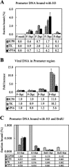
ChIP analysis of H3 histone associated with viral gene promoter regions during HSV-1 lytic infection of Sy5y cells. Isolated nuclei of infected cells were assayed by ChIP using histone H3 antibody. (A and B) The values for ChIP samples at the genomic region of interest were determined by quantitative PCR, normalized to GAPDH, and then compared to input values (total viral DNAs). (C) For the double ChIP experiment, the viral DNA levels for ChIP samples were determined by quantitative PCR and then compared to input viral DNA values. Unlike the single ChIP data, the double ChIP data were not normalized to GAPDH. GAPDH was used as a positive control for the double ChIP experiment. Error bars indicate standard deviations.
FIG. 4.
FIG. 4.
Free and DNA-bound H3 histone protein profiles from sucrose gradient centrifugation. At 3 or 6 hpi Sy5y cells were harvested and cell lysates fractionated on 5 to 50% (wt/vol) sucrose gradients. Viral DNA, cell (GAPDH) DNA, and H3 histone protein levels were measured by real-time PCR (A and B) and Western blotting (C), respectively. The percentage of total gradient histone protein in Western blot fractions was determined by densitometry (D).
FIG. 5.
FIG. 5.
A model for the temporal association of HSV-1 DNA and histones during lytic infection. 1. Naked linear virion DNA is released from capsids at the nuclear pore and circularizes in the nucleus. 2. It rapidly associates with histones and transcription is initiated. 3. After viral replication initiates, newly replicated viral DNA is not associated with histones, even though they appear to be available. 4. Newly replicated naked viral DNA is packaged into C capsids, which mature into virions on export from the nucleus and the cell.

References

    1. Berger, S. L. 2002. Histone modifications in transcriptional regulation. Curr. Opin. Genet. Dev. 12142-148. - PubMed
    1. Booy, F. P., W. W. Newcomb, B. L. Trus, J. C. Brown, T. S. Baker, and A. C. Steven. 1991. Liquid-crystalline, phage-like packing of encapsidated DNA in herpes simplex virus. Cell 641007-1015. - PMC - PubMed
    1. Deshmane, S. L., and N. W. Fraser. 1989. During latency, herpes simplex virus type 1 DNA is associated with nucleosomes in a chromatin structure. J. Virol. 63943-947. - PMC - PubMed
    1. Farnsworth, A., T. W. Wisner, M. Webb, R. Roller, G. Cohen, R. Eisenberg, and D. C. Johnson. 2007. Herpes simplex virus glycoproteins gB and gH function in fusion between the virion envelope and the outer nuclear membrane. Proc. Natl. Acad. Sci. USA 10410187-11092. - PMC - PubMed
    1. Gibson, W., and B. Roizman. 1971. Compartmentalization of spermine and spermidine in the herpes simplex virion. Proc. Natl. Acad. Sci. USA 682818-2821. - PMC - PubMed

Publication types

LinkOut - more resources