. 2008 Mar;82(6):3090-8.
doi: 10.1128/JVI.01479-07. Epub 2007 Dec 26.

RNA structure modulates splicing efficiency at the human immunodeficiency virus type 1 major splice donor

Affiliations

RNA structure modulates splicing efficiency at the human immunodeficiency virus type 1 major splice donor

Truus E M Abbink et al. J Virol. 2008 Mar.

Abstract

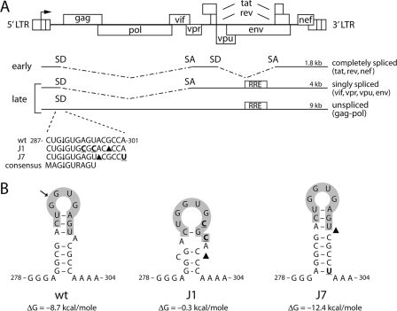
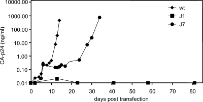
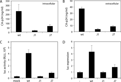
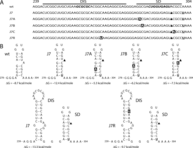
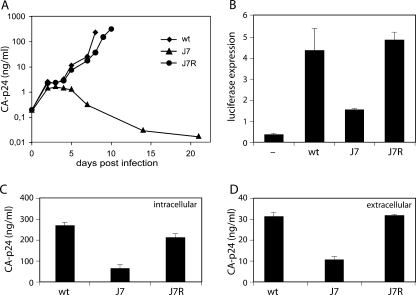
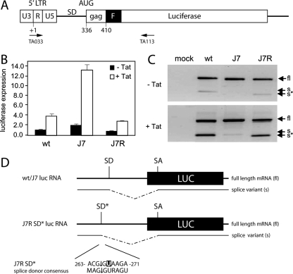
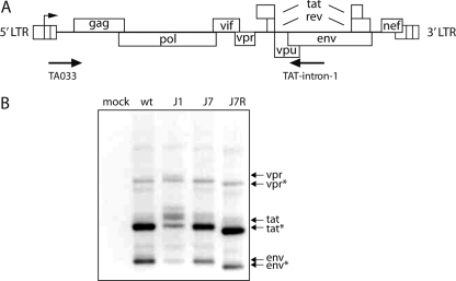
The untranslated leader of the human immunodeficiency virus type 1 (HIV-1) RNA genome encodes essential sequence and structural motifs that control various replication steps. The 5' splice site or splice donor (SD) is embedded in a semistable hairpin, but the function of this structure is unknown. We stabilized this SD hairpin by creating an additional base pair and demonstrated a severe HIV-1 replication defect. A splicing defect was apparent in RNA analyses of virus-infected cells and cells transfected with appropriate reporter constructs. We selected multiple virus revertants in search for interesting second-site escape pathways. Most revertants acquired an additional mutation that modulated the stability of the mutant SD hairpin. One revertant acquired a single nucleotide change in the upstream DIS hairpin. We demonstrate that a novel SD site is created by this upstream mutation, which obviously reduces the number of leader nucleotides that are included in spliced HIV-1 transcripts. These results suggest a novel role of RNA structure in the regulation of HIV-1 splicing.


Figures

FIG. 1.
FIG. 1.
HIV-1 SD mutants. (A) The HIV-1 DNA genome and the three classes of spliced mRNAs are shown. The 5′ and 3′ LTRs are divided into three segments (U3, R, and U5). Transcription starts at the U3-R border (arrow). SD and SA sites are shown in a simplified scheme. The RRE is required to export the unspliced and singly spliced mRNAs from the nucleus to the cytoplasm. All spliced mRNAs are spliced at the 5′ major SD. The wt and mutant (J1 and J7) sequences of the major 5′ SD site are shown. Mutated nucleotides are indicated in bold and underlined, and deletions are indicated with black triangles. ↓, cleavage site. The consensus sequence is indicated underneath, with M indicating an A or C nucleotide and R indicating a purine. (B) The J1 and J7 mutations affect the SD hairpin structure. ΔG values were determined with the Mfold program and are indicated underneath the RNA structure. The J1 hairpin is unlikely to fold, since its ΔG value is close to zero.
FIG. 2.
FIG. 2.
Replication of wt and SD mutant viruses. SupT1 cells were transfected with 250 ng of wt or mutant molecular clones. Virus production was measured in the culture medium by CA-p24 ELISA at several days posttransfection.
FIG. 3.
FIG. 3.
Characterization of wt and SD mutant viruses. (A and B) SD mutations decrease CA-p24 expression intracellularly (A) and extracellularly (B). C33A cells were transfected with wt or mutant proviral clones and lysed at 2 days posttransfection, and CA-p24 was determined by ELISA in the lysates and culture medium. (C) Infectivity of SD mutant viruses is severely decreased. TZM-bl indicator cells that express luciferase from a 5′ LTR promoter were infected with the indicated viruses. Luciferase activity was measured at 2 days postinfection. RLU, relative light units. (D) Tat production is greatly reduced by SD mutations. Tat protein production was determined in cotransfection assays with the indicated wt or mutant molecular clones and an LTR-luciferase expression construct. The basal expression level was determined in the absence of proviral DNA (lane labeled with a dash).
FIG. 4.
FIG. 4.
Forced evolution of the J7 SD mutant yields two classes of virus revertants. (A) Six cultures of SupT1 cells were transfected with the J7 proviral clone. Over time, replicating viruses were observed in all cultures, and viruses were passaged onto fresh cells at the peak of infection. In addition, chromosomal DNA was isolated from infected cells at the time of passage, and the leader region of the integrated proviral genome was sequenced, of which only the DIS-SD region is depicted. The J7 mutations (indicated in bold and underlined; black triangles indicate the deletion) remained present. Nucleotide changes acquired during evolution are shown in white in black boxes. Nucleotide positions are indicated. The J7A mutation (G283A) was selected in three cultures. (B) Effects of J7 variants on DIS and SD hairpins. The J7A, J7B, and J7C mutations destabilize the SD hairpin by disrupting a base pair. The ΔG values are indicated below the RNA structures. The J7R reversion leaves the stabilized SD hairpin intact but slightly destabilizes the DIS hairpin by altering a G-C to a G-U base pair. The ΔG values of the wt and J7R DIS hairpins are indicated.
FIG. 5.
FIG. 5.
The J7R mutation restores protein production and virus replication. (A) Replication of wt, J7, and J7R mutant viruses. SupT1 cells were infected with equal amounts of viruses. CA-p24 production was measured in the culture medium at several days postinfection. (B) Tat expression is restored to wt levels by the J7R mutation. See the legend to Fig. 3D for details. (C and D) CA-p24 expression in C33A cells (C) and in culture medium (D) at 2 days posttransfection.
FIG. 6.
FIG. 6.
Effect of J7R mutation on gene expression. (A) Luciferase reporter. See the legend to Fig. 1 for details. Primers used for RT-PCR analysis are indicated. (B) wt and mutant HIV-1 leader RNA-driven luciferase expression. Luciferase constructs were transfected into C33A cells, and pcDNA3 (white bars) or pcDNA3-Tat (black bars) was cotransfected. pRL-CMV was cotransfected as an internal control. Transfections were performed in triplicate. The graph shows normalized firefly luciferase activities. Similar results were obtained in three independent experiments. (C) Analysis of luciferase mRNA expressed by the wt and mutant reporter constructs. Transfection assays were performed as described for panel B. Total RNA was isolated at 2 days posttransfection and reverse transcribed with primer TA113. The cDNA was subsequently amplified with the TA033 and TA113 primers. The PCR products were applied to agarose gels. Arrows indicate PCR products derived from the unspliced, full-length (fl), and spliced (s) luciferase mRNAs. The J7R lane shows a shorter splice product (s*). In the presence of Tat, the ratio between fl and s or s* products is altered. A control reaction in which reverse transcriptase was omitted from the RT-PCR did not yield any amplified products (results not shown). (D) The J7R mutation results in the formation of an SD* site that results in splicing at nt 265 of the leader RNA. The fl, s, and s* RT-PCR products were cloned and sequenced. All wt and J7 mutant s products were spliced at the 5′ major SD, whereas the J7R mutant s* was spliced at the alternative major splice site (SD*) in the DIS region (nt 265). The sequence surrounding the J7R mutation constitutes an SD consensus sequence. For both s and s* mRNAs, the 3′ SA site is located at nt 569 of the luciferase transcript.
FIG. 7.
FIG. 7.
The alternative SD* is used as a 5′ splice site in J7R viral RNA. (A) The HIV-1 DNA genome and the positions of oligonucleotide primers used for RT-PCR are shown. The Tat-intron-1 primer is located in the env region. RT-PCR is able to detect all spliced mRNAs of the 4-kb class (singly spliced; also see Fig. 1). (B) Identification of wt and mutant splice variants. C33A cells were transfected with proviral constructs. Total RNA was isolated at 2 days posttransfection and reverse transcribed. The cDNA was amplified and analyzed via agarose gel electrophoresis. The J7R mutation resulted in a population of smaller mRNAs than those observed for the wt and J7 mutant viruses. Sequence analysis indicated that the alternative SD* was used for splicing in all clones (eight of eight clones analyzed).

References

    1. Abbink, T. E. M., and B. Berkhout. 2003. A novel long distance base-pairing interaction in human immunodeficiency virus type 1 RNA occludes the gag start codon. J. Biol. Chem. 27811601-11611. - PubMed
    1. Abbink, T. E. M., M. Ooms, P. C. J. Haasnoot, and B. Berkhout. 2005. The HIV-1 leader RNA conformational switch regulates RNA dimerization but does not regulate mRNA translation. Biochemistry 449058-9066. - PubMed
    1. Amendt, B. A., Z.-H. Si, and C. M. Stoltzfus. 1995. Presence of exon splicing silencers within human immunodeficiency virus type 1 tat exon 2 and tat-rev exon 3: evidence for inhibition mediated by cellular factors. Mol. Cell. Biol. 154606-4615. - PMC - PubMed
    1. Beerens, N., and B. Berkhout. 2002. The tRNA primer activation signal in the HIV-1 genome is important for initiation and processive elongation of reverse transcription. J. Virol. 762329-2339. - PMC - PubMed
    1. Beerens, N., F. Groot, and B. Berkhout. 2001. Initiation of HIV-1 reverse transcription is regulated by a primer activation signal. J. Biol. Chem. 27631247-31256. - PubMed

Publication types

LinkOut - more resources