. 2007 Dec 26;27(52):14365-74.
doi: 10.1523/JNEUROSCI.4549-07.2007.

Using imagination to understand the neural basis of episodic memory

Affiliations

Using imagination to understand the neural basis of episodic memory

Demis Hassabis et al. J Neurosci. .

Abstract

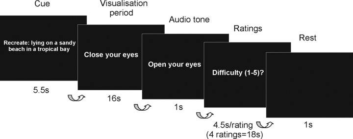
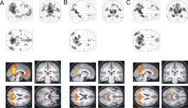
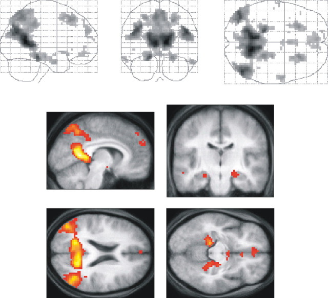
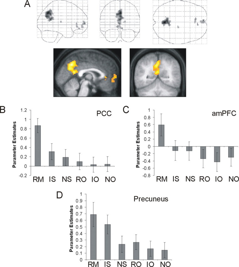
Functional MRI (fMRI) studies investigating the neural basis of episodic memory recall, and the related task of thinking about plausible personal future events, have revealed a consistent network of associated brain regions. Surprisingly little, however, is understood about the contributions individual brain areas make to the overall recollective experience. To examine this, we used a novel fMRI paradigm in which subjects had to imagine fictitious experiences. In contrast to future thinking, this results in experiences that are not explicitly temporal in nature or as reliant on self-processing. By using previously imagined fictitious experiences as a comparison for episodic memories, we identified the neural basis of a key process engaged in common, namely scene construction, involving the generation, maintenance and visualization of complex spatial contexts. This was associated with activations in a distributed network, including hippocampus, parahippocampal gyrus, and retrosplenial cortex. Importantly, we disambiguated these common effects from episodic memory-specific responses in anterior medial prefrontal cortex, posterior cingulate cortex and precuneus. These latter regions may support self-schema and familiarity processes, and contribute to the brain's ability to distinguish real from imaginary memories. We conclude that scene construction constitutes a common process underlying episodic memory and imagination of fictitious experiences, and suggest it may partially account for the similar brain networks implicated in navigation, episodic future thinking, and the default mode. We suggest that additional brain regions are co-opted into this core network in a task-specific manner to support functions such as episodic memory that may have additional requirements.


Figures

Figure 1.
Figure 1.
Experimental design. A text cue, prefixed by an instruction keyword denoting trial type [“Recall,” “Recreate,” “Imagine” (see Materials and Methods)], was presented for 5.5 s describing the scene or object to be visualized. Subjects were then instructed to close their eyes and begin visualizing the scene or object in as much detail as possible for the entire 16 s duration. A simple audio tone, played through headphones and lasting 1 s, indicated the end of the visualization period at which point the subject opened their eyes. Subjects then used a five-button MR-compatible box to rate their just visualized scene or object across four ratings on five-point scales: difficulty, vividness, coherence, and memory. Subjects were given 4.5 s to respond per rating, resulting in an overall rating period of 18 s. This was followed by a 1 s rest period in which a blank screen was presented before the start of the next trial.
Figure 2.
Figure 2.
Comparison of the main scene conditions with their respective object baselines. The top row shows sagittal, coronal, and axial images from a “glass brain,” which enables one to appreciate activations in all locations and levels in the brain simultaneously. The bottom row shows activations on a selection of relevant sagittal, coronal, and axial sections from the averaged structural MRI scan of the 21 subjects, at a threshold of p < 0.001 uncorrected. A, RM > RO, This contrast reveals the well established network for episodic memory retrieval that includes bilateral hippocampi, parahippocampal gyrus, retrosplenial and posterior parietal cortices, right thalamus, middle temporal cortices, and medial prefrontal cortex. Table 3 details the coordinates of all the activation peaks. B, NS > NO, This contrast reveals a similar network for imagining new fictitious experiences that includes right hippocampus, parahippocampal gyrus, retrosplenial and posterior parietal cortices, and ventromedial prefrontal cortex. Table 4 details the coordinates of all the activation peaks. C, IS > IO, This contrast also reveals a similar network for recalling imagined fictitious experiences previously created in a prescan interview that includes right hippocampus, parahippocampal gyrus, retrosplenial and posterior parietal cortices, and medial prefrontal cortex. Table 5 details the coordinates of all the activation peaks.
Figure 3.
Figure 3.
Brain areas in common to the three scene conditions. A conjunction analysis revealed the brain regions activated in common by the three scene conditions and therefore likely involved in “scene construction,” the primary process these three conditions have in common. This network included bilateral hippocampi, parahippocampal gyrus, retrosplenial and posterior parietal cortices, middle temporal cortices, and medial prefrontal cortex. Table 6 details the coordinates of all the activation peaks. Views of this distributed brain network are also shown in the bottom on a selection of relevant sagittal, coronal, and axial sections from the averaged structural MRI scan of the 21 subjects, at a threshold of p < 0.001 uncorrected.
Figure 4.
Figure 4.
Comparison of real and imagined memories. A, Contrasting recall of real memories to the recall of imaginary memories while controlling for externally versus internally generated stimuli and creativity processes, (RM − RO) > (IS − IO), revealed that the precuneus, PCC, and amPFC were preferentially engaged for real memories. Table 7 details the coordinates of all the activation peaks. Bottom, Views of these brain regions on a sagittal and coronal section from the averaged structural MRI scan of the 21 subjects, at a threshold of p < 0.001 uncorrected. B–D, Condition-specific parameter estimates (betas) in arbitrary units at the peak voxels. Error bars represent the SE. RM, Recall of real memories; IS, recall of previously imagined scenes; NS, constructing new fictitious scenes; RO, recall of previously seen objects; IO, recall of previously imagined objects; NO, imagining new objects. B, At the peak voxel in PCC. C, At the peak voxel in amPFC. D, At the peak voxel in the precuneus.

References

    1. Addis DR, Wong AT, Schacter DL. Remembering the past and imagining the future: common and distinct neural substrates during event construction and elaboration. Neuropsychologia. 2007;45:1363–1377. - PMC - PubMed
    1. Amodio DM, Frith CD. Meeting of minds: the medial frontal cortex and social cognition. Nat Rev Neurosci. 2006;7:268–277. - PubMed
    1. Atance CM, O'Neill DK. Episodic future thinking. Trends Cogn Sci. 2001;5:533–539. - PubMed
    1. Bartlett FC. Cambridge: Cambridge UP; 1932. Remembering.
    1. Buckner RL, Carroll DC. Self-projection and the brain. Trends Cogn Sci. 2007;11:49–57. - PubMed

Publication types