. 2007 Dec 26;27(52):14459-69.
doi: 10.1523/JNEUROSCI.4701-07.2007.

A critical function for beta-amyloid precursor protein in neuronal migration revealed by in utero RNA interference

Affiliations

A critical function for beta-amyloid precursor protein in neuronal migration revealed by in utero RNA interference

Tracy L Young-Pearse et al. J Neurosci. .

Abstract

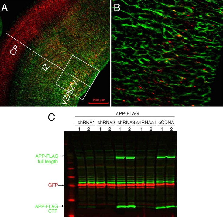
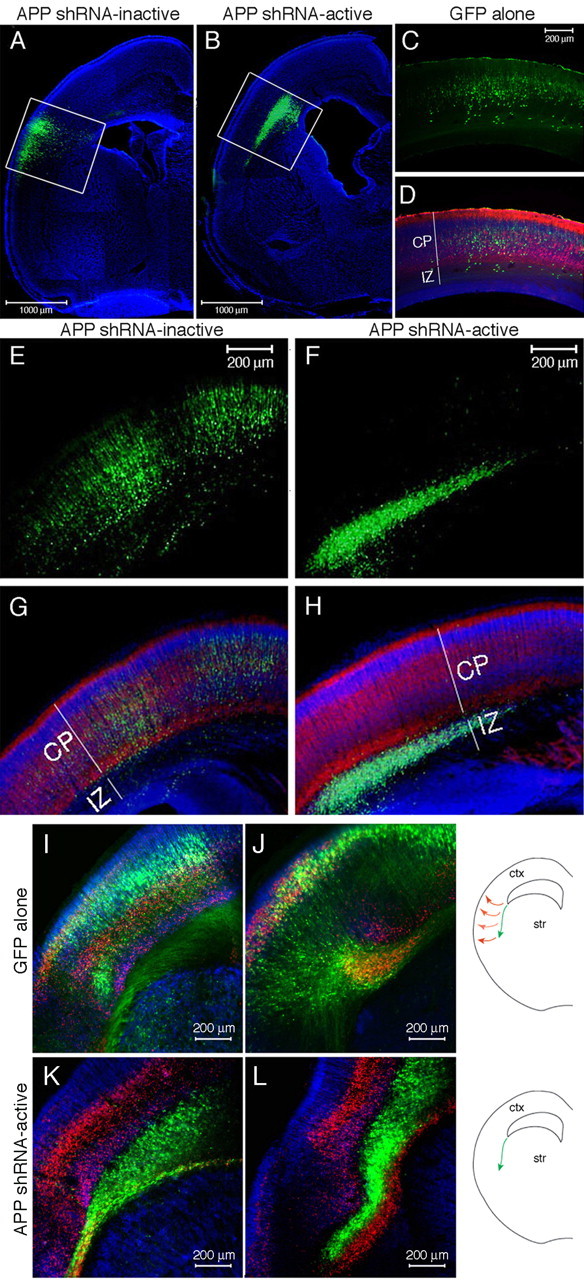
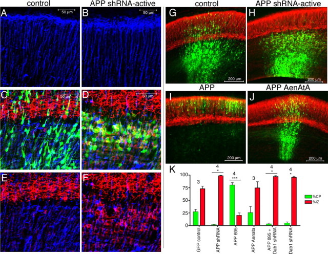
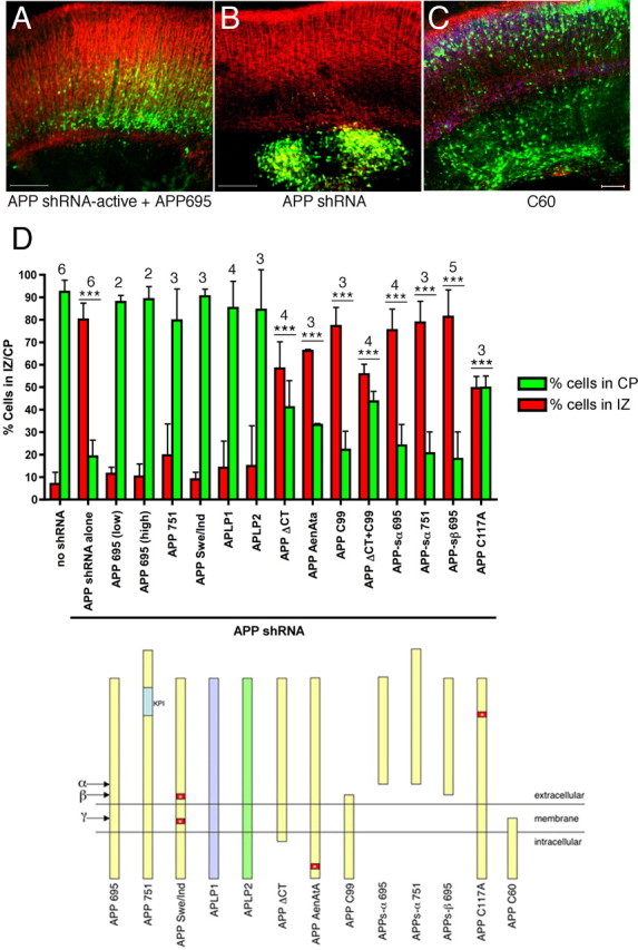
Physiological processing of the beta-amyloid precursor protein (APP) generates amyloid beta-protein, which can assemble into oligomers that mediate synaptic failure in Alzheimer's disease. Two decades of research have led to human trials of compounds that chronically target this processing, and yet the normal function of APP in vivo remains unclear. We used the method of in utero electroporation of shRNA constructs into the developing cortex to acutely knock down APP in rodents. This approach revealed that neuronal precursor cells in embryonic cortex require APP to migrate correctly into the nascent cortical plate. cDNAs encoding human APP or its homologues, amyloid precursor-like protein 1 (APLP1) or APLP2, fully rescued the shRNA-mediated migration defect. Analysis of an array of mutations and deletions in APP revealed that both the extracellular and cytoplasmic domains of APP are required for efficient rescue. Whereas knock-down of APP inhibited cortical plate entry, overexpression of APP caused accelerated migration of cells past the cortical plate boundary, confirming that normal APP levels are required for correct neuronal migration. In addition, we found that Disabled-1 (Dab1), an adaptor protein with a well established role in cortical cell migration, acts downstream of APP for this function in cortical plate entry. We conclude that full-length APP functions as an important factor for proper migration of neuronal precursors into the cortical plate during the development of the mammalian brain.


Figures

Figure 1.
Figure 1.
Generation of shRNA constructs targeting endogenous rodent APP in embryonic cortex. A, B, Coronal sections of E15 mouse cortex immunostained for APP (red) and nestin (green). Magnified view of IZ/SVZ/VZ (box in A) is shown in B. SVZ, Subventricular zone; VZ, ventricular zone. C, FLAG-tagged murine APP construct was cotransfected into CHO cells with an empty vector or with 3 different shRNA constructs targeting rodent APP (shRNA1, shRNA2, shRNA3), along with a GFP construct to control for transfection efficiency. Transfections in duplicate wells are shown. 48 h post-transfection, cells were lysed and Western blotted for FLAG (green) and GFP (red). shRNAall, Mix of all three shRNAs.
Figure 2.
Figure 2.
Electroporation of APP shRNA in embryonic rat cortex. DNA encoding GFP and APP shRNA was coelectroporated into one lateral ventricle of an E13 rat and harvested 6 d later. Cells of the ventricular zone lining the ventricle are electroporated. A–H, Location of the electroporated cells (green) is shown 6 d after electroporation of GFP alone (C, D), GFP with an inactive shRNA (A, E, G), or GFP with an active shRNA targeting APP (B, F, H). White boxes delineate electroporated regions (A, B) that are magnified (E–H). I–L, Electroporation of the corticostriatal boundary with GFP alone (I, J) or APP shRNA-active (K, L). A–H, Immunostaining for MAP2 (red) denotes the differentiated neurons of the cortical plate. Nuclei are stained with DAPI (blue). I–L, Immunostaining for MAP2 is shown in blue, and Tbr-1 is shown in red. Str, Striatum; ctx, cortex.
Figure 3.
Figure 3.
Early effects of acutely altering APP levels in embryonic cortex. A–J, Electroporation of GFP alone (A, C, E, G) or coelectroporation of GFP plus active APP shRNA (B, D, F, H), GFP plus APP (I), or GFP plus mutant APP AenAtA (J) into E13 cortex and harvesting 3 d later. Electroporated cells are green (GFP). Immunostaining for BLBP (A–F) is blue and MAP2 is red (C–J). A, B, BLBP immunostaining in upper cortical plate in electroporated region. C–F, MAP2 with BLBP double staining at the cortical plate/intermediate zone boundary. K, Quantification of cells in the IZ and CP for each condition shown in G–J. Error bars indicate SD; *p < 0.05; ***p < 0.001, percentage of cells in IZ and CP relative to the corresponding values in GFP control. Numbers above bars show the number of independent brains analyzed for each condition.
Figure 4.
Figure 4.
Cells electroporated with active APP shRNA remain trapped in the intermediate zone and form a heterotopia. Cortices were coelectroporated at E13 with GFP and APP shRNA-active (A, C, E, F, G, H, I) or with control DNA (B, D) and harvested 30 d postnatally. Brightfield image of a coronal section of a cortex electroporated with active APP shRNA (A). Arrows point to a heterotopia only present in the electroporated hemisphere. B–I, Electroporated cells are shown in green. Location and morphology of control electroporated cells in the CP (B, D) and of APP shRNA-active electroporated cells in the IZ (C, E). MAP2 (red) and NeuN (blue) immunostaining of APP shRNA electroporated cells in the IZ (H) and CP (F) at P30. G, I, GFAP immunostaining (red) in the heterotopia.
Figure 5.
Figure 5.
Rescue of APP shRNA-induced migration defects. Embryos were electroporated at E13, harvested after 6 d in utero, and brain sections immunostained for MAP2 (red). A, B, Representative sections from APP shRNA-active plus APP695 (A) and APP shRNA-active alone (B) are shown. Scale bars, 200 μm. C, Image of section from an E13 embryo electroporated with only the C-terminal 60 aa of APP (C60) and GFP (green). Immunostaining for MAP2 shown in red and for NeuN in blue. Scale bar, 100 μm. D, Quantification (top graph) of cells in the IZ and CP for each of the rescue constructs diagramed below. Error bars indicate SD; ***p < 0.001, percentage of cells in IZ and CP relative to the corresponding values with control DNA (no shRNA). The numbers above the bars show the number of independent brains analyzed for each condition.
Figure 6.
Figure 6.
APP and Disabled-1 functionally interact to affect neuronal migration in the cortical plate. A–D, Embryos were electroporated at E13, harvested after 6 d in utero, and brain sections immunostained for MAP2 (blue) and Tbr1 (red). E, Quantification of cells in the IZ and CP for each of the indicated constructs; n = 3 independent brains for each condition. Error bars indicate SD; **p < 0.01.

References

    1. Anliker B, Muller U. The functions of mammalian amyloid precursor protein and related amyloid precursor-like proteins. Neurodegener Dis. 2006;3:239–246. - PubMed
    1. Araki W, Kitaguchi N, Tokushima Y, Ishii K, Aratake H, Shimohama S, Nakamura S, Kimura J. Trophic effect of β-amyloid precursor protein on cerebral cortical neurons in culture. Biochem Biophys Res Commun. 1991;181:265–271. - PubMed
    1. Bai J, Ramos RL, Ackman JB, Thomas AM, Lee RV, LoTurco JJ. RNAi reveals doublecortin is required for radial migration in rat neocortex. Nat Neurosci. 2003;6:1277–1283. - PubMed
    1. Bayer SA, Altman J. Development of the endopiriform nucleus and the claustrum in the rat brain. Neuroscience. 1991;45:391–412. - PubMed
    1. Bayer SA, Altman J, Russo RJ, Dai XF, Simmons JA. Cell migration in the rat embryonic neocortex. J Comp Neurol. 1991;307:499–516. - PubMed

Publication types

MeSH terms

Substances