. 2008 Mar;28(5):1841-50.
doi: 10.1128/MCB.01468-07. Epub 2007 Dec 26.

ATP binding by monarch-1/NLRP12 is critical for its inhibitory function

Affiliations

ATP binding by monarch-1/NLRP12 is critical for its inhibitory function

Zhengmao Ye et al. Mol Cell Biol. 2008 Mar.

Abstract

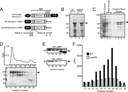
The recently discovered nucleotide binding domain-leucine rich repeat (NLR) gene family is conserved from plants to mammals, and several members are associated with human autoinflammatory or immunodeficiency disorders. This family is defined by a central nucleotide binding domain that contains the highly conserved Walker A and Walker B motifs. Although the nucleotide binding domain is a defining feature of this family, it has not been extensively studied in its purified form. In this report, we show that purified Monarch-1/NLRP12, an NLR protein that negatively regulates NF-kappaB signaling, specifically binds ATP and exhibits ATP hydrolysis activity. Intact Walker A/B motifs are required for this activity. These motifs are also required for Monarch-1 to undergo self-oligomerization, Toll-like receptor- or CD40L-activated association with NF-kappaB-inducing kinase (NIK) and interleukin-1 receptor-associated kinase 1 (IRAK-1), degradation of NIK, and inhibition of IRAK-1 phosphorylation. The stable expression of a Walker A/B mutant in THP-1 monocytes results in increased production of proinflammatory cytokines and chemokines to an extent comparable to that in cells in which Monarch-1 is silenced via short hairpin RNA. The results of this study are consistent with a model wherein ATP binding regulates the anti-inflammatory activity of Monarch-1.


Figures

FIG. 1.
FIG. 1.
The generation and purification of Monarch-1-NBD fusion proteins. (A) Schematic diagram of domain structure of full-length Monarch-1 and WT and mutA/B Monarch-1-NBD fusion proteins. The positions of the Walker A and Walker B motifs are depicted as solid bars within the NACHT domain. The conserved amino acids within both motifs are shown, and the underlined residues were substituted with alanine by site-directed mutagenesis. (B) WT Monarch-1-NBD and mut A/B Monarch-1-NBD were transformed into E. coli, and soluble bacterial extracts were analyzed by SDS-PAGE and Coomassie blue staining. Arrowhead, Monarch-1-NBD fusion protein; U, uninduced; I, IPTG-induced. The sizes of molecular weight markers (in thousands) are indicated on the left. (C) Bacterial lysates were passed over an amylose resin column targeting MBP. Eluted proteins were resolved by SDS-PAGE and visualized with Coomassie blue stain. (D) Eluates from the amylose resin column were further purified by size exclusion chromatography. The absorbance profile of size exclusion purification is depicted (upper panel), and the size exclusion fractions were analyzed by SDS-PAGE and visualized by Coomassie blue staining (lower panel). (E) Western blots from size exclusion fractions 17 to 21 were probed with an anti-MBP or anti-His antibody to verify the intactness of the N terminus and C terminus, respectively, of the Monarch-1-NBD fusion protein. (F) Size exclusion fractions from WT or mutA/B Monarch-1-NBD were tested for ATP binding activity using [γ-35S]ATP, and specific binding was normalized to the protein concentration of each fraction.
FIG. 2.
FIG. 2.
Purified Monarch-1-NBD fusion protein specifically binds ATP. (A) Gel filtration fractions 18 to 19 (depicted in Fig. 1) were pooled and further purified by cobalt metal affinity chromatography. The purity of the final purification product was assessed by SDS-PAGE and Coomassie blue staining. Arrowhead, Monarch-1-NBD fusion protein. The sizes of molecular weight markers (in thousands) are indicated on the left. (B) Homologous competition assays were performed to assess the ATP binding affinity of Monarch-1-NBD. (C) Specificity of ATP binding was determined by incubating Monarch-1-NBD fusion protein with [γ-35S]ATP and 10 μM of the indicated unlabeled competitor nucleotide. Significance of the difference from control samples was estimated by Student's t test. “None” indicates no nucleotide. A double asterisk signifies a P value of <0.000003. A single asterisk signifies a P value of 0.011 for GTP or 0.0085 for CTP.
FIG. 3.
FIG. 3.
Mammalian cell-derived recombinant Monarch-1ΔLRR binds and hydrolyzes ATP. (A) Diagram depicting full-length Monarch-1 and the WT and mutA/B Monarch-1ΔLRR proteins produced in HEK293EBNA cells. (B) Soluble extracts of HEK293EBNA cells expressing WT and mutA/B Monarch-1ΔLRR were enriched for Monarch-1ΔLRR using a cobalt metal affinity column (lanes 1 and 2) and further purified over an anti-Flag affinity matrix (lanes 3 and 4). The purity was evaluated by Coomassie blue staining. The sizes of molecular weight markers (in thousands) are indicated on the left. (C) The double-purified Monarch-1ΔLRR proteins were analyzed by Western blotting using anti-His and anti-Flag antibodies. (D) ATP binding activity of purified WT and mutA/B Monarch-1ΔLRR was determined by incubating 500 ng purified protein with [γ-35S]ATP. Error bars represent the standard deviations of ATP binding measurements in triplicate. (E) The ATP binding affinity of WT Monarch-1ΔLRR was determined by homologous competition binding assays. (F) The nucleotide binding preference of WT Monarch-1ΔLRR was determined by incubating WT Monarch-1ΔLRR with [γ-35S]ATP and increasing concentrations of unlabeled nucleotide. Significance of the differences from control samples (nucleotide concentrations of 10−9 M) was estimated by Student's t test. A double asterisk signifies a P value of <0.0005. A single asterisk signifies a P value of <0.02. (G) The ATPase activity of purified WT or mutA/B Monarch-1ΔLRR was measured by visualizing the conversion of [α-32P]ATP to [α-32P]ADP by TLC. HEK293EBNA lysate and purified bovine serum albumin (BSA) were used as positive and negative controls, respectively. Arrowhead, Monarch-1-NBD fusion protein.
FIG. 4.
FIG. 4.
Nucleotide binding is required for Monarch-1 self association. HEK293T cells were transfected with full-length forms of HA- or Flag-tagged WT or mutA/B Monarch-1. Flag-tagged proteins were immunoprecipitated (IP) with an anti-Flag antibody, and Western blots (IB) were probed with anti-HA antibody to detect self-associated Monarch-1.
FIG. 5.
FIG. 5.
Nucleotide binding is required for Monarch-1 to suppress NIK-mediated p100 processing. (A) HEK293T cells were cotransfected with NIK and HA-tagged WT or mutA/B Monarch-1. Cell lysates were immunoprecipitated (IP) with anti-NIK antibodies, and Western blots (IB) were probed with anti-HA antibodies to detect Monarch-1. ctrl lg, isotype control antibody. (B) THP-WT or THP-mutA/B cells were stimulated with CD40L for indicated times. Endogenous NIK was immunoprecipitated, and Western blots were probed with anti-HA to detect coprecipitating Monarch-1. Control immunoprecipitations were performed with an isotype control antibody to monitor specificity. Control Western blottings were performed to monitor the expression of Monarch-1 and NIK in cellular lysates. (C) THP-EV, THP-WT, or THP-mutA/B cells were stimulated with Pam3Cys4 for 18 h to induce p100 expression. The cells were then treated with CD40L for an additional 5 h to induce p100 cleavage to p52. Cells were fractionated into nuclear and cytoplasmic fractions, and proteins from each fraction were separated by SDS-PAGE. Western blots were probed with anti-p100 to detect p100 and its cleaved form, p52. Anti-HA was used to monitor Monarch-1 expression. These results are representative of at least five separate experiments.
FIG. 6.
FIG. 6.
Nucleotide binding by Monarch-1 is required for the suppression of IRAK-1 hyperphosphorylation. (A) THP-WT or THP-mutA/B cells were stimulated for the indicated times with Pam3Cys4. Endogenous IRAK-1 was immunoprecipitated (IP), and Western blots (IB) were probed with anti-HA to detect Monarch-1. Control samples were immunoprecipitated with an isotype-matched antibody. Control Western blottings were performed on cellular lysates to monitor the levels of Monarch-1 and IRAK-1. (B) THP-EV, THP-WT, and THP-mutA/B cells were stimulated with Pam3Cys4 for the indicated times. Lysates were separated by SDS-PAGE, and Western blots were probed with anti-IRAK-1 antibodies. Control Western blottings were performed and probed with anti-HA to monitor Monarch-1 expression. These results are representative of at least five separate experiments.
FIG. 7.
FIG. 7.
Nucleotide binding is required for Monarch-1-mediated suppression of proinflammatory cytokine and chemokine production. THP-EV, THP-WT, THP-mutA/B, and THP-shMon were stimulated with Pam3Cys4 for 18 h and then CD40L for an additional 5 h. Cell culture supernatants were harvested, and cytokine/chemokine levels determined by ELISA.

References

    1. Aksentijevich, I., M. Nowak, M. Mallah, J. J. Chae, W. T. Watford, S. R. Hofmann, L. Stein, R. Russo, D. Goldsmith, P. Dent, H. F. Rosenberg, F. Austin, E. F. Remmers, J. E. Balow, Jr., S. Rosenzweig, H. Komarow, N. G. Shoham, G. Wood, J. Jones, N. Mangra, H. Carrero, B. S. Adams, T. L. Moore, K. Schikler, H. Hoffman, D. J. Lovell, R. Lipnick, K. Barron, J. J. O'Shea, D. L. Kastner, and R. Goldbach-Mansky. 2002. De novo CIAS1 mutations, cytokine activation, and evidence for genetic heterogeneity in patients with neonatal-onset multisystem inflammatory disease (NOMID): a new member of the expanding family of pyrin-associated autoinflammatory diseases. Arthritis Rheum. 463340-3348. - PMC - PubMed
    1. Albrecht, M., and F. L. Takken. 2006. Update on the domain architectures of NLRs and R proteins. Biochem. Biophys. Res. Commun. 339459-462. - PubMed
    1. Beutler, B. 2001. Autoimmunity and apoptosis: the Crohn's connection. Immunity 155-14. - PubMed
    1. Duncan, J. A., D. T. Bergstralh, Y. Wang, S. B. Willingham, Z. Ye, A. G. Zimmermann, and J. P. Ting. 2007. Cryopyrin/NALP3 binds ATP/dATP, is an ATPase, and requires ATP binding to mediate inflammatory signaling. Proc. Natl. Acad. Sci. USA 1048041-8046. - PMC - PubMed
    1. Hoffman, H. M., J. L. Mueller, D. H. Broide, A. A. Wanderer, and R. D. Kolodner. 2001. Mutation of a new gene encoding a putative pyrin-like protein causes familial cold autoinflammatory syndrome and Muckle-Wells syndrome. Nat. Genet. 29301-305. - PMC - PubMed

Publication types

MeSH terms

LinkOut - more resources