. 2007 Dec 12;1(3):e96.
doi: 10.1371/journal.pntd.0000096.

Pediatric measles vaccine expressing a dengue antigen induces durable serotype-specific neutralizing antibodies to dengue virus

Affiliations

Pediatric measles vaccine expressing a dengue antigen induces durable serotype-specific neutralizing antibodies to dengue virus

Samantha Brandler et al. PLoS Negl Trop Dis. .

Abstract

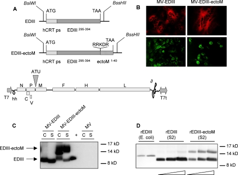
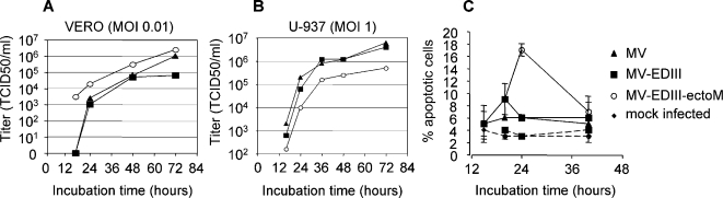
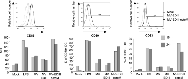
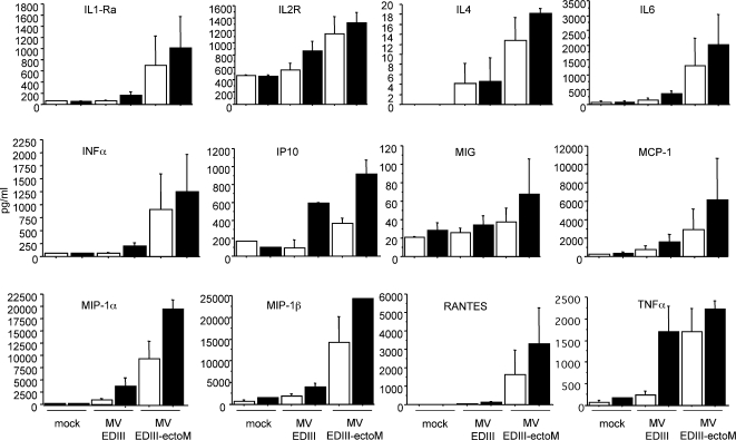
Dengue disease is an increasing global health problem that threatens one-third of the world's population. Despite decades of efforts, no licensed vaccine against dengue is available. With the aim to develop an affordable vaccine that could be used in young populations living in tropical areas, we evaluated a new strategy based on the expression of a minimal dengue antigen by a vector derived from pediatric live-attenuated Schwarz measles vaccine (MV). As a proof-of-concept, we inserted into the MV vector a sequence encoding a minimal combined dengue antigen composed of the envelope domain III (EDIII) fused to the ectodomain of the membrane protein (ectoM) from DV serotype-1. Immunization of mice susceptible to MV resulted in a long-term production of DV1 serotype-specific neutralizing antibodies. The presence of ectoM was critical to the immunogenicity of inserted EDIII. The adjuvant capacity of ectoM correlated with its ability to promote the maturation of dendritic cells and the secretion of proinflammatory and antiviral cytokines and chemokines involved in adaptive immunity. The protective efficacy of this vaccine should be studied in non-human primates. A combined measles-dengue vaccine might provide a one-shot approach to immunize children against both diseases where they co-exist.


Conflict of interest statement

The authors have declared that no competing interests exist.

Figures

Figure 1
Figure 1. Expression of DV1 EDIII and EDIII-ectoM by recombinant MV vector.
(A) Schematic representation of DV1 EDIII and EDIII-ectoM constructs and of recombinant MV vector. The human calreticulin peptide signal sequence (hCRT ps), the DV1 envelope E domain III (EDIII) and the M ectodomain (ectoM) are indicated. Original furin-like cleavage site RRDKR and amino acid positions are indicated. The EDIII and EDIII-ectoM sequences were cloned into the ATU position 1 of MV vector using BsiWI/BssHII sites. The MV genes are indicated as follows: nucleoprotein (N), phosphoprotein and V/C accessory proteins (PVC), matrix (M) fusion (F), hemagglutinin (H) and polymerase (L). T7 RNA polymerase promoter (T7), T7 RNA polymerase terminator (T7t), hepatitis delta virus ribozyme (∂), hammerhead ribozyme (hh). (B) Detection of DV1 EDIII in Vero cells infected for 24h with MV-EDIII or MV-EDIII-ectoM. EDIII was detected in two different experiments using either anti-EDIII 4E11 antibody (red label) or anti-DV1-HMAF (green label). (C) DV1 EDIII expression in cell lysates (C) and supernatants (S) of Vero cells infected by MV-EDIII and MV-EDIII-ectoM analyzed by western blot (cell lysates are 20 times more concentrated than supernatants). (+ ) positive control (5 ng of DV1 EDIII produced in drosophila S2 cells). DV1 EDIII was probed with the 4E11 mouse monoclonal anti-DV1 EDIII. (D) Western blot analysis of recombinant rEDIII protein produced in E.coli (100 ng) and recombinant rEDIII or rEDIII-ectoM proteins produced in drosophila S2 cells supernatants (0.5, 1 and 2 µg/well). DV1 EDIII was probed with 4E11 mouse monoclonal anti-DV1 EDIII.
Figure 2
Figure 2. In vitro replication and cytopathic effects induced by recombinant MV-DV.
(A, B) Growth kinetics of recombinant MV-EDIII and MV-EDIII-ectoM viruses compared with standard MV on Vero and U-937 cells (MOI 0.01 and 1, respectively). Cell-associated virus titers are indicated in TCID50. (C) Apoptosis of DCs infected by recombinant viruses (MOI 10). MV (triangles), MV-EDIII (squares), MV-EDIII-ectoM (open circles), mock infected (diamonds). UV inactivated viruses (same symbols with dotted lines). The percentage of apoptotic cells represents the percentage of Annexin-V positive and propidium iodide negative cells (determined using a flow cytometer). Values are means ± SE from two independent experiments, each performed in duplicate.
Figure 3
Figure 3. Neutralization tests in presence of recombinant rEDIII protein or synthetic ectoM peptide.
The neutralization activity of immune sera collected before DV exposure (diamonds and triangles, 1/100 dilution) and post DV exposure (open squares 1/1000 dilution) was tested in presence of increasing doses of recombinant EDIII (produced in drosophila cells) or ectoM synthetic peptide. Pools of sera were preincubated with rEDIII or ectoM for 1h at 37°C before FRNT75 titration on Vero cells (50 FFU DV1 FGA89 for 2h at 37°C).
Figure 4
Figure 4. Detection of DV1 EDIII in human DCs incubated for 17h with MV-EDIII or MV-EDIII-ectoM (MOI 1).
EDIII was detected using anti-EDIII 4E11 antibody (red label). Anti-human MHC-II dimer primary antibody was used as a DC marker (green label) (kindly provided by Neefjed J.). Cells nuclei were labeled with DAPI.
Figure 5
Figure 5. Expression of CD86, CD80, CD83 cell surface molecules on human DCs incubated in presence of MV-EDIII and MV-EDIII-ectoM (MOI of 1), or mock treated.
The expression of cell surface markers was analyzed after 15h and 24h incubation by flow cytometry. Data shown are representative of three individual experiments. Thin light-grey line represent staining of mock-treated DC, dashed grey lines represent staining of MV-EDIII-infected DCs, and thick black line represent staining of MV-EDIII-ectoM-infected DCs.
Figure 6
Figure 6. Cytokine and chemokine secretion by human DC incubated in presence of MV-EDIII and MV-EDIII-ectoM (MOI 1).
The supernatants harvested at 16h (white bars) and 24h (black bars) after infection were analyzed with a multiplex assay to measure the concentration of cytokines and chemokines. The values are means ± SE from four independent experiments.

Similar articles

Cited by

References

    1. Mackenzie JS, Gubler DJ, Petersen LR. Emerging flaviviruses: the spread and resurgence of Japanese encephalitis, West Nile and dengue viruses. Nat Med. 2004;10:S98–109. - PubMed
    1. Catteau A, Kalinina O, Wagner MC, Deubel V, Courageot MP, et al. Dengue virus M protein contains a proapoptotic sequence referred to as ApoptoM. J Gen Virol. 2003;84:2781–2793. - PubMed
    1. Halstead SB, Nimmannitya S, Cohen SN. Observations related to pathogenesis of dengue hemorrhagic fever. IV. Relation of disease severity to antibody response and virus recovered. Yale J Biol Med. 1970;42:311–328. - PMC - PubMed
    1. Halstead SB, O'Rourke EJ. Antibody-enhanced dengue virus infection in primate leukocytes. Nature. 1977;265:739–741. - PubMed
    1. Modis Y, Ogata S, Clements D, Harrison SC. A ligand-binding pocket in the dengue virus envelope glycoprotein. Proc Natl Acad Sci U S A. 2003;100:6986–6991. - PMC - PubMed

Publication types

MeSH terms