. 2008 Mar;47(3):908-18.
doi: 10.1002/hep.22110.

Aberrant methylation of multiple tumor suppressor genes in aging liver, chronic hepatitis, and hepatocellular carcinoma

Affiliations

Aberrant methylation of multiple tumor suppressor genes in aging liver, chronic hepatitis, and hepatocellular carcinoma

Naoshi Nishida et al. Hepatology. 2008 Mar.

Abstract

Aberrant DNA methylation is an important epigenetic alteration in hepatocellular carcinoma (HCC). However, the molecular processes underlying the methylator phenotype and the contribution of hepatitis viruses are poorly understood. The current study is a comprehensive methylation analysis of human liver tissue specimens. A total of 176 liver tissues, including 77 pairs of HCCs and matching noncancerous liver and 22 normal livers, were analyzed for methylation. Methylation of 19 epigenetic markers was quantified, and the results were correlated with different disease states and the presence or absence of hepatitis B virus (HBV) and hepatitis C virus (HCV) infections. Based on methylation profiles, the 19 loci were categorized into 3 groups. Normal liver tissues showed methylation primarily in group 1 loci (HIC-1, CASP8, GSTP1, SOCS1, RASSF1A, p16, APC), which was significantly higher than group 2 (CDH1, RUNX3, RIZ1, SFRP2, MINT31) and group 3 markers (COX2, MINT1, CACNA1G, RASSF2, MINT2, Reprimo, DCC) (P < 0.0001). Noncancerous livers demonstrated increased methylation in both group 1 and group 2 loci. Methylation was significantly more abundant in HCV-positive livers compared with normal liver tissues. Conversely, HCC showed frequent methylation at each locus investigated in all 3 groups. However, the group 3 loci showed more dense and frequent methylation in HCV-positive cancers compared with both HBV-positive cancers and virus-negative cancers (P < 0.0001).

Conclusion: Methylation in HCC is frequent but occurs in a gene-specific and disease-specific manner. Methylation profiling allowed us to determine that aberrant methylation is commonly present in normal aging livers, and sequentially progresses with advancing stages of chronic viral infection. Finally, our data provide evidence that HCV infection may accelerate the methylation process and suggests a continuum of increasing methylation with persistent viral infection and carcinogenesis in the liver.


Conflict of interest statement

Potential conflict of interest: Nothing to report.

Figures

Fig. 1
Fig. 1
Distribution of percent methylation levels (box and whiskers plots) and their frequencies (gray bar) at each of the 19 methylation loci (A, B, and C), and overall distribution of methylation level expressed as Z-scores in 3 groups (D, E, and F) as observed in normal liver (A, D), noncancerous liver from HCC patients (B, E), and HCC (C, F). Red boxes and whiskers plots denote 75% and 95% distribution, respectively, and the red lines in the boxes show the median values. *Statistically significant by ANOVA and post-hoc comparisons. (D) Differences were significant between group 1 versus group 2, and group 1 versus group 3 [ANOVA; F(2, 415) = 46.7; P < 0.0001, Kruskal-Wallis test; P < 0.0001]. (E) Differences were significant between every pair [ANOVA; F(2, 1460) = 153.5, P < 0.0001, Kruskal-Wallis test; P < 0.0001]. (F) Differences were significant between every pair [ANOVA; F(2, 1536) = 207.4, P < 0.0001, Kruskal-Wallis test; P < 0.0001]
Fig. 2
Fig. 2
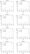
Correlation between methylation densities at 7 group 1 loci (HIC-1, CASP8, GSTP1, SOCS1, RASSF1A, p16, and APC) and 1 group 2 locus (SFRP 2) in normal liver. The r value of Pearson correlation test and ρ value of Spearman correlation test were calculated.
Fig. 3
Fig. 3
Distribution of methylation at various subgroups at methylation loci in noncancerous liver tissues of HCC patients and their corresponding HCCs classified according to virus status. Samples were sorted in ascending order based on the number of methylated loci. A hatched square denotes a locus with methylation (>2% methylation density), and a solid square denotes a locus with ≥25% methylation. An open square indicates no methylation.
Fig. 4
Fig. 4
Classification of HCC cases according to differences in methylation between HCCs and their matching noncancerous tissues using hierarchical clustering analysis. Green trees represent group 1 clusters, and red trees represent group 2 clusters. A solid gray circle represents HBV-positive, solid black represent HCV-positive, and an open circle indicates virus-negative cases.
Fig. 5
Fig. 5
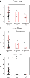
Distribution of methylation density (box and whiskers plots) of group 1 (A), group 2 (B), and group 3 loci (C) in HCC classified as HCV-related, HBV-related, and virus-negative. Each density was expressed as a Z-score. Red boxes and whiskers denote 75% and 95% distributions, respectively, and the red lines in the boxes showed median values. *Differences were significant by ANOVA and post hoc comparisons. Methylation of group 1 loci were significantly higher in HCV-related HCC than in virus-negative (A) [ANOVA; F(2, 522) = 4.34, P = 0.0134, Kruskal-Wallis test; P = 0.0168]. Similarly, methylation was prominent in HCV-related and HBV-related HCC compared with non-B non-C–related HCC at group 2 loci. (B) [ANOVA; F(2, 372) = 12.17, P < 0.0001, Kruskal-Wallis test; P < 0.0001], and methylation of group 3 was prominent in HCV-related HCC compared with HBV-related and virus-negative HCC. (C) [ANOVA; F(2, 522) = 18.31, P < 0.0001, Kruskal-Wallis test, P < 0.0001].

References

    1. Issa JP. CpG island methylator phenotype in cancer. Nat Rev Cancer. 2004;4:988–993. - PubMed
    1. Ushijima T. Detection and interpretation of altered methylation patterns in cancer cells. Nat Rev Cancer. 2005;5:223–231. - PubMed
    1. Baylin SB, Ohm JE. Epigenetic gene silencing in cancer: a mechanism for early oncogenic pathway addiction? Nat Rev Cancer. 2006;6:107–116. - PubMed
    1. Yu J, Ni M, Xu J, Zhang H, Gao B, Gu J, et al. Methylation profiling of twenty promoter-CpG islands of genes which may contribute to hepatocellular carcinogenesis. BMC Cancer. 2002;2:29. - PMC - PubMed
    1. Lee S, Lee HJ, Kim JH, Lee HS, Jang JJ, Kang GH. Aberrant CpG island hypermethylation along multistep hepatocarcinogenesis. Am J Pathol. 2003;163:1371–1378. - PMC - PubMed

Publication types