

Macrophage activation by endogenous danger signals
- PMID: 18161744
- PMCID: PMC2724989
- DOI: 10.1002/path.2284
Macrophage activation by endogenous danger signals
Abstract
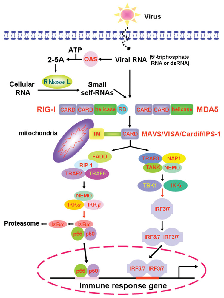
Macrophages are cells that function as a first line of defence against invading microorganisms. One of the hallmarks of macrophages is their ability to become activated in response to exogenous 'danger signals'. Most microbes have molecular patterns (PAMPS) that are recognized by macrophages and trigger this activation response. There are many aspects of the activation response to PAMPS that are recapitulated when macrophages encounter endogenous danger signals. In response to damaged or stressed self, macrophages undergo physiological changes that include the initiation of signal transduction cascades from germline-encoded receptors, resulting in the elaboration of chemokines, cytokines and toxic mediators. This response to endogenous mediators can enhance inflammation, and thereby contribute to autoimmune pathologies. Often the overall inflammatory response is the result of cooperative activation signals from both exogenous and endogenous signals. Macrophage activation plays a critical role, not only in the initiation of the inflammatory response but also in the resolution of this response. The clearance of granulocytes and the elaboration of anti-inflammatory mediators by macrophages contribute to the dissolution of the inflammatory response. Thus, macrophages are a key player in the initiation, propagation and resolution of inflammation. This review summarizes our understanding of the role of macrophages in inflammation. We pay particular attention to the endogenous danger signals that macrophages may encounter and the responses that these signals induce. The molecular mechanisms responsible for these responses and the diseases that result from inappropriately controlled macrophage activation are also examined.
2007 Pathological Society of Great Britain and Ireland
Conflict of interest statement
No conflicts of interest were declared.
Figures
References
-
- Adams DO, Hamilton TA. The cell biology of macrophage activation. Annu Rev Immunol. 1984;2:283–318. - PubMed
-
- Mege JL, Meghari S, Honstettre A, Capo C, Raoult D. The two faces of interleukin 10 in human infectious diseases. Lancet Infect Dis. 2006;6:557–569. - PubMed
-
- O’Garra A, Vieira P. T(H)1 cells control themselves by producing interleukin-10. Nat Rev Immunol. 2007;7:425–428. - PubMed
-
- Shen HM, Pervaiz S. TNF receptor superfamily-induced cell death: redox-dependent execution. FASEB J. 2006;20:1589–1598. - PubMed
Publication types
MeSH terms
Substances
Grants and funding
LinkOut - more resources
Full Text Sources
Other Literature Sources
