. 2008 Feb 15;68(3):392-408.
doi: 10.1002/dneu.20596.

Inhibition of Müller glial cell division blocks regeneration of the light-damaged zebrafish retina

Affiliations

Inhibition of Müller glial cell division blocks regeneration of the light-damaged zebrafish retina

Ryan Thummel et al. Dev Neurobiol. .

Abstract

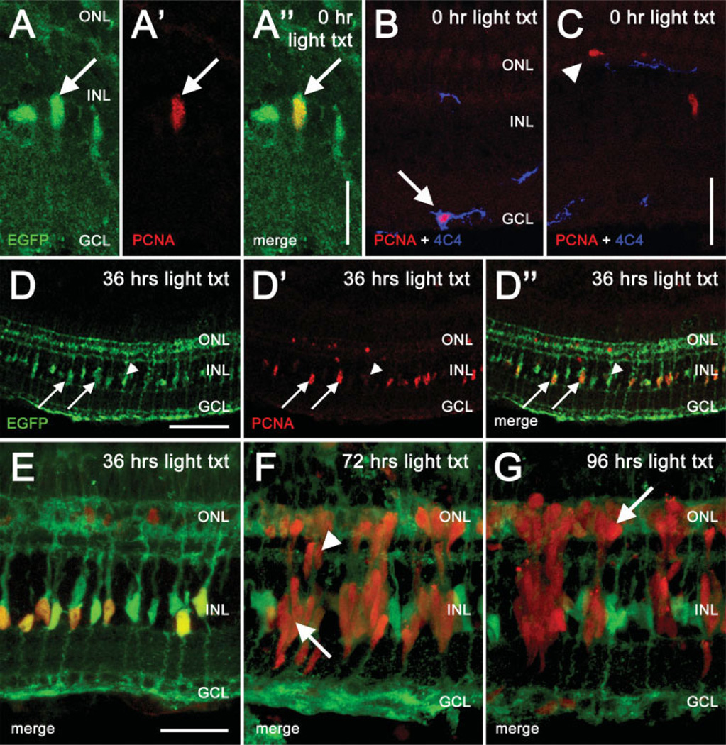
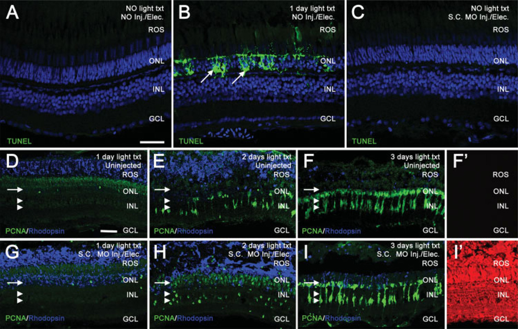
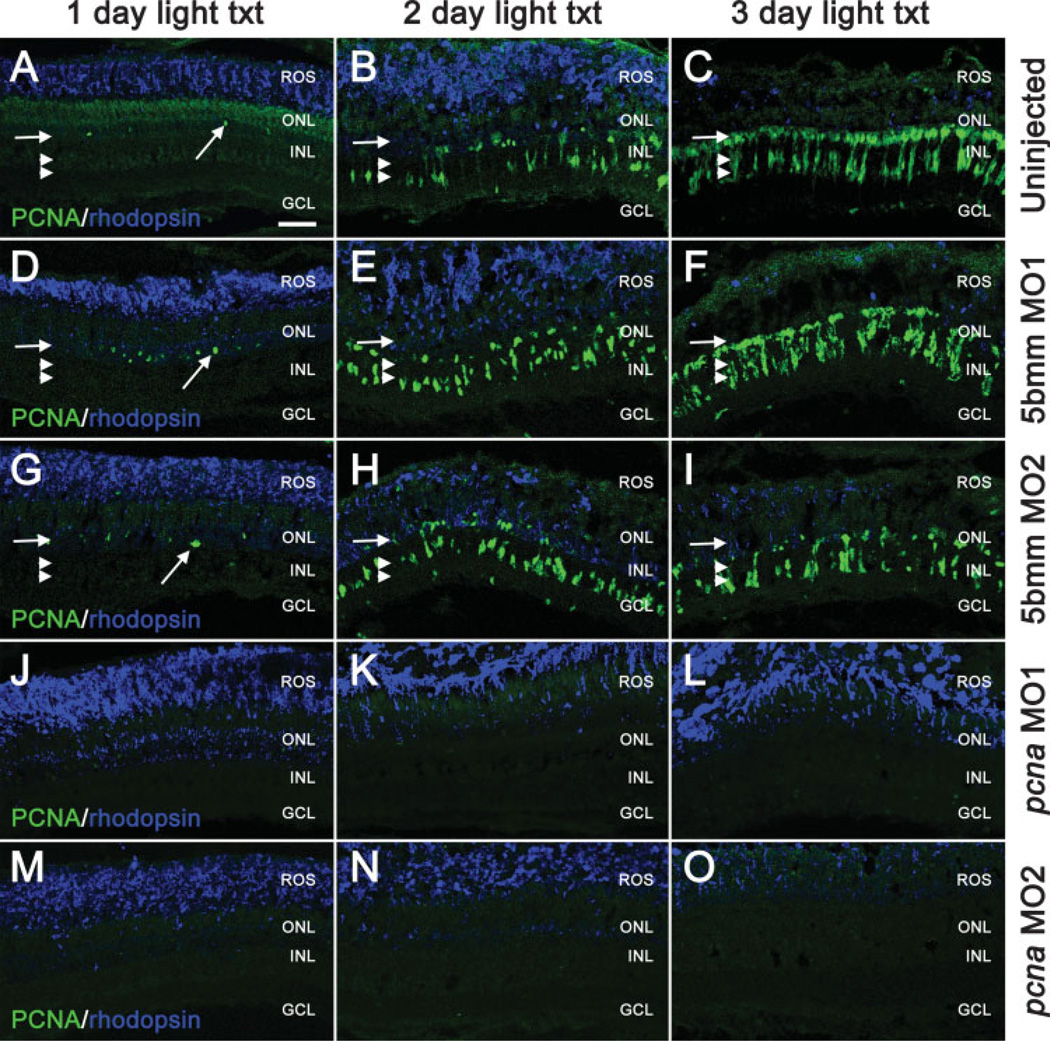
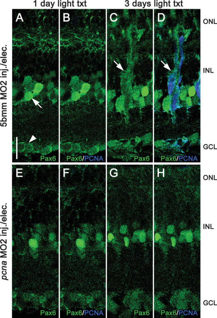
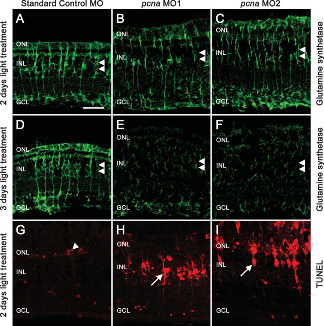
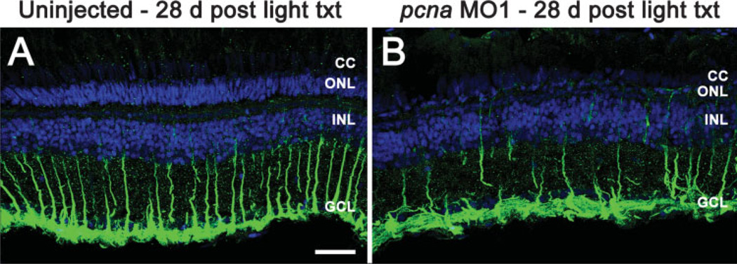
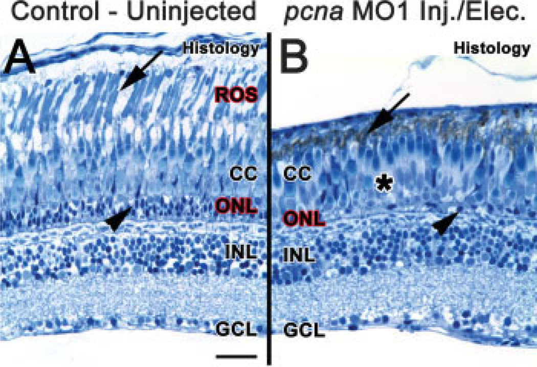
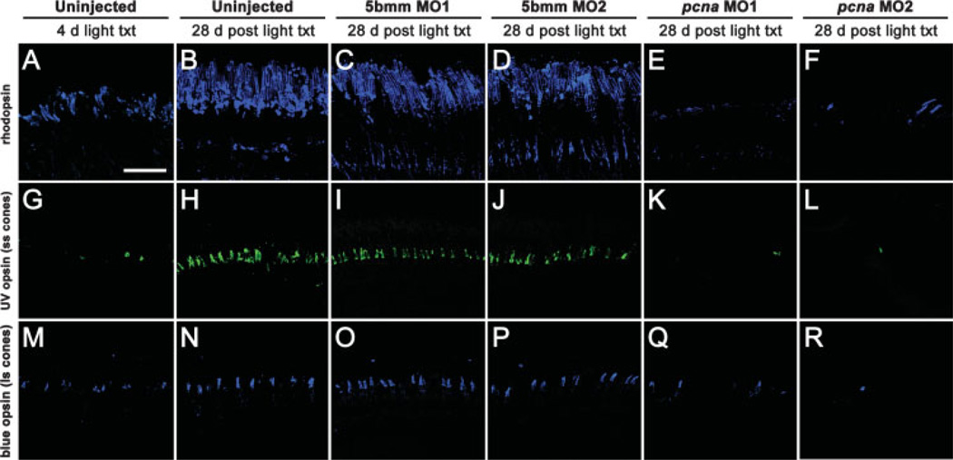
The adult zebrafish retina possesses a robust regenerative response. In the light-damaged retina, Müller glial cell divisions precede regeneration of rod and cone photoreceptors. Neuronal progenitors, which arise from the Müller glia, continue to divide and use the Müller glial cell processes to migrate to the outer nuclear layer and replace the lost photoreceptors. We tested the necessity of Müller glial cell division for photoreceptor regeneration. As knockdown tools were unavailable for use in the adult zebrafish retina, we developed a method to conditionally inhibit the expression of specific proteins by in vivo electroporation of morpholinos. We determined that two separate morpholinos targeted against the proliferating cell nuclear antigen (PCNA) mRNA reduced PCNA protein levels. Furthermore, injection and in vivo electroporation of PCNA morpholinos immediately prior to starting intense light exposure inhibited both Müller glial cell proliferation and neuronal progenitor marker Pax6 expression. PCNA knockdown additionally resulted in decreased expression of glutamine synthetase in Müller glia and Müller glial cell death, while amacrine and ganglion cells were unaffected. Finally, histological and immunological methods showed that long-term effects of PCNA knockdown resulted in decreased numbers of Müller glia and the failure to regenerate rod photoreceptors, short single cones, and long single cones. These data suggest that Müller glial cell division is necessary for proper photoreceptor regeneration in the light-damaged zebrafish retina and are consistent with the Müller glia serving as the source of neuronal progenitor cells in regenerating teleost retinas.


Figures

Figure 1
Figure 1
Localization of PCNA in the Tg(gfap:EGFP)nt11 albino zebrafish retina during light treatment. EGFP expression in the Müller glial cells in the Tg(gfap:EGFP)nt11 albino zebrafish was examined relative to PCNA immunolocalization during the intense light treatment. In the control (0 h, panels A and B), PCNA expression was rarely observed and was restricted to three cell types: EGFP-expressing Müller glial cells in the INL (panels A′ and A″, arrow), 4C4-positive microglia cells located near the GCL (panel B, arrow), and resident rod precursor cells in the ONL (panel C, arrowhead). At 36 h (panels D and E), PCNA expression is detected in large numbers of the INL nuclei and it colocalizes with the EGFP expression in the Müller glial cells (panels D–D″, arrows). Not all Müller glial cells are PCNA-positive (panels D–D″, arrowhead). At 72 h, the EGFP-expressing Müller glial cells no longer colabel with PCNA (panel F, arrow). However, PCNA-positive neuronal progenitors have begun to migrate from the INL to the ONL along the Muüller glial processes (panel F, arrowhead). At 96 h, large numbers of PCNA-positive progenitors populate the ONL (panel G, arrow). These results suggest the initial Müller cell proliferation event, which occurs subsequent to the light-induced photoreceptor damage, produces a population of neuronal progenitor cells that continue to proliferate. Scale bars: panel A″, 12 µm (panels A–A″); panel C (B–C), 25 µm; panel D, 100 µm (D–D″); panel E, 25 µm (E–G).
Figure 2
Figure 2
In vivo electroporation of morpholinos into the zebrafish retina does not cause cell death or adversely affect the regeneration response. TUNEL assays were used to compare cell death between uninjected, 24 h light-treated, and electroporated retinas (panels A–C). TO-PRO-3 (shown in blue) was used to stain nuclei. In uninjected, nonlight treated retinas, TUNEL labeling (shown in green) was not observed (panel A). In contrast, retinas light-treated for 24 h exhibited large amounts of TUNEL-positive nuclei in the ONL, representing dying photoreceptors (panel B, arrows). Non light-treated retinas electroporated with a Standard Control morpholino exhibited no detectable TUNEL labeling 24 h following electroporation (panel C), similar to uninjected control retinas. Uninjected retinas (panels D–F) and Standard Control electroporated retinas (panels G–I) were analyzed for PCNA (shown in green) and rhodopsin (shown in blue) immunolocalization. Panels D–F show the normal regenerative response at 1, 2, and 3 days of light, respectively. Rhodopsin decreases as rod photoreceptors degenerate. PCNA-positive, Müller glial derived, neuronal progenitors increase over the timecourse in both the INL (panels D–F, double arrowheads) and the ONL (panels D–F, arrow). Panel F′ shows background levels of red autofluorescence in uninjected retinas at 3 days of light treatment. Panels G–I show the normal regenerative response in retinas electroporated with a Standard Control morpholino. Similar to uninjected retinas, rhodopsin decreases and PCNA increases throughout the timecourse in both the INL (panels G–I, double arrowheads) and the ONL (panels G–I, arrow), indicating that electroporation does not adversely affect the regenerative response. Panel I′ shows a high level of lissamine-tagged Standard Control morpholino in all retinal layers at 3 days of light treatment. Scale bars: panel A, 25 µm (A–C); panel D, 25 µm (D–I′).
Figure 3
Figure 3
Injection and in vivo electroporation of pcna morpholino during constant light exposure knocks down PCNA levels. Dark-adapted adult albino zebrafish were injected and electroporated with either the pcna morpholino 1 (pcna MO1), pcna morpholino 2 (pcna MO2), 5-base mismatch morpholino 1 (5bmm MO1), or 5-base mismatch morpholino 2 (5bmm MO2) and exposed to constant intense light. Retinas were harvested at 1, 2, and 3 days into the light treatment and immunolabeled with PCNA (shown in green) and rhodopsin (shown in blue). In the uninjected control retinas (panels A–C) and the 5-base mismatch control retinas (panels D–I), light exposure results in a progressive accumulation of PCNA-positive cells within the INL and ONL and a loss of rhodopsin. Arrows and double arrowheads indicate the relative positions of the PCNA-positive cells in the ONL and in INL, respectively. In contrast, the pcna morphant retinas lack any PCNA-positive cells throughout the light exposure (Panels J–O). Scale bar: panel A, 25 µm (A–O).
Figure 4
Figure 4
pcna morphant retinas lack Pax6-positive neuronal progenitors. Pax6 immunolocalization (shown in green) was performed to test whether neuronal progenitors are generated in pcna morphant retinas. Pax6 labels retinal neuronal progenitors and mature amacrine and ganglion cells (Bernardos et al., 2007). At 1 day of light treatment, 5-base mismatch control retinas (Panels A and B) exhibited Pax6 immunolocalization in mature amacrine (panel A, arrow) and ganglion cells (panel A, arrowhead). At 3 days of light treatment, following Müller glial cell division, 5-base mismatch control retinas exhibited Pax6 expression in Muüller glial-derived neuronal progenitors (panel C, arrow), which colabeled with PCNA (panel D, shown in blue). Similar to control retinas, pcna morphant retinas (panels E–H) exhibited Pax6-positive mature amacrine and ganglion cells at 1 day (panels E and F) and 3 days (panels G and H) of light treatment. However, the large and diffusely stained Pax6-postitive neuronal precursors were never detected in pcna morphant retinas (panels G and H). Scale bar: panel A, 10 µm (A–I).
Figure 5
Figure 5
PCNA knockdown results in decreased glutamine synthetase levels, loss of Müller glial processes, and Müller glial cell death. Dark-adapted adult albino zebrafish were injected and electroporated with the Standard Control morpholino, pcna morpholino 1 (pcna MO1), or pcna morpholino 2 (pcna MO2), and placed in constant light. Retinas were harvested after either 2 or 3 days and processed to immunolocalize glutamine synthetase (green), which labels Müller glial cell nuclei housed in the INL (panels A–F, double arrowheads) and Müller glial cell processes extending to the ONL and GCL. The retinas injected and electroporated with the Standard Control morpholino exhibit robust glutamine synthetase expression in the Muüler cells (Panels A and D). The pcna morphant retinas exhibited a disruption of Müller glial cell morphology and a reduction of glutamine synthetase expression (Panels B, C, E, and F). To test for Muüller glia cell death, the retinas were immunolabeled with TUNEL (Panels G–I; red). The TUNEL-positive cells (arrowheads) in the control retina were primarily restricted to the outer nuclear layer (ONL), which is consistent with light-damaged photoreceptors (Panel G). However, pcna morphant retinas exhibited many TUNEL-positive cells in the INL (Panels H and I, arrows), which possess processes like Muüller glial cells. Scale bar: panel A, 25 µm (A–I).
Figure 6
Figure 6
PCNA knockdown results in a reduced number of Müller glial cells 28 days post light treatment. To determine the number of Müller glial cells, GFAP (green) was immunolocalized at 28 days following constant light treatment. Uninjected control retinas (panel A) exhibited Müller glial cell processes extending from the inner nuclear layer (INL) beyond the ganglion cell layer (GCL) to the inner limiting membrane. In contrast, pcna morphant retinas contained fewer numbers of GFAP-positive Müller glial cell processes (panel B). TO-PRO-3 counter-staining of retinas labeled cell nuclei in blue. Scale bar: panel A, 25 µm (A–B).
Figure 7
Figure 7
PCNA knockdown results in a reduction of ROS and ONL nuclei 28 days post light treatment. At 28 days following constant light treatment, histological sections of uninjected retinas (panel A) showed a regenerated outer nuclear layer (ONL, arrowhead), cone cell layer (CC) and rod outer segments (ROS, arrow). In contrast, histological sections from pcna morphant retinas (panel B) showed an absence of distinguishable ROS (arrow), very few nuclei present in the ONL (arrowhead), a disorganized cone cell layer, and large sections absent of long single cones (asterisk). Scale bar: panel A, 25 µm (A–B).
Figure 8
Figure 8
PCNA knockdown results in a reduction and disorganization of photoreceptors 28 days post light treatment. Rhodopsin immunolabeled rod outer segments, UV opsin immunolabeled short single cone outer segments (ss cones), and blue opsin immunolabeled long single cone outer segments (ls cones). Following 4 days of constant light treatment, uninjected control retinas exhibited reduced numbers and disorganized rod (panel A) and cone outer segments (panels G and M). However, by 28 days post light treatment, uninjected retinas regenerated both rod and cone outer segments (panels B, H, and N). 5-base mismatch control retinas, using either 5-base mismatch morpholino 1 (5bmm MO1) or 5-base mismatch morpholino 2 (5bmm MO2), showed a similar regeneration of rod and cone photoreceptors at 28 days post light treatment (panels C, D, I, J, O, and P). In contrast, pcna morphant retinas, using either pcna morpholino 1 (pcna MO1) or pcna morpholino 2 (pcna MO2), exhibited greatly reduced numbers of rod outer segments (panels E and F, arrows). In addition, cone photoreceptors were also present in greatly reduced numbers in pcna morphant retinas (panels K, L, Q, and R). Scale bar: panel A, 50 µm (A–R).

Similar articles

Cited by

References

    1. Bernardos RL, Barthel LK, Meyers JR, Raymond PA. Late-stage neuronal progenitors in the retina are radial Muller glia that function as retinal stem cells. J Neurosci. 2007;27:7028–7040. - PMC - PubMed
    1. Cameron DA, Gentile KL, Middleton FA, Yurco P. Gene expression profiles of intact and regenerating zebrafish retina. Mol Vis. 2005;11:775–791. - PubMed
    1. Choi HJ, Choi YH, Yee SB, Im E, Jung JH, Kim ND. Ircinin-1 induces cell cycle arrest and apoptosis in SK-MEL-2 human melanoma cells. Mol Carcinog. 2005;44:162–173. - PubMed
    1. Das AV, Mallya KB, Zhao X, Ahmad F, Bhattacharya S, Thoreson WB, Hegde GV, et al. Neural stem cell properties of Muller glia in the mammalian retina: Regulation by Notch and Wnt signaling. Dev Biol. 2006;299:283–302. - PubMed
    1. Detrich HW, Westerfield M, Zon LI. Methods in Cell Biology: The Zebrafish Biology. New York: Academic Press; 1999.

Publication types

MeSH terms

LinkOut - more resources