. 2007 Dec;5(12):e329.
doi: 10.1371/journal.pbio.0050329.

GATA3-driven Th2 responses inhibit TGF-beta1-induced FOXP3 expression and the formation of regulatory T cells

Affiliations

GATA3-driven Th2 responses inhibit TGF-beta1-induced FOXP3 expression and the formation of regulatory T cells

Pierre-Yves Mantel et al. PLoS Biol. 2007 Dec.

Abstract

Transcription factors act in concert to induce lineage commitment towards Th1, Th2, or T regulatory (Treg) cells, and their counter-regulatory mechanisms were shown to be critical for polarization between Th1 and Th2 phenotypes. FOXP3 is an essential transcription factor for natural, thymus-derived (nTreg) and inducible Treg (iTreg) commitment; however, the mechanisms regulating its expression are as yet unknown. We describe a mechanism controlling iTreg polarization, which is overruled by the Th2 differentiation pathway. We demonstrated that interleukin 4 (IL-4) present at the time of T cell priming inhibits FOXP3. This inhibitory mechanism was also confirmed in Th2 cells and in T cells of transgenic mice overexpressing GATA-3 in T cells, which are shown to be deficient in transforming growth factor (TGF)-beta-mediated FOXP3 induction. This inhibition is mediated by direct binding of GATA3 to the FOXP3 promoter, which represses its transactivation process. Therefore, this study provides a new understanding of tolerance development, controlled by a type 2 immune response. IL-4 treatment in mice reduces iTreg cell frequency, highlighting that therapeutic approaches that target IL-4 or GATA3 might provide new preventive strategies facilitating tolerance induction particularly in Th2-mediated diseases, such as allergy.


Conflict of interest statement

Competing interests. The authors have declared that no competing interests exist.

Figures

Figure 1
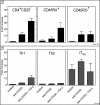
Figure 1. Th2 Cells Cannot Induce FOXP3 Expression
(A) Human T cells were activated with plate-bound anti-CD3/CD28 with or without TGF-β as indicated on the x-axis of (B). Cells were harvested after 5 days and FOXP3 mRNA was quantified by real-time PCR. Bars show the mean ± SD of 4 independent experiments. (B) In vitro differentiated Th1, Th2, or iTreg cells were activated with anti-CD3/CD28, TGF-β, or anti-IL-4 as indicated. The phenotype of these cells was confirmed by FACS and proliferation analysis (Figure S1). Bars show the mean ± SD of four independent experiments.
Figure 2
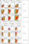
Figure 2. Th2 Cells Do Not Express FOXP3
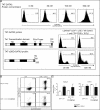
(A) Intracellular FACS analysis of FOXP3 expression in Th1, Th2, or iTreg-differentiated cells (two rounds, phenotype see Figure S1), rested or activated, with or without TGF-β. FOXP3 expression was measured after 5 d in culture. The dot blots are representative of three independent experiments. (B) Shows the same experimental setup, but naturally occurring Th2 cells were analyzed. Data are representative of three independent experiments.
Figure 3
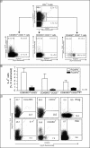
Figure 3. Th2- or IL-4–Producing Cells Lack FOXP3
(A) FACS analysis of intracellular FOXP3 and IL-4 expression following PMA/Ionomycin stimulation. CD4+ T cells were gated on the basis of CD45RO and CD25 surface expression (upper panel), and gated cells are shown below for the CD45RO+CD25 (A, left panel), the CD45RO+CD25+ (right panel), and the CD45ROCD25 subsets (central panel). A statistical analysis of eight independent donors after subtraction of the isotype control are shown in (B). The dotted gray line indicates the IC background level. The error bars show the error of the mean. (C) Similarly, a Th2 clone (BR8), CRTH2+ Th2 cells, IL-4-secreting cells, and memory T cells (CD45RO) were stained for FOXP3 and IL-4. Data are representative of three independent experiments.
Figure 4
Figure 4. FOXP3 Induction During the Differentiation Process
(A) Human CD4+CD45RA+ T cells were activated with plate-bound anti-CD3/CD28 in the presence of TGF-β (5 ng/ml) or IL-4 (25 ng/ml). The cells were harvested at different time points, and mRNA was quantified by real-time PCR for FOXP3 and GATA3 expression. Bars show the mean ± SD of three independent experiments. (B) Intracellular GATA3 and FOXP3 staining is shown after exposure of CD4+CD45RA+ T cells to differentiating conditions as in part A of the figure. Data are representative of three independent experiments.
Figure 5
Figure 5. Effect of IL-4 on FOXP3 Induction
(A) A statistical analysis was performed with six donors on day 5 (TGF-β (10 ng/ml) and with or without IL-4 (100 ng/ml)); Shown is the mean, and error bars indicated the SD of six donors. Statistical analysis was performed using the Dunnett test. Statistical significance is indicated by asterisks (*p ≤ 0.05, **p ≤ 0.01, Dunnett). (B) CD4+CD45RA+ cells were activated in the presence of a constant concentration of TGF-β (5 ng/ml) with an increasing concentration of IL-4, as indicated. Cells were harvested for mRNA quantification after 5 d. (C) CD4+CD45RA+ cells were stimulated in vitro with plate-bound anti-CD3/CD28, TGF-β (10ng/ml), and IL-4 (100 ng/ml) as indicated. After 1 h, cell lysates were prepared and analyzed by Western blot for phosphorylated SMAD2 and STAT6. Total STAT6 and GAPDH served as internal control. (D) Intracellular GATA3 and FOXP3 staining are shown after exposure of CD4+CD45RA+ T cells to IL-4 as described for panel B. Data are representative of three independent experiments.
Figure 6
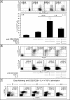
Figure 6. IL-4 Inhibits TGF-β–Mediated iTreg Commitment
CFSE-labeled CD4+CD45RA+ cells were activated with plate-bound anti-CD3/CD28, TGF-β, and IL-4, as indicated. After 5 d, cells were analyzed by flow cytometry (A) and results of six independent experiments are shown in the bar graph below (B). Statistical significance (one-way Anova, Newman-Keuls) is indicated by asterisks (**p ≤ 0.01, ***p ≤ 0.001). (C) Kinetic analysis of intracellular GATA3 and FOXP3 staining is shown in panel C following exposure of CD4+CD45RA+ T cells to anti-CD3/28, IL-4 and TGF-β. Data are representative of three independent experiments.
Figure 7
Figure 7. GATA3 Acts as a Negative Regulator of FOXP3 Expression
(A) Human naive CD4+CDRA+ T cells were transduced with 0, 20, 100, and 500 nM of TAT-GATA3 protein, and intracellular presence of GATA3 was analyzed using FACS following anti-CD3/CD28 activation of the cells. GFP-positive cells were gated and analyzed for intracellular FOXP3 expression following a 2-d incubation period (lower panel). Data are representative of four independent experiments. (B) CD4+CD25 T cells were isolated from D011.10 and D011.10xCD2-GATA3 mice and treated with OVA and TGF-β for 96 h. Surface CD4 and intracellular FOXP3 were measured by FACS. These data are representative of three independent experiments. (C) The cells treated as in (B) were harvested and mRNA was quantified by real-time PCR for SMAD7 and TGF-β expression. Bars show the mean ± SD of three independent experiments.
Figure 8
Figure 8. GATA3 Represses the Human FOXP3 Promoter
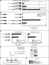
(A) Jurkat, Th2 cells, and human primary CD4 cells were transfected with an empty vector (pGL3 basic) or a vector containing the putative FOXP3 promoter region fused to the luciferase reporter gene. Bars show the mean ± SD of arbitrary light units normalized for renilla luciferase of four independent experiments; samples were measured in triplicates. (B) Naive CD4 T cells were transfected with the FOXP3 promoter reporter construct together with a GATA3 expression vector or an empty vector. Bars show the mean ± SD of three independent experiments. (C) Naive (left panel) or memory (right panel) CD4 T cells were transfected with wild-type or a GATA3 mutated 511-FOXP3 promoter reporter construct and activated with PMA and ionomycin. Bars show the mean ± SD of arbitrary light units normalized for renilla luciferase of eight independent experiments; samples were measured in triplicates. (D) Nuclear extracts were prepared from HEK cells transfected with GATA3 or an empty vector, (E) Th1, Th2, or iTreg cells and binding factors precipitated using biotinylated oligonucleotides. The oligonucleotides–transcription factor complexes were separated on a SDS-PAGE gel. The amounts of GATA3 protein in the precipitates were assessed by immunoblotting with anti-GATA3 mAb. Total nuclear extracts were also run as controls. Data are representative of three different experiments. (F) iTreg or Th2 cells were analyzed by ChIP for GATA3 binding to the FOXP3 promoter. The “input” represents PCR amplification of the total sample, which was not subjected to any precipitation. Results are representative of three independent experiments.

References

    1. Kurata H, Lee HJ, O'Garra A, Arai N. Ectopic expression of activated Stat6 induces the expression of Th2-specific cytokines and transcription factors in developing Th1 cells. Immunity. 1999;11:677–688. - PubMed
    1. Ouyang W, Ranganath SH, Weindel K, Bhattacharya D, Murphy TL, et al. Inhibition of Th1 development mediated by GATA-3 through an IL-4-independent mechanism. Immunity. 1998;9:745–755. - PubMed
    1. Zhu J, Yamane H, Cote-Sierra J, Guo L, Paul WE. GATA-3 promotes Th2 responses through three different mechanisms: induction of Th2 cytokine production, selective growth of Th2 cells and inhibition of Th1 cell-specific factors. Cell Res. 2006;16:3–10. - PubMed
    1. Usui T, Preiss JC, Kanno Y, Yao ZJ, Bream JH, et al. T-bet regulates Th1 responses through essential effects on GATA-3 function rather than on IFNG gene acetylation and transcription. J Exp Med. 2006;203:755–766. - PMC - PubMed
    1. Hwang ES, Szabo SJ, Schwartzberg PL, Glimcher LH. T helper cell fate specified by kinase-mediated interaction of T-bet with GATA-3. Science. 2005;307:430–433. - PubMed

Publication types

MeSH terms

LinkOut - more resources