. 2008 Apr;149(4):1757-72.
doi: 10.1210/en.2007-1205. Epub 2007 Dec 27.

Degradation of MEPE, DMP1, and release of SIBLING ASARM-peptides (minhibins): ASARM-peptide(s) are directly responsible for defective mineralization in HYP

Affiliations

Degradation of MEPE, DMP1, and release of SIBLING ASARM-peptides (minhibins): ASARM-peptide(s) are directly responsible for defective mineralization in HYP

Aline Martin et al. Endocrinology. 2008 Apr.

Abstract

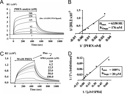
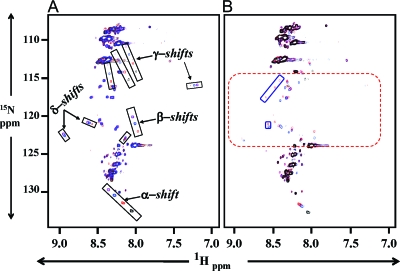
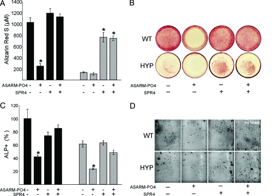
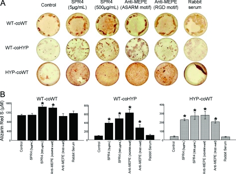
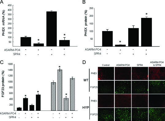
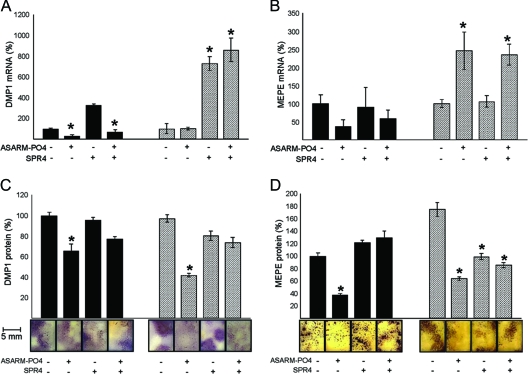
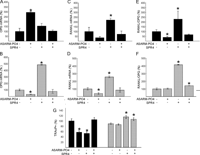
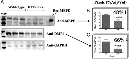
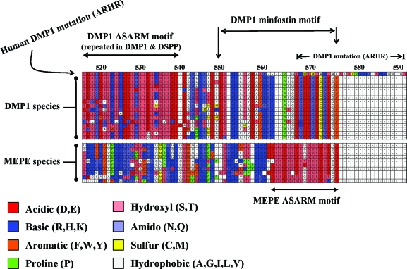
Mutations in PHEX (phosphate-regulating gene with homologies to endopeptidases on the X chromosome) and DMP1 (dentin matrix protein 1) result in X-linked hypophosphatemic rickets (HYP) and autosomal-recessive hypophosphatemic-rickets (ARHR), respectively. Specific binding of PHEX to matrix extracellular phosphoglycoprotein (MEPE) regulates the release of small protease-resistant MEPE peptides [acidic serine- and aspartate-rich MEPE-associated motif (ASARM) peptides]. ASARM peptides are potent inhibitors of mineralization (minhibins) that also occur in DMP1 [MEPE-related small integrin-binding ligand, N-linked glycoprotein (SIBLING) protein]. It is not known whether these peptides are directly responsible for the mineralization defect. We therefore used a bone marrow stromal cell (BMSC) coculture model, ASARM peptides, anti-ASARM antibodies, and a small synthetic PHEX peptide (SPR4; 4.2 kDa) to examine this. Surface plasmon resonance (SPR) and two-dimensional (1)H/(15)N nuclear magnetic resonance demonstrated specific binding of SPR4 peptide to ASARM peptide. When cultured individually for 21 d, HYP BMSCs displayed reduced mineralization compared with wild type (WT) (-87%, P < 0.05). When cocultured, both HYP and WT cells failed to mineralize. However, cocultures (HYP and WT) or monocultures of HYP BMSCs treated with SPR4 peptide or anti-ASARM neutralizing antibodies mineralized normally. WT BMSCs treated with ASARM peptide also failed to mineralize properly without SPR4 peptide or anti-ASARM neutralizing antibodies. ASARM peptide treatment decreased PHEX mRNA and protein (-80%, P < 0.05) and SPR4 peptide cotreatment reversed this by binding ASARM peptide. SPR4 peptide also reversed ASARM peptide-mediated changes in expression of key osteoclast and osteoblast differentiation genes. Western blots of HYP calvariae and BMSCs revealed massive degradation of both MEPE and DMP1 protein compared with the WT. We conclude that degradation of MEPE and DMP-1 and release of ASARM peptides are chiefly responsible for the HYP mineralization defect and changes in osteoblast-osteoclast differentiation.


Figures

Figure 1
Figure 1
SPR experiments reveal that sPHEX interacts directly with ASARM-PO4 peptide (A and B) and that this interaction is inhibited by the PHEX SPR4 Peptide (C and D). A, The indicated concentrations of sPHEX were flowed over a captured ASARM-PO4 surface during the period indicated by the bar, and the SPR response was recorded as a function of time in the displayed sensorgram. Notice that the binding is concentration dependent and saturable. B, Linear transformation of sensorgram in A with x- and y-coordinates plotted as inverse values for plasmon RU and sPHEX protein concentration 1/[nm PHEX]. C, The indicated concentrations of SPR4 peptide were flowed over the ASARM-PO4 surface with a constant concentration of sPHEX (50 nm). A concentration-dependent inhibition of the ASARM-PO4-PHEX interaction by SPR4 is observed. Random control SPR5 peptide had no effect. D, Linear transformation of sensorgram C with x- and y-coordinates plotted as inverse values for plasmon RU and SPR4 peptide concentration 1/[μm SPR4]. Each Biacore SPR experiment was independently performed three times for all data points (n = 3). For graph A (PHEX binding to ASARM-PO4), a KDapp of 176 ± 40 nm and a Bmax of 6338 ± 616 (sd) occurs. For graph B (inhibition of PHEX ASARM-PO4 binding by SPR4), a KIapp of 20 ± 11 μm was measured.
Figure 2
Figure 2
Two-dimensional 1H/15N NMR shows PHEX-SPR4 peptide (SPR4) binds directly to ASARM peptide. A, Overlaid NMR spectra of 15N isotopically labeled SPR4 peptide with no ASARM peptide (black), 1 molar equivalent ASARM-PO4 peptide (red), 3 molar equivalents ASARM-PO4 peptide (blue), and 6 molar equivalents (purple). The ASARM-PO4 peptide is nonisotopically labeled; thus, only the SPR4 peptide 1H/15N shifts are visible in the overlaid spectra. The major shifts are boxed and labeled as α, β, γ, and δ groups, respectively. A dose-dependent increase in shift occurs as illustrated by the black, red, blue, and purple peaks within each highlighted box. This demonstrates specific binding of SPR4 peptide to ASARM-PO4 peptide. B, The SPR4 PHEX peptide also binds nonphosphorylated ASARM peptide. An NMR spectrum of SPR4 in the presence of nonphosphorylated and phosphorylated ASARM peptide is shown in B. The overlaid spectra consist of SPR4 peptide without ASARM peptide (black spectrum), 1 molar equivalent of nonphosphorylated ASARM peptide (blue spectrum), and 1 molar equivalent of ASARM-PO4 peptide (red). The dashed red box highlights the region where many peaks appear as a result of the interaction between SPR4 and the peptide portion of ASARM. Solid red boxes enclose peaks unique to the phosphorylated peptide and reflect changes in SPR4 that are specific to interactions with the phosphate moiety at an equivalent 1:1 ratio. Increasing the concentration of ASARM peptide mimics the changes in the ppm positions observed with ASARM-PO4, but the same degree of change requires more ASARM to achieve compared with ASARM-PO4. The peaks in the blue boxes are considered unique because they were not observed at the highest concentration analyzed. The results indicate that peptide-peptide interactions between SPR4 and ASARM largely determine the bound conformation, whereas the phosphate moieties stabilize the interaction, increasing the binding strength.
Figure 3
Figure 3
Effects of incubation with ASARM-PO4 and/or SPR4 peptides on BMSCs from WT (black bars) or HYP (striped bars) in monoculture. A, ARS concentration; B, Representative photomicrographs of ARS staining; C, number of ALP-positive cells, represented as a percentage of total surface and relative to the untreated WT; D, representative photomicrographs of cells stained for ALP. The data represent the mean ± sem of triplicates from a representative experiment of three separate experiments that were not significantly different (Mann-Whitney U test). *, P < 0.05 vs. matched control cells.
Figure 4
Figure 4
Corrected mineralization after incubating BMSCs (cocultures; HYP and WT) with SPR4 peptide and anti-MEPE antibodies. WT cells were either cocultured with WT BMSCs (WT-coWT) or HYP (WT-coHYP), and HYP cells were cultured with WT cells (HYP-coWT). A, Representative photomicrographs of ARS staining; B, ARS concentration. The data represent the mean ± sem of triplicates from a representative experiment of three separate experiments that were not significantly different (Mann-Whitney U test). *, P < 0.05 vs. matched control.
Figure 5
Figure 5
BMSCs treated with ASARM-PO4 and SPR4 peptides show altered expression of PHEX and FGF-23. Incubation with ASARM-PO4 and/or SPR4 peptides on BMSCs from WT (black bars) or HYP (striped bars) are shown. A, Phex mRNA levels at d 3; B, number of cells immunostained for PHEX at d 14; C, number of cells immunostained for FGF23 at d 14. The data represent the mean ± sem of triplicates from a representative experiment of three separate experiments that were not significantly different (Mann-Whitney U test). Results are expressed as percentage of values for WT untreated cells. *, P < 0.05 vs. matched control cells. D, Representative photomicrographs of PHEX and FGF23 immunostaining (×20).
Figure 6
Figure 6
BMSCs treated with ASARM-PO4 and SPR4 peptides show altered expression of DMP1 and MEPE. Incubation with ASARM-PO4 and/or SPR4 peptides of BMSCs from WT (black bars) or HYP (striped bars). A, DMP1 mRNA levels at d 3; B, DMP1 immunostained surfaces at d 14 (bar graphs and representative photomicrographs); C, MEPE mRNA levels at d 3; D, MEPE-immunostained surfaces at d 14 (bar graphs and representative photomicrographs). The data represent the mean ± sem of triplicates from a representative experiment of three separate experiments that were not significantly different (Mann-Whitney U test). Results are expressed as percentage of values for WT untreated cells *, P < 0.05 vs. matched control cells.
Figure 7
Figure 7
BMSCs incubated with ASARM-PO4 peptide or SPR4 peptide showed altered osteoclastogenesis. Incubation with ASARM-PO4 and/or SPR4 peptides of BMSCs from WT (black bars) or HYP (striped bars) on osteoclastogenesis are shown. A and B, OPG mRNA levels at d 3 for WT (A) and HYP (B); C and D, RANKL mRNA levels at d 3 for WT (C) and HYP (D); E and F, RANKL/OPG mRNA ratio at d 3 for WT (E) and HYP (F); G, TRAcP-immunostained surfaces. The data represent the mean ± sem of triplicates from a representative experiment of three separate experiments that were not significantly different (Mann-Whitney U test). Results are expressed as percentage of values for WT untreated cells. *, P < 0.05 vs. matched control cells.
Figure 8
Figure 8
MEPE and DMP1 proteins are degraded in HYP mice calvariae. A, Chemiluminescent Western blots of HYP and WT calvariae screened with MEPE, DMP1, and GAPDH antibodies as indicated; B, adjusted pixel intensity of anti-MEPE blot; C, adjusted pixel intensity of anti-DMP1 blot. WT are left and HYP right on respective histograms. Calvariae from five male WT and HYP mice were used (n = 5; HYP and WT). Three separate representative calvariae are shown for WT and HYP.
Figure 9
Figure 9
Clustal alignment of MEPE and DMP1 COOH-terminal regions with ASARM motifs. Alignments are ordered from top to bottom as follows: for DMP1, mutated human DMP1 (ARHR), normal human, chimpanzee, macaque-primate, bush baby, cat, elephant, cow, rabbit, hedgehog, tenrec, squirrel, opossum, duck-billed platypus, guinea pig, mouse, and rat; for MEPE, human, chimpanzee, macaque-primate, rhesus-primate, bat, dog, cow, squirrel, rat, and mouse. There are two key regions, an ASARM motif region (DMP1 and MEPE) and a minfostin motif region (DMP1). In both DMP1 and MEPE, the ASARM motif is highlighted and consists of acidic aspartate (D) and glutamate residues (E) plus hydroxyl serines (S). Several serine residues are likely phosphorylated and play key physiological roles. Note within the MEPE ASARM motif that there are also two highly conserved hydrophobic glycine residues (G). One of these residues is penultimate and occurs in all species. The DMP1 ASARM motif is repeated throughout the C-terminal region of the molecule. A region at the very COOH-terminal tip of DMP1 is highly conserved. This region is labeled as a minfostin motif in the diagram. This is because a mutation extending and altering the primary sequence of this motif results in ARHR. Cathepsin B and K sites are also highly conserved in DMP1 and MEPE and would result in the release of free protease-resistant ASARM peptides. Amino acid residues are colored according to chemical type, and a key is included below the figure. Numbers above the clustal alignment are amino acid residues for DMP1.

Similar articles

Cited by

References

    1. Francis F, Hennig S, Korn B, Reinhardt R, de Jong D, Poustka A, Lehrach H, Rowe PS, Goulding JN, Summerfield T, Mountford RC, Read AP, Popowska E, Pronicka E, Davies KE, O’Riordan JL, Econs MJ, Nesbitt T, Drezner MK, Oudet C, Pannetier S, Hanauer A, Strom TM, Meindl A, Lorenz B, Cagnoli M, Mohnike KL, Murken J, Meitinger T 1995 A gene (PEX) with homologies to endopeptidases is mutated in patients with X-linked hypophosphatemic rickets. The HYP Consortium. Nat Genet 11:130–136
    1. Prie D, Beck L, Urena P, Friedlander G 2005 Recent findings in phosphate homeostasis. Curr Opin Nephrol Hypertens 14:318–324 - PubMed
    1. Rowe PSN 2004 The wrickkened-pathways of FGF23, MEPE and PHEX. Crit Rev Oral Biol Med 15:264–281 - PMC - PubMed
    1. Turner AJ, Tanzawa K 1997 Mammalian membrane metallopeptidases: NEP, ECE, KELL, and PEX. FASEB J 11:355–364 - PubMed
    1. Rowe PSN, Garrett IR, Schwarz PM, Carnes DL, Lafer EM, Mundy GR, Gutierrez GE 2005 Surface plasmon resonance (SPR) confirms MEPE binds to PHEX via the MEPE-ASARM motif: a model for impaired mineralization in X-linked rickets (HYP). Bone 36:33–46 - PMC - PubMed

Publication types

MeSH terms