. 2008 Mar;19(3):865-76.
doi: 10.1091/mbc.e07-06-0604. Epub 2007 Dec 27.

Isc1p plays a key role in hydrogen peroxide resistance and chronological lifespan through modulation of iron levels and apoptosis

Affiliations

Isc1p plays a key role in hydrogen peroxide resistance and chronological lifespan through modulation of iron levels and apoptosis

Teresa Almeida et al. Mol Biol Cell. 2008 Mar.

Abstract

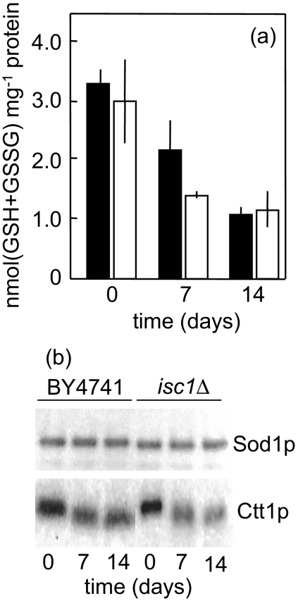
The inositolphosphosphingolipid phospholipase C (Isc1p) of Saccharomyces cerevisiae belongs to the family of neutral sphingomyelinases that generates the bioactive sphingolipid ceramide. In this work the role of Isc1p in oxidative stress resistance and chronological lifespan was investigated. Loss of Isc1p resulted in a higher sensitivity to hydrogen peroxide that was associated with an increase in oxidative stress markers, namely intracellular oxidation, protein carbonylation, and lipid peroxidation. Microarray analysis showed that Isc1p deficiency up-regulated the iron regulon leading to increased levels of iron, which is known to catalyze the production of the highly reactive hydroxyl radicals via the Fenton reaction. In agreement, iron chelation suppressed hydrogen peroxide sensitivity of isc1Delta mutants. Cells lacking Isc1p also displayed a shortened chronological lifespan associated with oxidative stress markers and aging of parental cells was correlated with a decrease in Isc1p activity. The analysis of DNA fragmentation and caspase-like activity showed that Isc1p deficiency increased apoptotic cell death associated with oxidative stress and aging. Furthermore, deletion of Yca1p metacaspase suppressed the oxidative stress sensitivity and premature aging phenotypes of isc1Delta mutants. These results indicate that Isc1p plays an important role in the regulation of cellular redox homeostasis, through modulation of iron levels, and of apoptosis.


Figures

Figure 1.
Figure 1.
Role of Isc1p in hydrogen peroxide resistance. (a) S. cerevisiae BY4741, isc1Δ, yca1Δ, and isc1Δyca1Δ mutant cells were grown in YPD medium to the exponential phase (OD600 = 0.6) and exposed to 1.5 mM H2O2 for 1 h. (b) S. cerevisiae BY4741 transformed with pYES2 or pYES2-ISC1 were grown in minimal medium with 2% galactose and treated with 10 mM H2O2 for 1 h. Controls (□) and treated cells (■) were plated on YPD 1.5% agar medium. Cell viability was expressed as the percentage of the colony-forming units (treated cells vs. nonstressed cells). Values are means ± SD of three independent experiments. **p < 0.01.
Figure 2.
Figure 2.
Effect of hydrogen peroxide on intracellular oxidation and oxidative damages. S. cerevisiae BY4741 (■) and isc1Δ mutant (□) cells were grown to exponential phase and treated with H2O2 for 60 min. (a) Intracellular oxidation. Cells were labeled with the molecular probe H2DCFDA and lysed as described in Materials and Methods. Data are expressed as the fluorescence ratio between 1.5 mM H2O2-treated and untreated cells. (b) Protein carbonylation. Proteins were derivatized with DNPH and slot-blotted into a PVDF membrane. Immunodetection was performed using an anti-DNP antibody, as described in Materials and Methods. Quantitative analysis of total protein carbonyl content was performed by densitometry using data taken from the same membrane. (c) Lipid peroxidation. Cellular extracts were prepared, and MDA was determined as described in Materials and Methods. Values are means ± SD of three independent experiments. **p < 0.01.
Figure 3.
Figure 3.
Functional categories of genes differentially expressed in isc1Δ mutant cells. S. cerevisiae BY4741 and isc1Δ mutant cells were grown to exponential phase, and RNA was isolated as described in Materials and Methods. Whole changes in the transcriptome were analyzed using Genefilters (Research Genetics). Genes up- or down-regulated in isc1Δ cells were sorted in groups according to Munich Information Center for Protein Sequences (MIPS) database. Down-regulated genes associated with protein biosynthesis (n = 87) were not included. See Supplementary Table S1 for all data.
Figure 4.
Figure 4.
Iron overload contributes to oxidative stress sensitivity of Isc1p-deficient cells. S. cerevisiae BY4741 (■) and isc1Δ (□) cells were grown on YPD media to exponential (log) or postdiauxic shift (PDS) phase. (a) Iron levels were quantified as described in Materials and Methods. (b) Cell lysates were separated by native PAGE, and MnSOD activity was detected in situ as described in Materials and Methods. (c) S. cerevisiae BY4741 CTH2-LacZ (■) and isc1Δ CTH2-LacZ (□) cells, expressing the consensus Aft1 binding sequences from CTH2 promoter fused to a LacZ reporter, were grown on minimal media to the log phase. β-galactosidase activity was measured in cells untreated or treated with BPS for 4 h. (d) H2O2 resistance. S. cerevisiae BY4741 and isc1Δ cells grown on YPD media to exponential phase and preincubated with 20 μM BPS for 4 h were treated with 1.5 mM H2O2 for 1 h. Cell viability was expressed as the percentage of the colony-forming units (H2O2 treated cells vs. nonstressed cells). (e) Intracellular oxidation. Cells pretreated with BPS were labeled with the molecular probe H2DCFDA, exposed to 1.5 mM H2O2 for 1 h, and lysed as described in Materials and Methods. Data are expressed as the fluorescence ratio between H2O2-treated and untreated cells. Values are means ± SD of three independent experiments. **p < 0.01.
Figure 5.
Figure 5.
Isc1p deficiency decreases chronological lifespan by a Yca1p-dependent mechanism. BY4741 (■), isc1Δ (□), yca1Δ (▴) and isc1Δ yca1Δ (○) cells were grown to the exponential (a) or postdiauxic (b) phase, washed twice with H2O, and kept in H2O at 26°C. The viability was determined by standard dilution plate counts and expressed as the percentage of the colony-forming units at time 0 h. Data are means ± SD of three independent experiments.
Figure 6.
Figure 6.
Intracellular oxidation and oxidative damages during chronological aging. Yeast cells were grown to postdiauxic phase, washed twice with H2O, and kept in H2O at 26°C. (a) Intracellular oxidation. S. cerevisiae BY4741 (■) and isc1Δ mutant (□) cells were labeled with the molecular probe H2DCFDA and lysed as described in Materials and Methods. Data are expressed in arbitrary units at indicated time. (b) Protein carbonylation. Protein extracts were prepared from S. cerevisiae BY4741 (■) and isc1Δ mutant (□) cells, derivatized with DNPH and slot-blotted into a PVDF membrane. Immunodetection was performed using an anti-DNP antibody, as described in Materials and Methods. Quantitative analysis of total protein carbonyl content was performed by densitometry using data taken from the same membrane. (c) Lipid peroxidation. Cellular extracts were prepared and MDA was determined as described in Materials and Methods. Values are means ± SD of three independent experiments. **p < 0.01.
Figure 7.
Figure 7.
Antioxidant defenses during chronological aging. S. cerevisiae BY4741 (■) and isc1Δ mutant (□) cells were grown to postdiauxic phase, washed twice with H2O, and kept in H2O at 26°C. (a) Total glutathione levels were determined as described in Materials and Methods. Values are means ± SD of three independent experiments. (b) Superoxide dismutase 1 (Sod1p) and Catalase T (Ctt1p) activity. Cells were lysed and proteins were separated by native PAGE. Enzyme activity was detected as described in Materials and Methods. A representative experiment is shown.
Figure 8.
Figure 8.
Isc1 specific activity and protein levels during chronological aging. (a) For Isc1p activity (□), S. cerevisiae BY4741 cells were grown in YPD medium to postdiauxic phase, washed twice with H2O, and kept in H2O at 26°C. Enzyme activity was determined as described in Materials and Methods and expressed as percentage of control (enzyme activity at time 0 h). For Isc1 protein levels (■), S. cerevisiae isc1Δ pYES2-ISC1-FLAG cells were grown in minimal medium with 2% galactose, washed twice with H2O, and kept in H2O. Proteins were isolated, separated by SDS-PAGE, and blotted into a membrane. Isc1p levels were determined by immunoblotting, using an anti-FLAG antibody, as described in Materials and Methods. Results were expressed as percentage of control (Isc1p level at time 0 h). Values are means ± SD. (b) A representative blot is shown (out of three independent experiments). A replica gel was stained with Coomassie blue (loading control; only a region of the gel is shown).
Figure 9.
Figure 9.
Apoptotic markers during oxidative stress. S. cerevisiae BY4741 and isc1Δ cells were grown to the exponential phase and treated with 1.5 mM H2O2 for 200 min. (a) DNA fragmentation was detected by the TUNEL assay as described in Materials and Methods. The nucleus was stained with PI. (b) Quantification of TUNEL-positive cells. Values are means ± SD of three independent experiments. **p < 0.01 (isc1Δ vs. BY4741). Bar, 5 μm.
Figure 10.
Figure 10.
Apoptotic markers during chronological aging. S. cerevisiae BY4741 (■), isc1Δ (□), and isc1Δyca1Δ (▨) cells were grown to the postdiauxic phase, washed twice with H2O, and kept in H2O at 26°C. (a) DNA fragmentation was detected by the TUNEL assay as described in Materials and Methods. The nucleus was stained with PI. (b) Quantification of TUNEL-positive cells. (c) Caspase-like or ASPase activity. Percentage of cells displaying caspase-like or ASPase activity was assessed by flow cytometric quantification of cells incubated with D2R, as described in Materials and Methods. A representative experiment for control (0 d) and 10-d old cells is shown in d. Values are means ± SD of three independent experiments. **p < 0.01 (isc1Δ vs. BY4741). Bar, 5 μm.
Figure 11.
Figure 11.
A model for the role of Isc1p in the regulation of iron uptake: implications during oxidative stress and cell aging. During oxidative stress induced by exogenous H2O2 or mitochondrial dysfunction in aged cells, iron catalyzes the conversion of H2O2 into hydroxyl radicals (Fenton reaction), leading to the accumulation of oxidative damages and to apoptotic cell death. Iron uptake genes are down-regulated by an Isc1p-dependent mediated pathway. In isc1Δ mutant cells, iron overload promotes the Fenton reaction and therefore increases apoptosis.

Similar articles

Cited by

References

    1. Abramova N. E., Cohen B. D., Sertil O., Kapoor R., Davies K. J., Lowry C. V. Regulatory mechanisms controlling expression of the DAN/TIR mannoprotein genes during anaerobic remodeling of the cell wall in Saccharomyces cerevisiae. Genetics. 2001;157:1169–1177. - PMC - PubMed
    1. Aerts A. M., Francois I. E., Bammens L., Cammue B. P., Smets B., Winderickx J., Accardo S., De Vos D. E., Thevissen K. Level of M(IP)2C sphingolipid affects plant defensin sensitivity, oxidative stress resistance and chronological life-span in yeast. FEBS Lett. 2006;580:1903–1907. - PubMed
    1. Almeida B., Sampaio-Marques B., Carvalho J., Silva M. T., Leao C., Rodrigues F., Ludovico P. An atypical active cell death process underlies the fungicidal activity of ciclopirox olamine against the yeast Saccharomyces cerevisiae. FEMS Yeast Res. 2007;7:404–412. - PubMed
    1. Ausubel F. A., Brent R., Kingston D., Moore D., Seidman J. G., Smith J. A., Struhl K. Current Protocols in Molecular Biology. New York: John Wiley and Sons; 1998.
    1. Belinha I., Amorim M. A., Rodrigues P., de Freitas V., Moradas-Ferreira P., Mateus N., Costa V. Quercetin increases oxidative stress resistance and longevity in Saccharomyces cerevisiae. J. Agric. Food Chem. 2007;55:2446–2451. - PubMed

Publication types

MeSH terms