. 2008 Mar;19(3):912-28.
doi: 10.1091/mbc.e07-06-0596. Epub 2007 Dec 27.

Caveolin-1 and -2 interact with connexin43 and regulate gap junctional intercellular communication in keratinocytes

Affiliations

Caveolin-1 and -2 interact with connexin43 and regulate gap junctional intercellular communication in keratinocytes

Stéphanie Langlois et al. Mol Biol Cell. 2008 Mar.

Abstract

Connexin43 (Cx43) has been reported to interact with caveolin (Cav)-1, but the role of this association and whether other members of the caveolin family bind Cx43 had yet to be established. In this study, we show that Cx43 coimmunoprecipitates and colocalizes with Cav-1 and Cav-2 in rat epidermal keratinocytes. The colocalization of Cx43 with Cav-1 was confirmed in keratinocytes from human epidermis in vivo. Our mutation and Far Western analyses revealed that the C-terminal tail of Cx43 is required for its association with Cavs and that the Cx43/Cav-1 interaction is direct. Our results indicate that newly synthesized Cx43 interacts with Cavs in the Golgi apparatus and that the Cx43/Cavs complex also exists at the plasma membrane in lipid rafts. Using overexpression and small interfering RNA approaches, we demonstrated that caveolins regulate gap junctional intercellular communication (GJIC) and that the presence of Cx43 in lipid raft domains may contribute to the mechanism modulating GJIC. Our results suggest that the Cx43/Cavs association occurs during exocytic transport, and they clearly indicate that caveolin regulates GJIC.


Figures
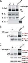
Figure 1.
Figure 1.
Cx43 coimmunoprecipitates with Cav-1 and Cav-2 in REKs. (A) WT REKs were infected with a vector encoding Cx43-GFP, or a control empty vector (V). Western blot showed that REKs express endogenous Cx43, Cav-1, and Cav-2, but not Cav-3 (positive control: muscle). (B) Both Cx43 (P0 and P forms) and Cx43-GFP were found in Cav-1 immunoprecipitates. (C) Cx43 (P0 and P forms) and Cx43-GFP, and Cav-1, coimmunoprecipitated with Cav-2. IP, immunoprecipitation; IB, immunoblotting.
Figure 2.
Figure 2.
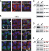
Cx43 mainly colocalizes with Cav-1 and -2 in intracellular compartments of REKs. (A) REKs or REKs overexpressing Cx43-GFP were fixed with formaldehyde and labeled with Cav-1 and -2. Confocal imaging of REKs overexpressing Cx43-GFP (A, green, top and middle) revealed Cx43 in gap junctions plaques at the cell surface (arrowheads) and in intracellular localization in a Golgi-like pattern (arrows). Cav-1 (red, top, arrow) and -2 (red, middle, arrows) were mainly detected in intracellular compartments in a Golgi-like pattern. Overlay images suggest that Cx43-GFP is colocalized (yellow, top and middle, arrows) with Cav-1 and -2 in the Golgi. Colocalization (yellow) of Cav-1 (green, bottom) with Cav-2 (red, bottom) was observed in intracellular compartments of REKs (arrow). (B) REKs or REKs overexpressing Cx43-GFP were fixed with methanol/acetone and labeled with Cav-1 and -2. Cx43-GFP (B, green, top and middle) was found in intracellular compartments (arrow) and at the plasma membrane (arrowheads), whereas Cav-1 (B, red, top) and -2 (B, red, middle) were mainly detected at the plasma membrane. Overlay images revealed that the majority of Cx43-GFP was not colocalized with Cav-1 and -2 at the plasma membrane. However, few Cx43-GFP plaques were colocalized with Cavs (yellow, arrowheads) at the plasma membrane. Colocalization (yellow, arrowheads) of Cav-1 (red, bottom) with Cav-2 (green, bottom) was observed at the plasma membrane of REKs. Blue, nuclei. Bars, 10 μm.
Figure 3.
Figure 3.
Cx43 colocalizes with Cav-1 in keratinocytes from human skin epidermis. Confocal imaging of human epidermis indicates that Cx43 (green, top) was detected in the vital layers (between dashed line and dotted line) and absent in the cornified layer (between dashed lines). Cav-1 staining (red, middle) was pronounced in basal and spinosum layers (between dashed line and dotted line), and it was absent in the cornified layer (between dashed lines). Overlay images suggest that Cx43 is colocalized with Cav-1 in the vital layers of human epidermis (yellow, arrowheads, bottom). Blue, nuclei. Bar, 10 μm.
Figure 4.
Figure 4.
Cx43 colocalizes with Cavs in the Golgi apparatus. (A) REKs overexpressing Cx43-GFP were labeled with TGN38 (red, left), and REKs were colabeled for Cav-1 (red, middle) and TGN38 (green) or with Cav-2 (red, right) and GPP130 (green). Overlay images show that Cx43-GFP, Cav-1, and Cav-2 were colocalized (yellow) with the resident Golgi proteins. REKs or REKs overexpressing Cx43-GFP were treated with BFA for 6 h before their fixation with formaldehyde and labeling with Cav-1 and -2. (B) Treatment with BFA resulted in a loss of most Cx43-GFP (green, top and middle) at gap junction plaques (arrowheads), compared with untreated cells (green, inset, top). Golgi localization of Cx43-GFP (green, top and middle), Cav-1 and -2 (red, top and middle) was lost in BFA-treated cells. Colocalization of Cav-1 (green, bottom) with -2 (red, bottom) in the Golgi was lost in REKs. Blue, nuclei. Bar, 10 μm. (C) Lysates from untreated and BFA-treated REKs were subjected to Western blot. BFA treatment dramatically reduced the P species of Cx43. β-Actin was used as a loading control. Treatment with BFA reduced the amount of Cx43 found in Cav-1 (D) and Cav-2 (E) immunoprecipitates, without affecting the amount of Cav-1 coimmunoprecipitating with Cav-2 (E). IP, immunoprecipitation; IB, immunoblotting.
Figure 5.
Figure 5.
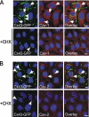
Newly synthesized Cx43 and Cavs colocalize in the Golgi apparatus. REKs overexpressing Cx43-GFP were treated with CHX for 6 h before their fixation with formaldehyde and labeling with Cav-1 (A) and -2 (B). Treatment with CHX resulted in a loss of Cx43-GFP in gap junction plaques (A and B, green, arrowheads). It also resulted in a loss of the intracellular pools of Cx43-GFP (A and B, green, arrows), Cav-1 (A, red, arrows), and Cav-2 (B, red, arrows) in the Golgi apparatus. Blue, nuclei. Bars, 10 μm.
Figure 6.
Figure 6.
A population of Cx43 interacts with Cavs at the plasma membrane in lipid rafts. WT REKs or REKs infected with Cx43-GFP, or the control empty vector (V), were treated with 10 mM MβC for 1 h. Untreated (A) and treated cells (B) were lysed in 1% Triton X-100 to obtain insoluble (I) and soluble (S) fractions, which were analyzed by Western blotting. EEA1 was used as a fractionation control. Note that Cx43, Cav-1, and Cav-2 were mainly found in the Triton-insoluble fractions (I), but that they were translocated to Triton-soluble fractions (S) after MβC treatment. (C) WT REKs were subjected to sucrose density gradient centrifugation. Twelve fractions were collected and analyzed by Western blot. Note that the fractions 4–6 (raft) are enriched in the P forms of Cx43, whereas the fractions 8–12 (nonraft) are enriched in the P0 form. (D) Fractions 4–6 (R) and fractions 8–12 (NR) were pooled and equal amounts of proteins were subjected to Western blotting. REKs were biotinylated (+), or subjected to mock biotinylation (−), and then subjected to coimmunoprecipitations by using Cav-1 and Cav-2 antibodies, as presented in the scheme (E). SDS extracts were collected, and an aliquot (2%) was analyzed by Western blot (F and G, lanes 1 and 2); the remainder of the lysates underwent neutravidin precipitation before SDS-PAGE (F and G, lanes 3 and 4). Cx43, Cav-1, and Cav-2 were recovered when REKs were treated with biotin (F and G, lane 4), but not when biotin was omitted (F and G, lane 3). The Cx43/Cav-1 and Cx43/Cav-2 complexes were recovered by anti-Cav-1 (F, lanes 5 and 6) and anti-Cav-2 IP (G, lanes 5 and 6), eluted from the antibodies, and aliquots (10%) were analyzed directly. The remainder of the immunoprecipitates underwent neutravidin precipitations followed by SDS-PAGE (F and G, lanes 7 and 8). Biotinylated Cx43 was coimmunoprecipitated with Cav-1 (F, top, lane 8) and Cav-2 (G, top, lane 8), and biotinylated Cav-1 was also present in Cav-2 immunoprecipitates (G, middle, lane 8). Lysates from untreated and MβC-treated WT REKs were subjected to Western blot (H) and IPs (I and J). Treatment with MβC reduced the amount of Cx43 found in Cav-1 (I) and Cav-2 (J) immunoprecipitates. IP, immunoprecipitation; IB, immunoblotting.
Figure 7.
Figure 7.
The C-terminal tail of Cx43 is required for its association with Cavs. (A) A predicted topology of GFP-tagged Cx43 depicting four transmembrane domains (M1–M4), two extracellular loops (EL-1 and EL-2), a cytoplasmic loop (CL), and an N terminus and a C terminus (CT) are shown. Similar to WT Cx43, the C-terminal domain of all the Cx43 mutants used in this study were fused to GFP. The G21R, G138R, and G60S are missense mutations present in the first transmembrane domain, the cytoplasmic loop, and the first extracellular loop, respectively. The fs260 mutant represents a frameshift where residues 260–305 are aberrant amino acid residues and in which the C-terminal domain is truncated. The Δ244 mutant is truncated after residue 243 in its C terminus. (B) REKs overexpressing Cx43-GFP or GFP-tagged Cx43 mutants were subjected to Western blot. β-Actin was used as a loading control. (C) Triton solubility of GFP-tagged Cx43 constructs in REKs overexpressing GFP-tagged WT Cx43 and mutants. Insoluble (I) and soluble (S) fractions were analyzed by Western blotting. EEA1 was used as a fractionation control. Amount of Cx43-GFP and GFP-tagged Cx43 mutants in each fraction was quantified, and data are expressed as percentage of Cx43 in Triton-soluble and -insoluble fractions (n = 3, *p < 0.05). Cx43-GFP and the G21R, G138R, and G60S mutants coimmunoprecipitated with Cav-1 (D) and Cav-2 (E), but not the Δ244 and fs260 mutants. An antibody directed against the N-terminal domain (NT1) of Cx43 was used to detect all the Cx43 constructs. If present in the Cavs immunoprecipitates, the Δ244 mutant would have been detected as a band just below the IgG heavy chain band, whereas the fs260 mutant would have been detected as a slightly higher band than the IgG band. IP, immunoprecipitation; IB, immunoblotting.
Figure 8.
Figure 8.
Cx43 can bind directly to Cav-1. Cav-1 was immunoprecipitated from two sets of Cav-1-transfected and untransfected 293T cells, and the immunoprecipitates were resolved by SDS-PAGE and transferred to nitrocellulose. The blot was probed with Cx43CT (Far Western [FW]), which bound to an ∼21- to 25-kDa doublet only in immunoprecipitates from Cav-1–expressing cells (red, top). Bound Cx43CT was detected using antibodies against the C-terminal domain of Cx43. The blot was reprobed with anti-Cav-1 antibody (green, middle) and overlay images revealed that the Cx43CT bound to the same band detected with anti-Cav-1 antibodies (yellow, bottom).
Figure 9.
Figure 9.
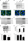
RNAi knockdown of Cav-1 and -2 expression reduces the pool of Cx43 present in lipid rafts and GJIC in REKs. WT REKs were infected with a retrovirus encoding a control (Ctl) RNAi, or an RNAi targeting Cav-1. (A) Western blot show that Cav-1 and Cav-2 expression was reduced by ∼80 and ∼40%, respectively, whereas Cx43 level remained unchanged in cells treated with Cav-1 siRNA. β-actin was used as a loading control. (B) Triton solubility of Cx43, Cav-1, and Cav-2. EEA1 was used as a fractionation control. S, soluble fractions; I, insoluble fractions. (C) Labeling of endogenous Cx43 by using an anti-Cx43 antibody obtained from Sigma (green) revealed its location within intracellular compartments (arrows) and at the plasma membrane (arrowheads) in both Ctl and Cavs-knockdown REKs. (D) Labeling of endogenous Cx43 by using the P4G9 anti-Cx43 antibody (green) mainly detected Cx43 at the plasma membrane (arrowheads) in both Ctl and Cavs-knockdown REKs. (E) Ctl and (F) Cavs-knockdown REKs were subjected to sucrose density gradient centrifugation. Twelve fractions were collected and analyzed by Western blot. (G) Fractions 4–6 (R) and fractions 8–12 (NR) were pooled, and equal amounts of proteins were submitted to Western blotting. (H) Percentage of Cx43 present in raft (fractions 4–6) and in nonraft fractions (8–12) were quantified when equal amounts of proteins were analyzed by Western blotting (n = 4, *p < 0.05 compared with the equivalent fractions in control cells). (I) Untreated and MβC-treated WT REKs were microinjected with Lucifer yellow. Data were expressed as percentage of microinjected cells that passed dye (n represents the number of microinjected cells from three independent experiments, *p < 0.05). (J) WT, Ctl REKs, or REKs that were infected with Cav-1 siRNA were microinjected with Lucifer yellow. Data are expressed as percentage of microinjected cells that passed dye (n represents the number of microinjected cells from three independent experiments, *p < 0.001 compared with WT and Ctl REKs).
Figure 10.
Figure 10.
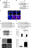
Overexpression of Cav-1 in 293T cells induces the translocation of Cx43 into lipid rafts and increases GJIC. 293T cells were transfected with Cav-1 or the empty vector (V), subjected to Western blotting, and probed for Cx43 and Cav-1 (A). β-Actin was used as a loading control. (B) Triton solubility of Cx43 and Cav-1. EEA1 was used as a fractionation control. S, soluble fraction; I, insoluble fractions. (C) 293T cells were transfected with Cav-1 or control empty vector (V) and labeled for Cx43 (P4G9 anti-Cx43 antibody, green) and Cav-1 (red). Confocal imaging of control 293T cells (top) revealed Cx43 at the cell surface (arrowheads) in the absence of Cav-1. No obvious difference was observed in Cx43 localization at the plasma membrane (arrowheads) in Cav-1–expressing 293T cells, compared with control cells (V). Overlay images suggest that a population of Cx43 at the plasma membrane colocalizes with Cav-1 (yellow, bottom, arrowheads). (D) Control empty vector (V) and (E) Cav-1–transfected 293T cells were subjected to sucrose density gradient centrifugation. Twelve fractions were collected and analyzed by Western blot. (F) Fractions 4–6 (R) and fractions 8–12 (NR) were pooled, and equal amounts of proteins were submitted to Western blotting. (G) Percentage of Cx43 present in raft (fractions 4–6) and in nonraft fractions (8–12) were quantified when equal amounts of proteins were analyzed by Western blotting (n = 3, *p < 0.05 compared with the equivalent fractions in cells transfected with the empty vector). (H) 293T cells were transfected with Cav-1-mRFP or control empty vector (V) and microinjected with Lucifer yellow. (I) Data are expressed as percentage of microinjected transfected cells that passed dye (n represents the number of microinjected cells from three independent experiments, *p < 0.05). Blue, nuclei. Bars, 10 μm.

Similar articles

Cited by

References

    1. Balijepalli R. C., Foell J. D., Hall D. D., Hell J. W., Kamp T. J. Localization of cardiac L-type Ca(2+) channels to a caveolar macromolecular signaling complex is required for beta(2)-adrenergic regulation. Proc. Natl. Acad. Sci. USA. 2006;103:7500–7505. - PMC - PubMed
    1. Barbuti A., Gravante B., Riolfo M., Milanesi R., Terragni B., DiFrancesco D. Localization of pacemaker channels in lipid rafts regulates channel kinetics. Circ. Res. 2004;94:1325–1331. - PubMed
    1. Barth K., Gentsch M., Blasche R., Pfuller A., Parshyna I., Koslowski R., Barth G., Kasper M. Distribution of caveolin-1 and connexin43 in normal and injured alveolar epithelial R3/1 cells. Histochem. Cell Biol. 2005;123:239–247. - PubMed
    1. Brainard A. M., Miller A. J., Martens J. R., England S. K. Maxi-K channels localize to caveolae in human myometrium: a role for an actin-channel-caveolin complex in the regulation of myometrial smooth muscle K+ current. Am. J. Physiol. 2005;289:C49–C57. - PubMed
    1. Brazer S. C., Singh B. B., Liu X., Swaim W., Ambudkar I. S. Caveolin-1 contributes to assembly of store-operated Ca2+ influx channels by regulating plasma membrane localization of TRPC1. J. Biol. Chem. 2003;278:27208–27215. - PMC - PubMed

Publication types

MeSH terms

LinkOut - more resources