. 2008 Feb;146(2):539-53.
doi: 10.1104/pp.107.112995. Epub 2007 Dec 27.

Specificity of RCN1-mediated protein phosphatase 2A regulation in meristem organization and stress response in roots

Affiliations

Specificity of RCN1-mediated protein phosphatase 2A regulation in meristem organization and stress response in roots

Joshua J Blakeslee et al. Plant Physiol. 2008 Feb.

Abstract

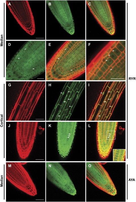
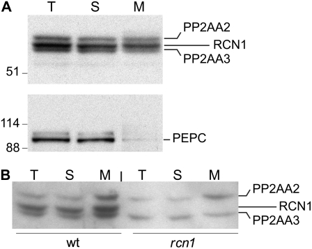
Protein dephosphorylation by the serine/threonine protein phosphatase 2A (PP2A) modulates a broad array of cellular functions. PP2A normally acts as a heterotrimeric holoenzyme complex comprising a catalytic subunit bound by regulatory A and B subunits. Characterization of the regulatory A subunit isoforms (ROOTS CURL IN NAPHTHYLPHTHALAMIC ACID1 [RCN1], PP2AA2, and PP2AA3) of Arabidopsis thaliana PP2A has shown that RCN1 plays a primary role in controlling root and hypocotyl PP2A activity in seedlings. Here we show that hypocotyl and root growth exhibit different requirements for RCN1-mediated regulation of PP2A activity. Roots of rcn1 mutant seedlings exhibit characteristic abnormalities in cell division patterns at the root apical meristem, as well as reduced growth under ionic, osmotic, and oxidative stress conditions. We constructed chimeric A subunit genes and found that restoration of normal root tip development in rcn1 plants requires both regulatory and coding sequences of RCN1, whereas the hypocotyl elongation defect of rcn1 plants can be complemented by either RCN1 or PP2AA3 transgenes. Furthermore, the RCN1 and PP2AA3 proteins exhibit ubiquitous subcellular localization patterns in seedlings and both associate with membrane compartments. Together, these results show that RCN1-containing PP2A has unique functions that cannot be attributed to isoform-specific expression and localization patterns. Postembryonic RCN1 function is required to maintain normal auxin distribution and stem cell function at the root apex. Our data show that RCN1-regulated phosphatase activity plays a unique role in regulating postembryonic root development and stress response.


Figures

Figure 1.
Figure 1.
RCN-YFP fusions supply regulatory A subunit function in yeast. RCN1-YFP and YFP-RCN1 fusions were expressed under control of the constitutive alcohol dehydrogenase promoter (Ammerer, 1983) in tpd3-1 yeast cells (van Zyl et al., 1992). A, Transformants carrying RCN-YFP fusions, an RCN1 construct, or the empty vector were streaked on duplicate YPD plates and incubated at 30°C or 37°C. The diagram at left indicates the construct carried by cells in the corresponding sectors on both plates. B, Cells carrying a native RCN1 construct, YFP-RCN1, RCN1-YFP, or a SUP35:GFP fusion (Satpute-Krishnan and Serio, 2005) were grown to early log phase and mounted for differential interference contrast (bottom panels) or fluorescence (top panels) microscopy. C, Protein extracts of cells carrying the constructs indicated were subjected to SDS-PAGE and immunoblotting, using anti-GFP antibodies to detect the fusion proteins. The positions of the RCN1-YFP (black arrows) and SUP35:GFP (asterisk) proteins are indicated at right. D, Cells carrying the constructs indicated were grown in liquid culture and 10-fold serial dilutions were spotted on plates containing YPD medium or YPD plus 600 mm NaCl. YPD plates were incubated at 30°C or 37°C and YPD NaCl plates were incubated at 30°C.
Figure 2.
Figure 2.
RCN1 and PP2AA3 fusions to YFP rescue hypocotyl elongation in the rcn1 mutant. A, The YFP coding sequence was fused at the N- or C-terminal end of the RCN1 gene (see “Materials and Methods”) to generate in-frame fusions within the genomic RCN1 sequence. In the RCNpro:YFP-PP2AA3 (RYA) construct, the RCN1 coding sequence was replaced with the PP2AA3 cDNA. The PP2AA3pro:YFP-PP2AA3 (AYA) construct was derived from RYA by substituting upstream sequences from PP2AA3 for the RCN1 regulatory region. All constructs were transformed into the rcn1-1 mutant. B, Transformant families segregating for a single YFP-RCN1 or RCN1-YFP transgene were scored for hypocotyl elongation and for YFP fluorescence after 5 d of growth in the dark. Values shown represent the average hypocotyl lengths for each class (YFP+ or YFP) relative to the wild-type control (Ws). Error bars indicate sd; n > 25 for all controls; n > 45 for all segregating families. C, Homozygous transformant families carrying the transgene constructs indicated were scored for hypocotyl elongation after 5 d growth in the dark. Values shown represent the average hypocotyl lengths for each family relative to the wild-type control (Ws). Error bars indicate sd; n > 25 for all controls; n > 45 for all families carrying YFP fusion constructs. rcn1 PZP is an rcn1 transgenic line carrying the empty vector. Lines rcn1 RYA-32 and rcn1 AYA-6 each carry two copies of the fusion T-DNA, whereas line rcn1 AYA16 carries three or more copies. All other lines carry the transgene construct in single copy. Levels of statistical significance as determined by Student's t test: *, P > 0.15 versus rcn1 and P < 10−6 versus Ws; **, P < 10−30 versus rcn1 and P < 0.05 versus Ws; ^, P < 10−30 versus rcn1 and P > 0.8 versus Ws.
Figure 3.
Figure 3.
Isoform specificity of RCN1 function in root tip organization. Median longitudinal sections of propidium iodide-stained 4-dpg root tips were captured using confocal laser microscopy, revealing normal meristem organization in wild-type (Ws) roots and highlighting the disorganization of columella and initial cells in rcn1 root tips. Examples of moderate (rcn1 moderate) and severe (rcn1 severe) rcn1 disorganization phenotypes are shown. The YFP-RCN1 fusion restores wild-type morphology in rcn1 plants carrying the RYR construct, whereas the YFP-PP2AA3 fusions carried in RYA and AYA transformants fail to fully rescue the rcn1 phenotype. Brackets indicate the region of the QC; asterisks indicate cells representative of clearly defined columellar cell files. Scale bars, 25 μm.
Figure 4.
Figure 4.
Postembryonic PP2A function maintains normal root tip development. Median longitudinal sections of propidium iodide-stained 4-dpg root tips were captured using confocal microscopy. One wild-type (A) and two representative rcn1-6 mutant (B and C) root tips grown in the absence of cantharidin plus one wild-type root tip grown in the presence of 10 μm cantharidin (D) are shown. Cantharidin-treated wild-type roots show abnormalities around the QC (brackets) and reduced columellar cell file numbers matching those of rcn1-6 mutant roots. Asterisks indicate cells representative of clearly defined columellar cell files. The AGL42-GFP reporter is expressed in QC cells in 4-dpg seedling roots (E), but its expression is severely reduced in seedlings grown in the presence of 10 μm cantharidin (F and G). Each FM4-64-stained seedling was scanned sequentially for GFP fluorescence (upper row) and FM4-64 fluorescence (overlay shown in lower row). Under the imaging conditions used, a low level of background fluorescence is detected in wild-type Col root tips grown in the presence of cantharidin (H). DR5-GUS reporter activity is reduced in roots of seedlings carrying the rcn1-1 mutation in both the Col (I versus J) and Ws (K versus L) genetic backgrounds (see “Materials and Methods”). Scale bars, 25 μm (A–D and I–L) and 20 μm (E–H).
Figure 5.
Figure 5.
Abundance and localization of YFP-RCN1 protein in seedling roots. Confocal microscopy reveals that the YFP-RCN1 fusion protein is abundant in all cell layers of the root tip (A–C) and shows cytoplasmic and perinuclear (arrowheads) localization (D–F) in cells of the apical meristem. In mature cortical cells (G–I), nuclear localization (arrowheads) is evident. Membrane association (arrows) is observed in both mature (G–I) and apical (J–L) cortical cells. The YFP-PP2AA3 fusion also exhibits ubiquitous accumulation in the root tip (M–O). Propidium iodide fluorescence (A, G, J, and M) and YFP fluorescence (B, D, H, K, and N) are overlaid (C, E, F, I, L, and O) in medial (A–F and M–O) and cortical (G–L) optical sections of 4-dpg roots of lines rcn1 RYR-80 (A–L) and rcn1 AYA-2 (M–O). Beam intensity was increased for imaging YFP fluorescence in line rcn1 AYA-2 (N and O; compare with Supplemental Fig. S4, E and F). Scale bars, 25 μm (A–C and G–O) and 10 μm (D–F).
Figure 6.
Figure 6.
Subcellular fractionation of native A subunit isoforms. Soluble and microsomal membrane fractions were prepared from whole wild-type seedlings (A) or roots of wild-type and rcn1 plants (B), and subjected to SDS-PAGE and immunoblotting analysis using anti-RCN1 (top) and anti-PEPC (bottom) antibodies. T, Total; S, soluble; M, microsomal membrane.
Figure 7.
Figure 7.
Stress sensitivity is increased in rcn1 seedlings. Wild-type (black symbols) and rcn1-1 mutant seedlings (white symbols) were transferred from standard medium to plates containing the indicated concentrations of NaCl (A), KCl (B), mannitol (C), and hydrogen peroxide (D). New growth (elongation from the point of transfer) was measured after 7 d of additional growth. E, Root diameter at the midpoint of the new growth segment was measured for plants grown on NaCl and mannitol. F, Overall root length was measured on wild-type, mutant, and complemented mutant seedlings (transgenic lines R1H9 and R2Q3; see “Materials and Methods”) transferred to NaCl- or hydrogen peroxide-containing plates as described above. For all panels, each value shown represents the average for 12 to 15 seedlings; error bars indicate sd. Asterisks indicate levels of statistical significance as determined by Student's t test: *, P < 0.002 for rcn1 versus wild type; **, P < 10−7 versus rcn1 and P > 0.2 versus Ws.

Similar articles

Cited by

References

    1. Ammerer G (1983) Expression of genes in yeast using the ADC1 promoter. Methods Enzymol 101 192–201 - PubMed
    1. Andrade MA, Bork P (1995) HEAT repeats in the Huntington's disease protein. Nat Genet 11 115–116 - PubMed
    1. Anthony RG, Henriques R, Helfer A, Meszaros T, Rios G, Testerink C, Munnik T, Deak M, Koncz C, Bogre L (2004) A protein kinase target of a PDK1 signalling pathway is involved in root hair growth in Arabidopsis. EMBO J 23 572–581 - PMC - PubMed
    1. Ausubel FM, Brent R, Kingston RE, Moore DD, Seidman JG, Smith JA, Struhl K (1992) Current Protocols in Molecular Biology. Wiley-Interscience, New York
    1. Bechtold N, Ellis H, Pelletier G (1993) In planta Agrobacterium-mediated gene transfer by infiltration of adult Arabidopsis thaliana plants. C R Acad Sci Ser III Sci Vie 316 1194–1199

Publication types

MeSH terms