. 2007 Dec 28:6:83.
doi: 10.1186/1476-4598-6-83.

BGLAP is expressed in pancreatic cancer cells and increases their growth and invasion

Affiliations

BGLAP is expressed in pancreatic cancer cells and increases their growth and invasion

Hany Kayed et al. Mol Cancer. .

Abstract

Background: Bone gamma-carboxyglutamate protein (BGLAP; osteocalcin) is a small, highly conserved molecule first identified in the mineralized matrix of bone. It has been implicated in the pathophysiology of various malignancies. In this study, we analyzed the expression and role of BGLAP in the normal human pancreas, chronic pancreatitis (CP), and pancreatic ductal adenocarcinoma (PDAC) using quantitative RT-PCR, immunohistochemistry, immunocytochemistry and enzyme immunoassays, as well as cell proliferation and invasion assays. Gene silencing was carried out using specific siRNA molecules.

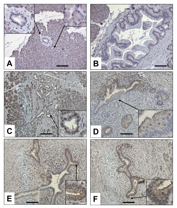
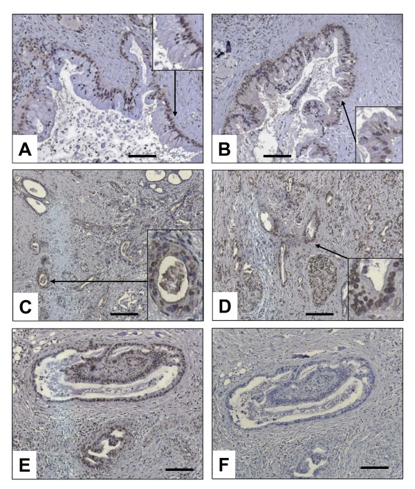
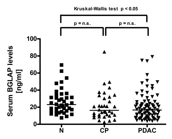
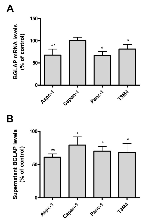
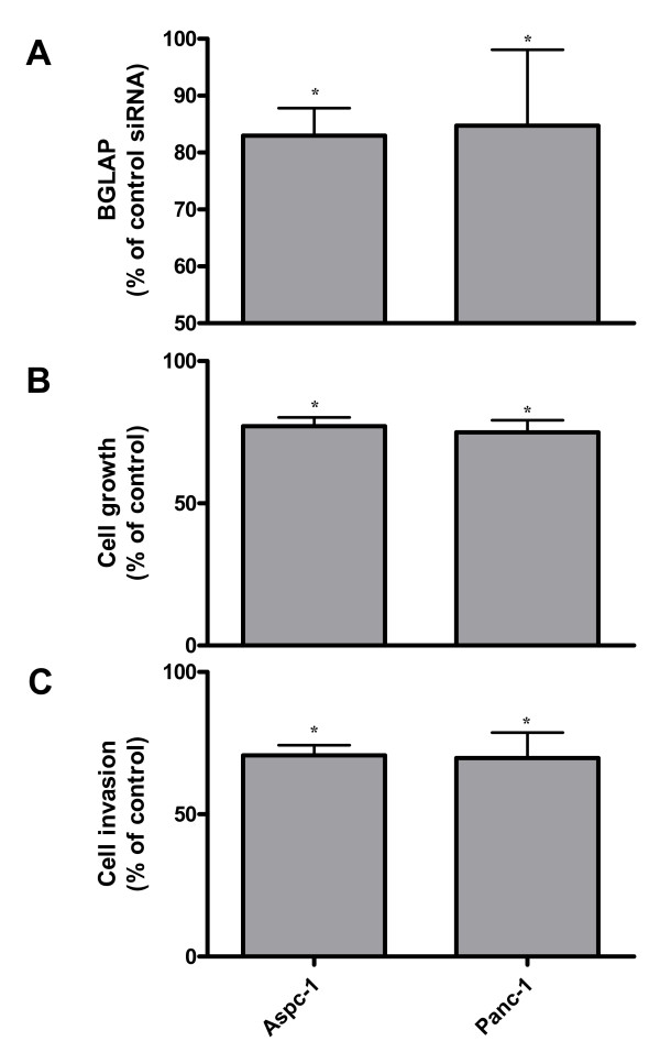
Results: Compared to the normal pancreas, BGLAP mRNA and protein levels were not significantly different in CP and PDAC tissues. BGLAP was faintly present in the cytoplasm of normal acinar cells but was strongly expressed in the cytoplasm and nuclei of tubular complexes and PanIN lesions of CP and PDAC tissues. Furthermore, BGLAP expression was found in the cancer cells in PDAC tissues as well as in 4 cultured pancreatic cancer cell lines. TNFalpha reduced BGLAP mRNA and protein expression levels in pancreatic cancer cell lines. In addition, BGLAP silencing led to reduction of both cell growth and invasion in those cells.

Conclusion: BGLAP is expressed in pancreatic cancer cells, where it potentially increases pancreatic cancer cell growth and invasion through autocrine and/or paracrine mechanisms.


Figures

Figure 1
Figure 1
Expression of BGLAP in pancreatic tissues. Quantitative RT-PCR analysis of mRNA levels for BGLAP in normal pancreatic, CP and PDAC tissue samples was carried out as described in the Methods section before (A) and after (B) normalization to Amy2A. RNA input was normalized to the average expression of the two housekeeping genes HPRT and cyclophilin B, and is presented as adjusted transcripts/10,000 CPB copies.
Figure 2
Figure 2
Localization of BGLAP in normal pancreatic and CP tissues by immunohistochemistry. Immunohistochemistry was performed using a specific BGLAP antibody as described in the Methods section. BGLAP was localized in the cytoplasm of the normal acinar cells but not in the normal ducts (A), and weakly localized in the PanIN1-2 lesions (B). In CP tissues, BGLAP was localized in the cytoplasm of the cells of the tubular complexes (C), PanIN1-2 lesions (D) and large ducts (E & F). Insets show high magnification of structures indicated by arrows. A magnification scale bar of 100 μm is shown.
Figure 3
Figure 3
Localization of BGLAP in PDAC tissues by immunohistochemistry. Immunohistochemistry was performed using a specific BGLAP antibody as described in the Methods section. BGLAP exhibited a cytoplasmic and occasionally nuclear localization high-grade PanIN lesions (A & B) and cancer cells (C-E). The specificity of the staining was performed using control IgG (F). Insets show high magnification of structures indicated by arrows. A magnification scale bar of 100 μm is shown.
Figure 4
Figure 4
Enyzme immunoassay of tissue samples. Enzyme immunoassay of BGLAP levels in normal pancreatic, CP and PDAC tissue samples was carried out as described in the Methods section. Horizontal bars represent median values.
Figure 5
Figure 5
Expression and localization of BGLAP in four cultured pancreatic cancer cell lines. (A) QRT-PCR analysis of mRNA levels for BGLAP in pancreatic cancer cells was carried out as described in the Methods section. RNA input was normalized to the average expression of the two housekeeping genes HPRT and cyclophilin B. Bars represent the mean values +/- SEM of 3 independent experiments as adjusted transcripts/10,000 CPB copies. (B) Enzyme immunoassay of BGLAP levels in cell culture supernatant of pancreatic cancer cell lines was carried out as described in the Methods section. Bars represent the mean values +/- SEM of 3 independent experiments in ng/ml. Localization of BGLAP in the cultured pancreatic cancer cell lines was performed by immunocytochemistry as described in the Methods section, using a specific BGLAP antibody (+). The specificity of the staining was confirmed using control IgG (-).
Figure 6
Figure 6
Effects of TNF-α on BGLAP expression in pancreatic cancer cells. (A) Pancreatic cancer cells were treated with the indicated dose of TNF-α for 48 h. Cell lysates and cell culture supernatants were collected for detection of BGLAP mRNA and protein expression, respectively, as described in the Methods section. Bars represent BGLAP mRNA (A) and protein expression levels (B) as a percentage of untreated cells and as determined by QRT-PCR and enzyme immunoassay, respectively. Data are presented as mean +/- SEM of three independent experiments (*: p < 0.05, **: p < 0.01).
Figure 7
Figure 7
The effects of BGLAP silencing on pancreatic cancer cell growth and invasion. (A) Aspc-1 and Panc-1 pancreatic cancer cells were subjected to BGLAP silencing using two different specific BGLAP siRNA molecules, and the effects of silencing were measured by enzyme immunoassay as described in the Methods section. (B) Cell growth and (C) invasion assays were performed as described in the Methods section. Data are presented as mean +/- SEM compared to the respective controls of three independent experiments (*: p < 0.05).

Similar articles

Cited by

References

    1. Pan LC, Price PA. The propeptide of rat bone gamma-carboxyglutamic acid protein shares homology with other vitamin K-dependent protein precursors. Proc Natl Acad Sci USA. 1985;82:6109–6113. doi: 10.1073/pnas.82.18.6109. - DOI - PMC - PubMed
    1. Price PA, Otsuka AA, Poser JW, Kristaponis J, Raman N. Characterization of a gamma-carboxyglutamic acid-containing protein from bone. Proc Natl Acad Sci USA. 1976;73:1447–1451. doi: 10.1073/pnas.73.5.1447. - DOI - PMC - PubMed
    1. Hauschka PV. Osteocalcin: the vitamin K-dependent Ca2+-binding protein of bone matrix. Haemostasis. 1986;16:258–272. - PubMed
    1. Ducy P, Desbois C, Boyce B, Pinero G, Story B, Dunstan C, Smith E, Bonadio J, Goldstein S, Gundberg C, et al. Increased bone formation in osteocalcin-deficient mice. Nature. 1996;382:448–452. doi: 10.1038/382448a0. - DOI - PubMed
    1. Boskey AL, Gadaleta S, Gundberg C, Doty SB, Ducy P, Karsenty G. Fourier transform infrared microspectroscopic analysis of bones of osteocalcin-deficient mice provides insight into the function of osteocalcin. Bone. 1998;23:187–196. doi: 10.1016/S8756-3282(98)00092-1. - DOI - PubMed

Publication types

MeSH terms