. 2008 Feb;44(2):361-9.
doi: 10.1016/j.yjmcc.2007.10.022. Epub 2007 Nov 12.

High glucose oxidizes SERCA cysteine-674 and prevents inhibition by nitric oxide of smooth muscle cell migration

Affiliations

High glucose oxidizes SERCA cysteine-674 and prevents inhibition by nitric oxide of smooth muscle cell migration

Xiaoyong Tong et al. J Mol Cell Cardiol. 2008 Feb.

Abstract

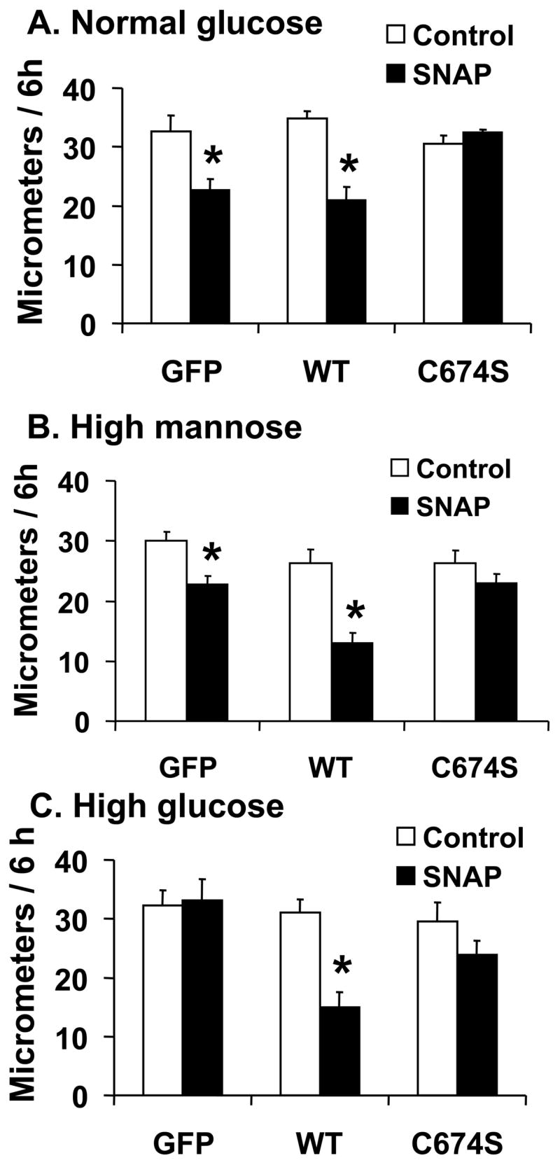
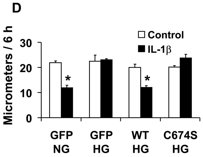
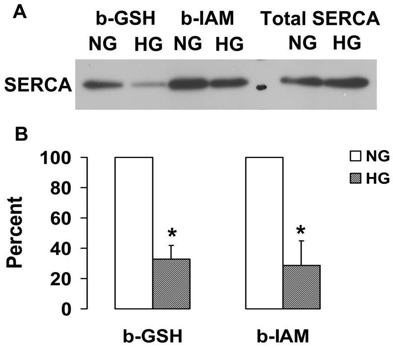
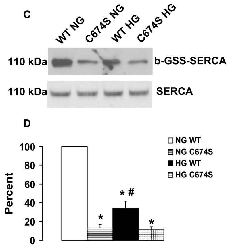
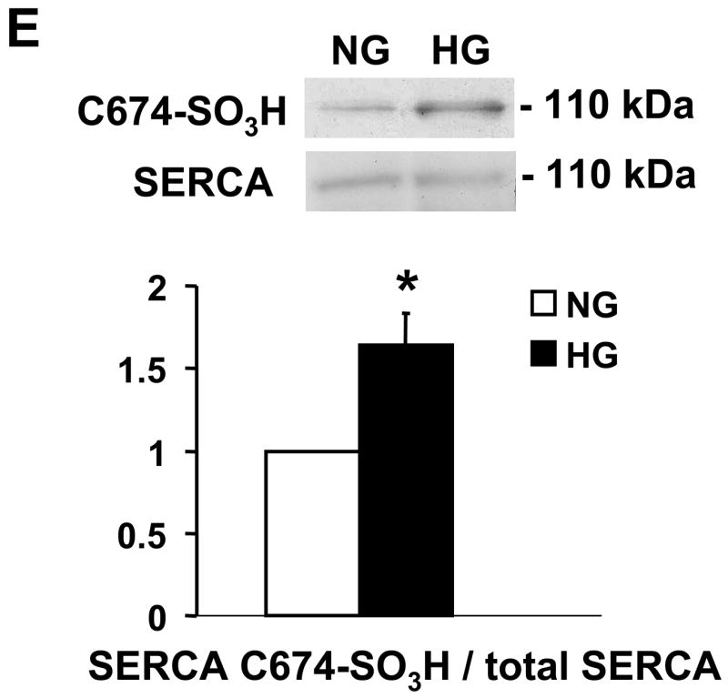
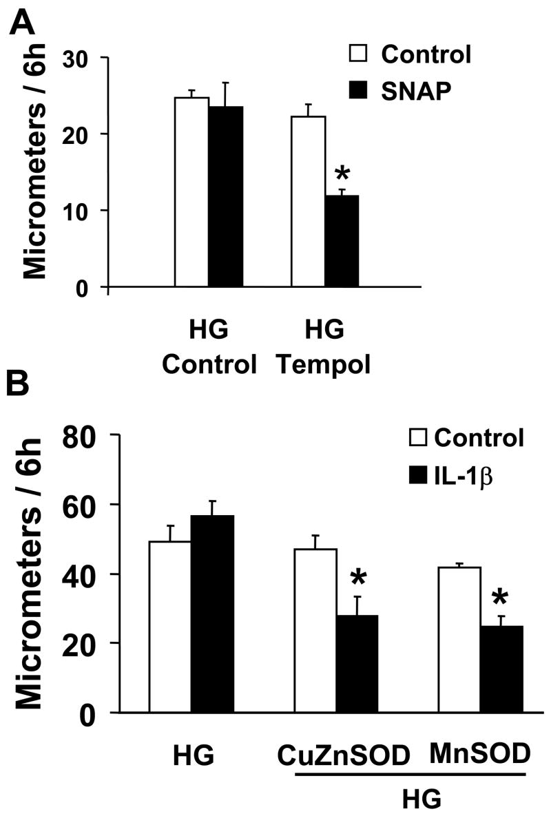
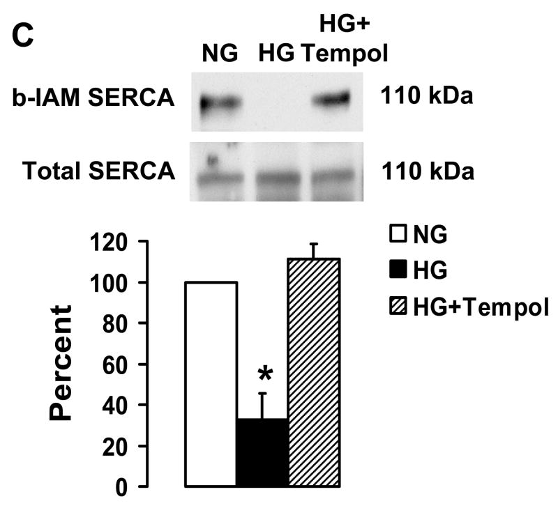
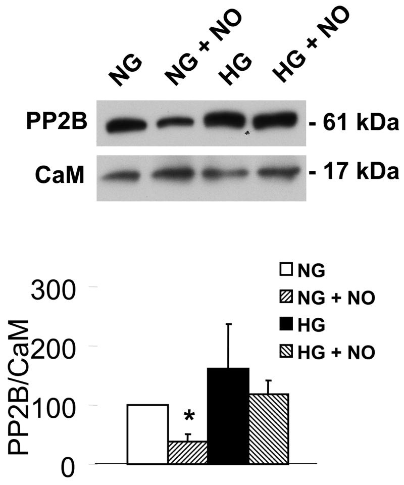
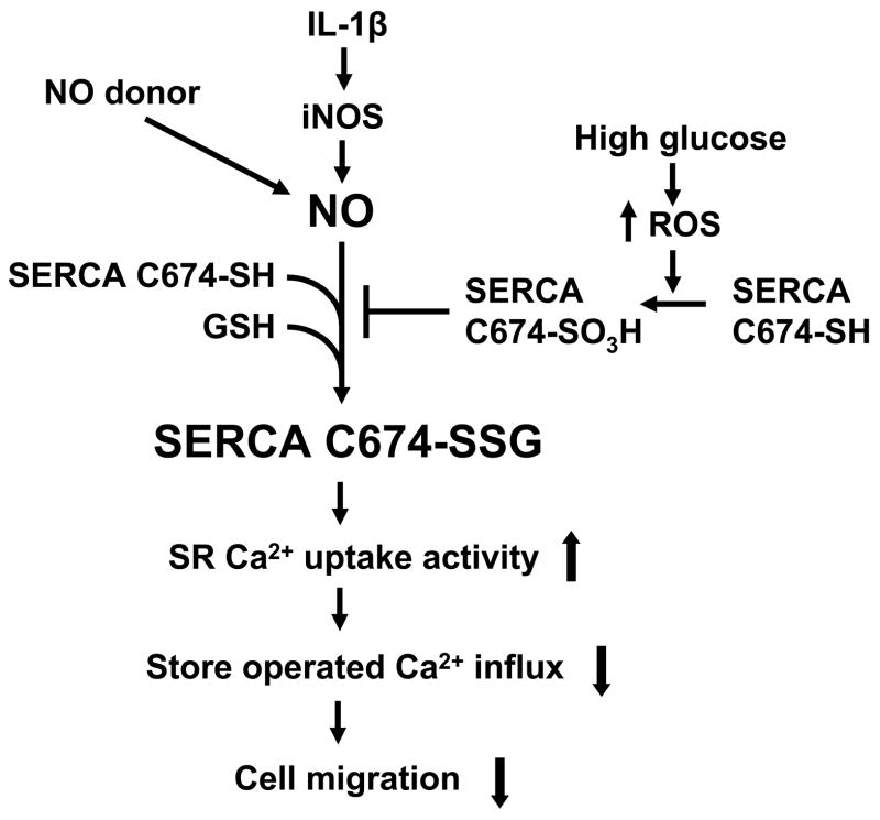
Nitric oxide (NO) causes S-glutathiolation of the reactive cysteine-674 in the sarcoplasmic/endoplasmic reticulum Ca(2+) ATPase (SERCA), thus increasing SERCA activity, and inhibiting Ca(2+) influx and migration of vascular smooth muscle cells (VSMC). Because increased VSMC migration contributes to accelerated neointimal growth and atherosclerosis in diabetes, the effect of culture of VSMC in high glucose (HG) was determined. Rat aortic VSMC were exposed to normal (5.5 mmol/L) or high (25 mmol/L) glucose for 3 days, and serum-induced cell migration during 6 h into a wounded cell monolayer was measured 5 min after adding the NO donor S-nitroso-N-acetylpenicillamine (SNAP) or 24 h after interleukin-1beta (IL-1beta) to express inducible nitric oxide synthase (iNOS). In normal glucose, SNAP or IL-1beta significantly inhibited migration in cells infected with adenovirus to express GFP or SERCA wild type (WT), but not with a C674S SERCA mutant. After HG, NO failed to inhibit migration, nor did it decrease calcium-dependent association of calmodulin with calcineurin, indicating that NO failed to decrease intracellular calcium levels via SERCA. In contrast, overexpression of SERCA WT, but not the SERCA C674S mutant, preserved the ability for NO to inhibit migration despite exposing the cells to HG. The antioxidant, Tempol, or overexpression of superoxide dismutase also prevented the effects of HG. Further studies showed that both biotinylated-iodoacetamide and NO-induced biotinylated glutathione labeling of SERCA C674 were decreased by HG, and a sequence-specific sulfonic acid antibody detected oxidation of the C674 SERCA thiol. These results indicate that failure of NO to inhibit migration in VSMC exposed to HG is due to oxidation of the SERCA reactive cysteine-674.


Figures

Figure 1
Figure 1
The effect of NO on the migration of rat aortic vascular smooth muscle cells (VSMC). A and B: NO donor SNAP significantly inhibited the migration of cells infected with either Ad-GFP or Ad-SERCA WT, but had no effect in cells infected with Ad-SERCA C674S in normal glucose (5.5 mmol/L) or high mannose (19.5 mmol/L plus glucose 5.5 mmol/L). C: In cells exposed to high glucose (25 mmol/L), SNAP did not inhibit migration. Overexpression of SERCA WT, but not SERCA C674S, preserved the ability of SNAP to inhibit migration. The results (A, B and C) are n=6 (mean ± SEM). *P<0.05, paired t-testbetween cells treated or not with SNAP. D: Cells were infected with Ad-WT or Ad-C674S SERCA for 2 d and then switched to medium containing NG or HG for an additional 3 days. Interleukin-1β (IL-1β, 5 ng/mL) was added 24 h before the migration assay to induce iNOS expression which releases NO. Ad-GFP served as a control. The results are n=5 (mean ± SEM). *P<0.05, paired t-test between cells treated or not with IL-1β.
Figure 1
Figure 1
The effect of NO on the migration of rat aortic vascular smooth muscle cells (VSMC). A and B: NO donor SNAP significantly inhibited the migration of cells infected with either Ad-GFP or Ad-SERCA WT, but had no effect in cells infected with Ad-SERCA C674S in normal glucose (5.5 mmol/L) or high mannose (19.5 mmol/L plus glucose 5.5 mmol/L). C: In cells exposed to high glucose (25 mmol/L), SNAP did not inhibit migration. Overexpression of SERCA WT, but not SERCA C674S, preserved the ability of SNAP to inhibit migration. The results (A, B and C) are n=6 (mean ± SEM). *P<0.05, paired t-testbetween cells treated or not with SNAP. D: Cells were infected with Ad-WT or Ad-C674S SERCA for 2 d and then switched to medium containing NG or HG for an additional 3 days. Interleukin-1β (IL-1β, 5 ng/mL) was added 24 h before the migration assay to induce iNOS expression which releases NO. Ad-GFP served as a control. The results are n=5 (mean ± SEM). *P<0.05, paired t-test between cells treated or not with IL-1β.
Figure 2
Figure 2
NO-induced biotinylated glutathione (b-GSH) and biotinylated iodoacetamide (b-IAM) labeling of SERCA and cysteine-674 sulfonic acid oxidation. A: b-GSH and b-IAM labeling of SERCA in uninfected VSMC exposed for 3 days to normal glucose (NG) or high glucose (HG). SERCA was detected with K30/A43 antibody. B: Bar graph shows summary of densitometry performed in three experiments indicating that both b-IAM labeling of SERCA and b-GSH binding to SERCA in uninfected VSMC exposed to HG were significantly less than that in NG. *P<0.05, paired t-test between cells treated with NG and HG. C. NO-induced S-glutathiolation of SERCA in VSMC infected with WT or C674S mutant SERCA exposed to NG or HG. SERCA was detected with IID8 910 antibody. D. Bar graph shows summary of 3 independent experiments indicating that NO-induced S-glutathiolation of SERCA was decreased in cells infected with SERCA WT exposed to HG compared to those exposed to NG (*P<0.05). Despite the fact that S-glutathiolation of SERCA was significantly decreased in cells infected with SERCA WT exposed to HG, the level remained significantly above that in cells infected with SERCA C674S mutant (# P<0.05). E: In membranes prepared from VSMC, a sequence-specific antibody against the SERCA cysteine-674 sulfonic acid shows increased detection in cells exposed to HG compared to cells exposed to NG. Total SERCA expression was not different. The bar graph in the lower panel summarizes 3 independent experiments showing that high glucose significantly increases the amount of the irreversible oxidation of SERCA expressed as the ratio of the densitometric values for detection with the SERCA C674-SO3H antibody with the total SERCA detected by the K30 antibody. *P<0.05, paired t-test.
Figure 2
Figure 2
NO-induced biotinylated glutathione (b-GSH) and biotinylated iodoacetamide (b-IAM) labeling of SERCA and cysteine-674 sulfonic acid oxidation. A: b-GSH and b-IAM labeling of SERCA in uninfected VSMC exposed for 3 days to normal glucose (NG) or high glucose (HG). SERCA was detected with K30/A43 antibody. B: Bar graph shows summary of densitometry performed in three experiments indicating that both b-IAM labeling of SERCA and b-GSH binding to SERCA in uninfected VSMC exposed to HG were significantly less than that in NG. *P<0.05, paired t-test between cells treated with NG and HG. C. NO-induced S-glutathiolation of SERCA in VSMC infected with WT or C674S mutant SERCA exposed to NG or HG. SERCA was detected with IID8 910 antibody. D. Bar graph shows summary of 3 independent experiments indicating that NO-induced S-glutathiolation of SERCA was decreased in cells infected with SERCA WT exposed to HG compared to those exposed to NG (*P<0.05). Despite the fact that S-glutathiolation of SERCA was significantly decreased in cells infected with SERCA WT exposed to HG, the level remained significantly above that in cells infected with SERCA C674S mutant (# P<0.05). E: In membranes prepared from VSMC, a sequence-specific antibody against the SERCA cysteine-674 sulfonic acid shows increased detection in cells exposed to HG compared to cells exposed to NG. Total SERCA expression was not different. The bar graph in the lower panel summarizes 3 independent experiments showing that high glucose significantly increases the amount of the irreversible oxidation of SERCA expressed as the ratio of the densitometric values for detection with the SERCA C674-SO3H antibody with the total SERCA detected by the K30 antibody. *P<0.05, paired t-test.
Figure 3
Figure 3
Tempol and overexpression of Cu/Zn SOD or MnSOD preserve NO-induced inhibition of migration in VSMC exposed to HG. A: Summary of cell migration in uninfected VSMC exposed to high glucose showing that treatment with Tempol has no effect on migration of cells in the absence of SNAP, but protects the ability of SNAP to inhibit migration of cells exposed to HG. N=3, *P<0.05, two-way ANOVA. B: Infection of cells with adenoviral vectors to overexpress Cu/Zn SOD and MnSOD preserved the ability of IL-1β induced iNOS to inhibit migration in VSMC exposed to HG. N=3, *P<0.05, two-way ANOVA. C: Tempol prevents the decrease in b-IAM labeling of SERCA in uninfected VSMC exposed to high glucose. The bar graph summarizes densitometry data from 3 independent experiments. *P<0.05, one-way ANOVA.
Figure 3
Figure 3
Tempol and overexpression of Cu/Zn SOD or MnSOD preserve NO-induced inhibition of migration in VSMC exposed to HG. A: Summary of cell migration in uninfected VSMC exposed to high glucose showing that treatment with Tempol has no effect on migration of cells in the absence of SNAP, but protects the ability of SNAP to inhibit migration of cells exposed to HG. N=3, *P<0.05, two-way ANOVA. B: Infection of cells with adenoviral vectors to overexpress Cu/Zn SOD and MnSOD preserved the ability of IL-1β induced iNOS to inhibit migration in VSMC exposed to HG. N=3, *P<0.05, two-way ANOVA. C: Tempol prevents the decrease in b-IAM labeling of SERCA in uninfected VSMC exposed to high glucose. The bar graph summarizes densitometry data from 3 independent experiments. *P<0.05, one-way ANOVA.
Figure 4
Figure 4
High glucose prevents the ability of NO to decrease calcium-dependent association of calmodulin (CaM) with PP2B. The upper panel shows that immuno-precipitated CaM is associated with less PP2B in VSMC treated with DETA-NONOate when the cells were exposed to NG, but not HG for 3 d. Immuno-blot showed that similar amounts of CaM were contained in the immuno-precipitate. The bar graph in the lower panel summarizes the ratio of the densitometric values detected by the PP2B antibody with that by the CaM antibody in 3 independent experiments. These data show that NO decreases the association of CaM-PP2B in NG, but not in HG. *P<0.05, two-way ANOVA.
Figure 5
Figure 5
The proposed mechanism of the regulation of NO of cell migration through redox regulation of SERCA2b in normal and high glucose. In normal glucose, NO released from exogenous NO donor or from iNOS induced by IL-1β induces the reversible S-glutathiolation of SERCA, predominantly on the most reactive thiol on cysteine 674, which increases SERCA activity and intracellular calcium uptake into SR/ER, decreases calcium influx, and inhibits cell migration. High glucose interrupts this cascade by the oxidation of the reactive thiol on SERCA cysteine 674, blocks the NO-induced S-glutathiolation of SERCA, and abolishes the inhibitory effect of NO on cell migration.

Similar articles

Cited by

References

    1. Yasunari K, Kohno M, Kano H, Yokokawa K, Minami M, Yoshikawa J. Mechanisms of action of troglitazone in the prevention of high glucose-induced migration and proliferation of cultured coronary smooth muscle cells. Circ Res. 1997;81:953–62. - PubMed
    1. Yasunari K, Kohno M, Kano H, Yokokawa K, Minami M, Yoshikawa J. Antioxidants improve impaired insulin-mediated glucose uptake and prevent migration and proliferation of cultured rabbit coronary smooth muscle cells induced by high glucose. Circ. 1999;99(10):1370–8. - PubMed
    1. Scherberich A, Campos-Toimil M, Ronde P, Takeda K, Beretz A. Migration of human vascular smooth muscle cells involves serum-dependent repeated cytosolic calcium transients. J Cell Sci. 2000;113:653–62. - PubMed
    1. Cohen RA, Weisbrod RM, Gericke M, Yaghoubi M, Bierl C, Bolotina VM. Mechanism of nitric oxide-induced vasodilatation. Refilling of intracellular stores by sarcoplasmic reticulum Ca2+ ATPase and inhibition of store-operated Ca2+ influx. Circ Res. 1999;84:210–9. - PubMed
    1. Touyz RM. Reactive oxygen species as mediators of calcium signaling by angiotensin II: implications in vascular physiology and pathophysiology. Antioxid Redox Signal. 2005;7(9–10):1302–14. - PubMed

Publication types

MeSH terms